Utilization of mural thrombus for local drug delivery into vascular tissue

a vascular tissue and mural thrombus technology, applied in the direction of drug compositions, extracellular fluid disorders, catheters, etc., can solve the problems of endovascular approach, aortic rupture and potential patient death, collagen degradation, etc., to facilitate the reduction of enzymatic degradation of proteins, promote cross-linking proteins, and stabilize the extracellular matrix

Inactive Publication Date: 2010-10-14
ENDOLOGIX LLC
View PDF105 Cites 33 Cited by
  • Summary
  • Abstract
  • Description
  • Claims
  • Application Information

AI Technical Summary

Benefits of technology

[0012]Some embodiments of the present disclosure are directed to a method for stabilizing an extracellular matrix in a wall of a blood vessel comprising advancing a delivery system to a treatment site positioned near a mural thrombus that covers at least a portion of the wall of the blood vessel. A delivery portion of the delivery device is advanced into the mural thrombus. A therapeutic agent is delivered through the delivery portion into the mural thrombus. The agent can transport from the mural thrombus into the extracellular matrix of the vessel wall by diffusion to facilitate reduction of enzymatic degradation of protein in the extracellular matrix by the action of the agent.
[0013]Additionally, some embodiments of the present disclosure are directed to a method for stabilizing an extracellular matrix layer in the vascular system of a body, comprising positioning a portion of a vascular catheter adjacent to or within a mural thrombus positioned adjacent to the extracellular matrix layer of a target region of the vascular system. A therapeutic agent is delivered in solution to the mural thrombus using the vascular catheter. The therapeutic agent can be transported to the extracellular matrix layer through the mural thrombus to promote the cross-linking protein in the extracellular matrix layer, thereby stabilizing the extracellular matrix.

Problems solved by technology

Degradation of the collagen structure can ultimately lead to aortic rupture and potential patient death.
However, the endovascular approach has two major challenges.
This approach may be reasonable in applications in the abdominal aorta but not suitable for application in the thoracic aortic due to the high flow rates and blood pressure acting on the balloon.
A second challenge is the presence of mural thrombus on the luminal surface of the diseased vessel.

Method used


Image

  • Utilization of mural thrombus for local drug delivery into vascular tissue
  • Utilization of mural thrombus for local drug delivery into vascular tissue
  • Utilization of mural thrombus for local drug delivery into vascular tissue

Examples


experiment 1

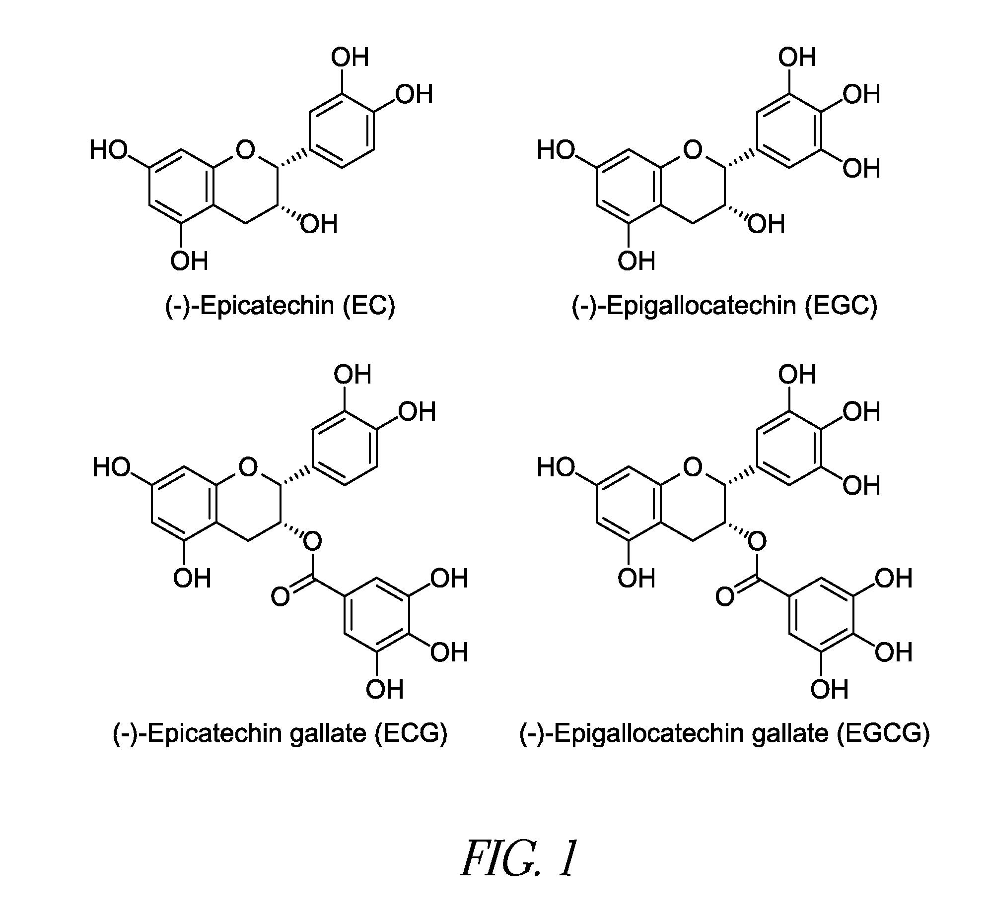
[0059]Model Development and Feasibility Testing: The initial experiment consisted of depositing a thrombus layer over a pericardium sample, injecting an aqueous solution of PPE into the thrombus at multiple sites and allowing the PPE time to react with the pericardium. The samples were examined for cross-linking of the collagen and changes to the thrombus associated with the PPE injection. All pericardium samples exhibited increased shrinkage temperature values indicative of increased collagen stabilization.

Shrinkage Temperature (° C.)123456AveSTDMAXMinRun 174.676.575.773.270.974.22.2176.570.9Run 273.472.870.770.870.170.171.31.4273.470.1Run 372.071.572.872.074.975.673.11.7175.671.5Run 475.177.276.876.473.873.875.51.5177.273.8Native69.367.768.669.168.568.968.70.5769.367.7

experiment 2

[0060]Solvent Delivery System: The second experiment used the thrombus pericardium system developed in the initial experiment. The aqueous PPE solution was replaced with an ethanol:water PPE solution. Controls for interaction of the pericardium with the ethanol water solution and thrombus were performed as part of this experiment. The 40:60 ethanol:water solution allowed for a greater PPE concentration (20% compared to 15% in water) but the increase in shrinkage temperatures observed were lower for the ethanol:water PPE solution than those found for PPE in water. The controls indicated no interactions between either the ethanol:water solution or the thrombus and the pericardium with respect to changes in the collagen shrinkage temperature.

Shrinkage Temperature (° C.)Treatment123456AveSTDMAXMinRun 1EtOH68.267.666.767.367.567.667.50.4968.266.7Run 2PPE70.071.071.470.070.970.470.60.5771.470.0Run 3PPE72.472.473.072.773.973.072.90.5673.972.4Run 4Blood68.268.168.267.667.868.168.00.2468.267...

experiment 4

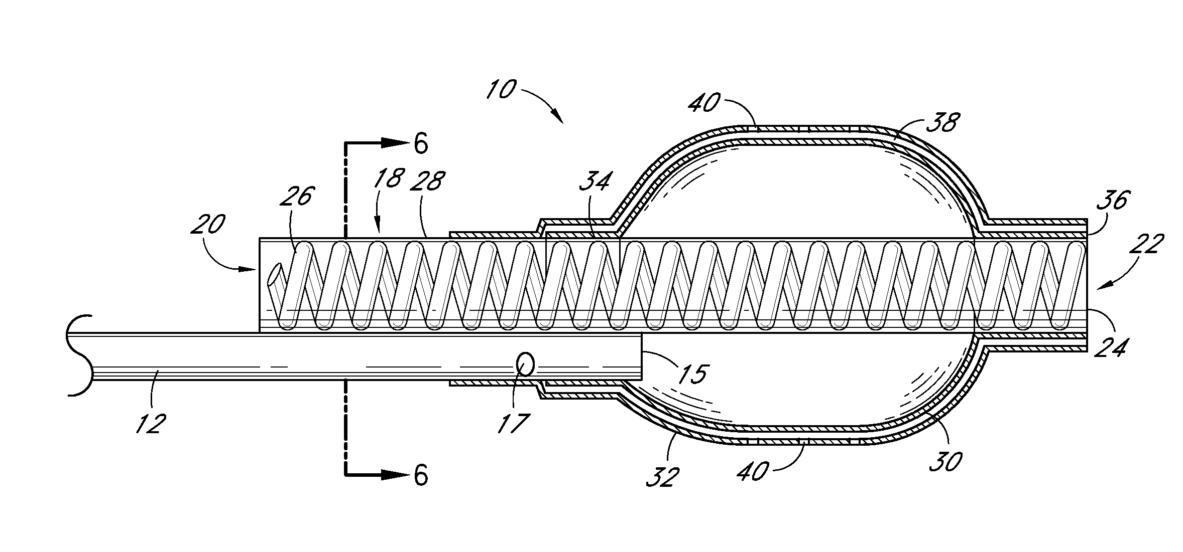
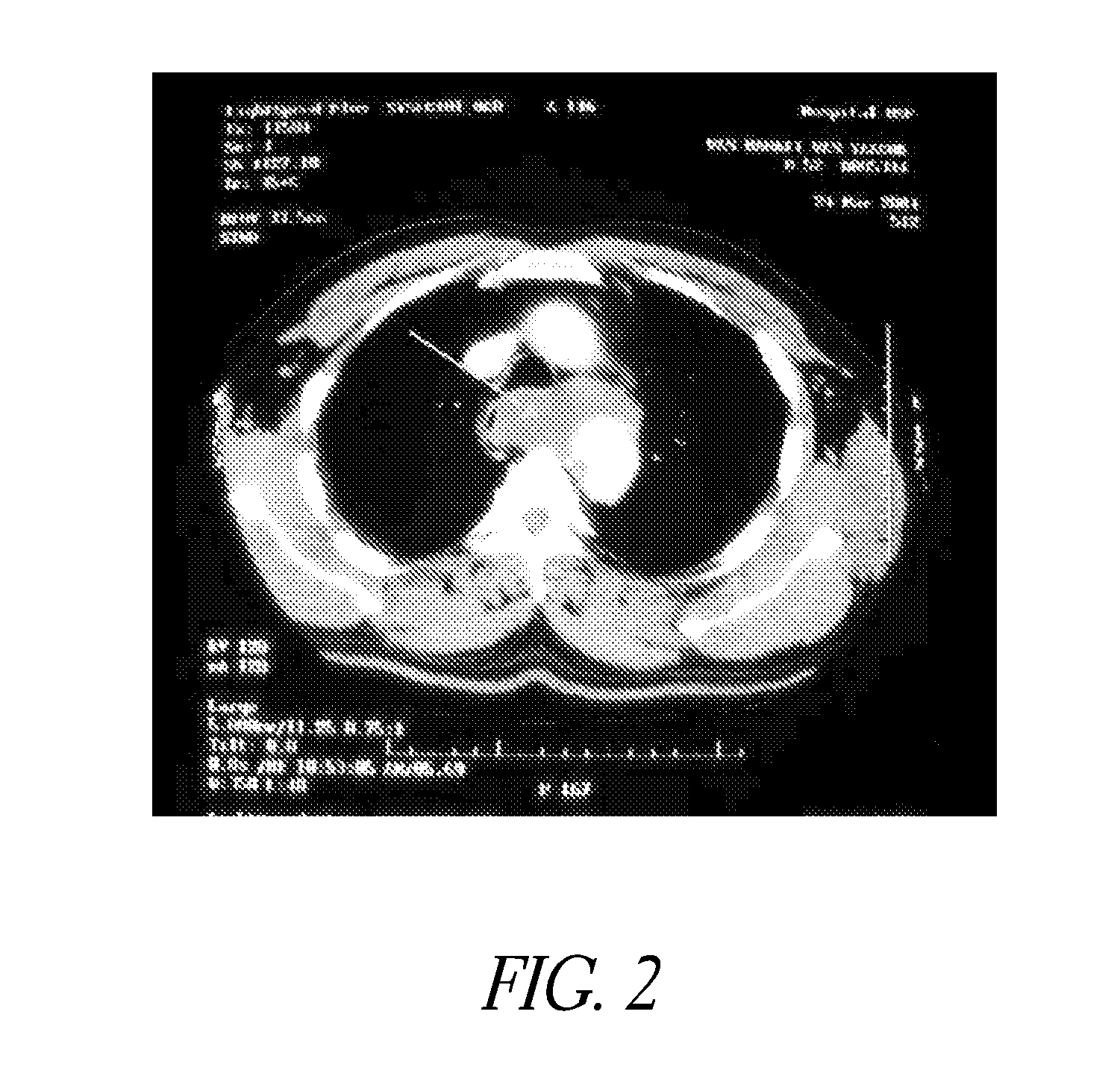
[0062]Evaluation of Delivery and Imaging Systems: The fourth experiment utilized samples similar to those used in the first three experiments. A delivery system was created consisting of a narrow gauge tube (5 Fr hollow catheter) with multiple holes with the end of the lumen plugged. Sufficient intact tubing was included to allow insertion of the irrigation portion of the catheter through the thrombus and into the interface between the thrombus and pericardium. A contrast media was used to image the injection of PPE in real time. A 19.4% PPE solution was used and diluted 50% with the contrast media. An open ended non-irrigation catheter and injections without contrast agent were also tested as controls. The contrast media had a positive impact on the collagen shrinkage temperature results. Samples treated with the PPE—contrast media solution had higher and more uniform increases in collagen shrinkage temperature than did the same catheters without the contrast media.

PPEShrinkage Tem...


PUM

PropertyMeasurementUnit
pHaaaaaaaaaa
pHaaaaaaaaaa
thickaaaaaaaaaa

Abstract

The present disclosure is directed to methods and systems for stabilizing an extracellular matrix in a wall of a blood vessel. The method comprises delivering a therapeutic agent into mural thrombus, which covers the wall of the blood vessel. The agent is transported from the mural thrombus into the extracellular matrix of the vessel wall by diffusion. The agent then acts to reduce the enzymatic degradation of protein in the extracellular matrix.

Description

CROSS-REFERENCE TO RELATED APPLICATIONS[0001]This application claims the benefit of U.S. Provisional Patent Application No. 61 / 168,199, filed on Apr. 9, 2009, the entire content of which is hereby incorporated by reference and should be considered part of this specification.[0002]This application is related to U.S. Provisional Patent Application No. 60 / 987,261, filed Nov. 12, 2007, U.S. Provisional Patent Application No. 61 / 012,356, filed Dec. 7, 2007, U.S. Provisional Patent Application No. 61 / 127,654, filed May 14, 2008, U.S. Provisional Patent Application No. 60 / 987,268, filed Nov. 12, 2007, U.S. Provisional Patent Application No. 61 / 012,579, filed Dec. 10, 2007, U.S. Provisional Patent Application No. 60 / 533,443, filed on Dec. 31, 2003, and U.S. patent application Ser. No. 12 / 269,677, filed Nov. 12, 2008, which are each hereby incorporated by reference in their entireties as if fully set forth herein.BACKGROUND OF THE DISCLOSURE[0003]1. Field of the Disclosure[0004]The present d...

Claims


Application Information

Patent Timeline
Patent Type & Authority Applications(United States)
IPC IPC(8): A61K31/7032A61M1/00A61K9/00A61K31/353A61P9/10A61P29/00A61P7/02
CPCA61K9/0024A61K31/353A61L2300/434A61L29/16A61L31/16A61K31/7032A61P7/02A61P9/10A61P29/00
Inventor SCHRECK, STEFAN G.BANKERT, CHARLES S.SCHANKERELI, KEMAL
Owner ENDOLOGIX LLC
Why Eureka
Try Eureka