Filovirus vectors and noninfectious filovirus-based particles
a technology of filovirus and filovirus, which is applied in the direction of viruses/bacteriophages, antibody medical ingredients, peptide sources, etc., can solve the problems of hampered identification of major determinants of ebola virus pathogenicity, reduce particle formation, enhance the hydrophobic association of proteins, and reduce the association with cellular membranes
- Summary
- Abstract
- Description
- Claims
- Application Information
AI Technical Summary
Benefits of technology
Problems solved by technology
Method used
Image
Examples
example 1
Generation of Transfectant Ebola Virus
Materials and Methods
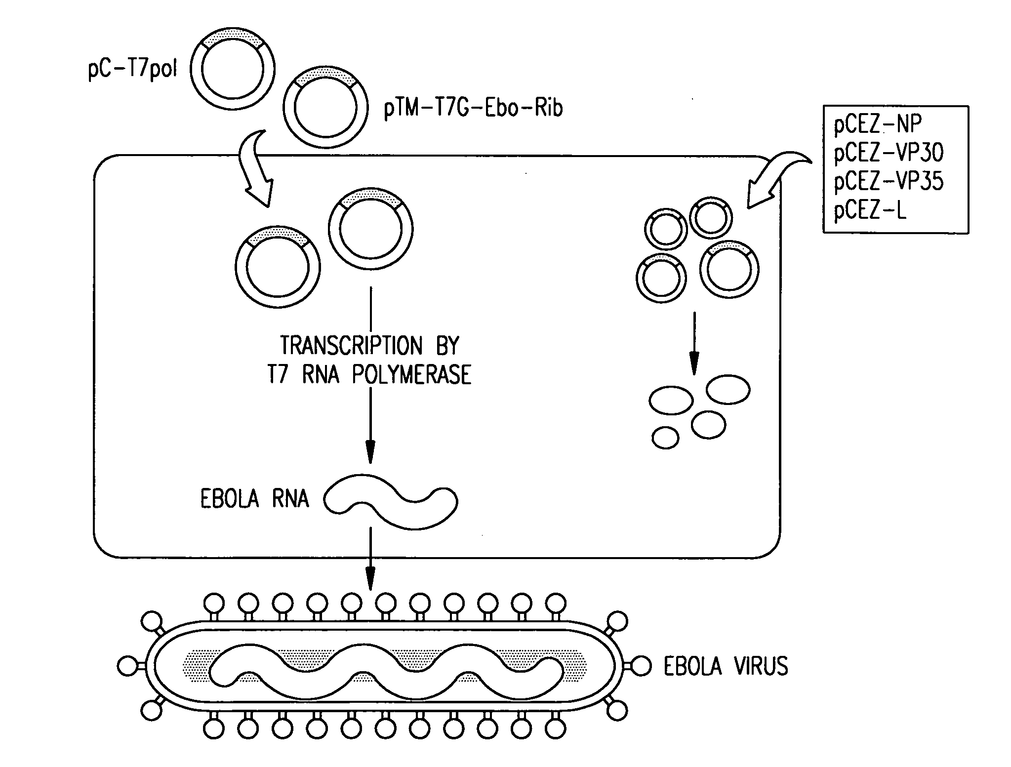
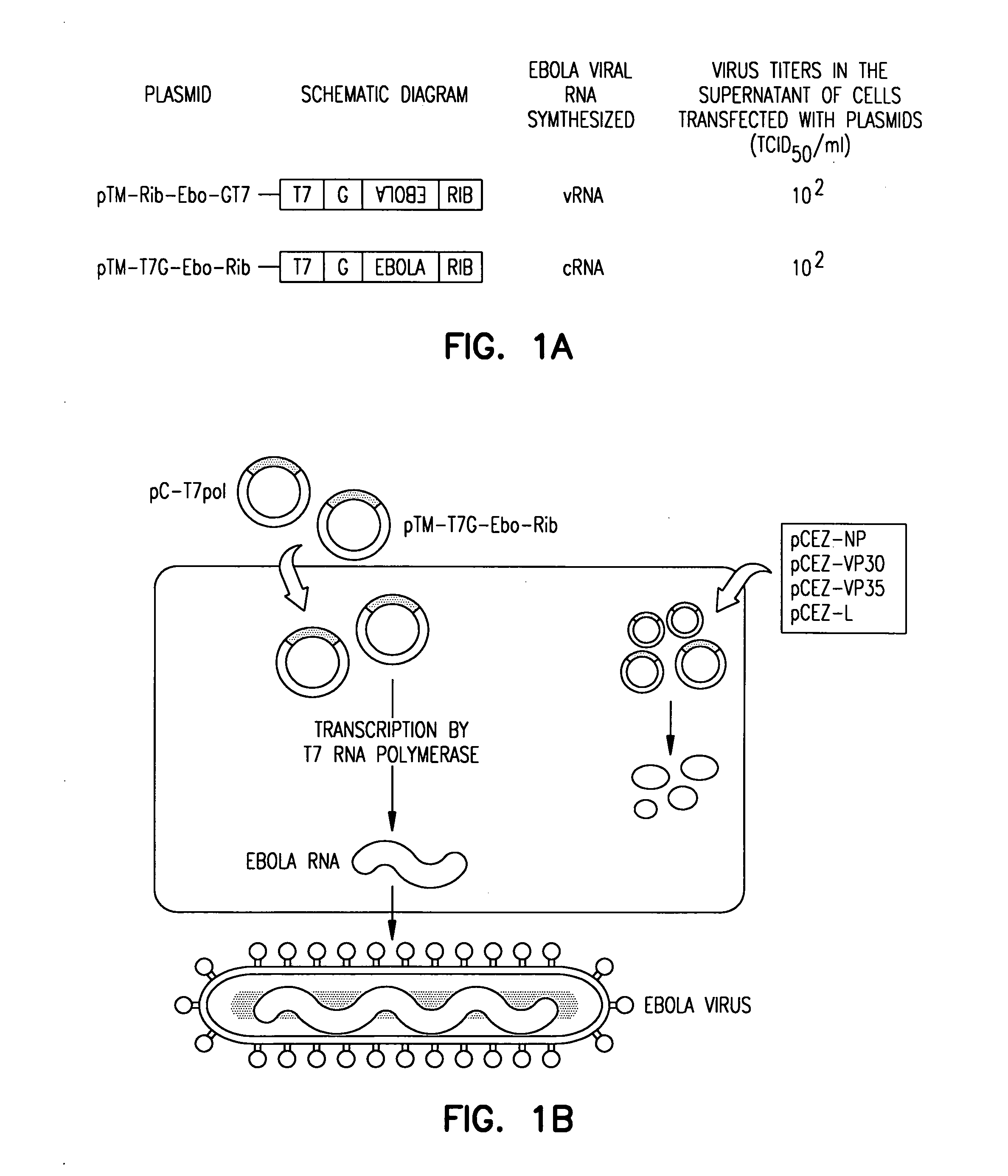
[0042]Efficiency of Virus Generation. To determine the efficiency of virus generation, Vero E6 cells were cotransfected with protein expression plasmids and the plasmid for Ebola virus vRNA or cRNA synthesis. Four days after transfection, the efficiency of virus generation was measured by determining the dose required to infect 50% of tissue culture cells (TCID50) per ml of supernatant. The data shown in FIG. 1A are representative results from three independent experiments. Experiments for the generation of Ebola virus as well as the characterization of recombinant Ebola virus were carried out in the BSL4 facility at the Canadian Science Centre for Human and Animal Health, Winnipeg, Canada. Cells were transfected with plasmids for the expression of the Ebola virus NP, L, VP30, and VP35 proteins, and with the plasmid for Ebola virus cRNA or vRNA synthesis, controlled by T7 RNA polymerase promoter and ribozyme sequences. T7 RN...
example 2
Generation of Noninfectious Ebola Particles
Materials and Methods
[0059]Cells. 293 and 293T human embryonic kidney cells were maintained in DMEM supplemented with 10% fetal calf serum, 2% L-glutamine, and penicillin-streptomycin solution (DMEM-FCS) (Sigma). The cells were grown at 37° C. in 5% CO2.
[0060]Construction of Plasmids. To generate cDNA constructs encoding the VP40 protein, primers were used that bind to the start and stop codons (positions 4479 and 5459 of the positive-sense antigenomic RNA) to reverse transcribe and PCR-amplify purified viral RNA (Titan RT-PCR Kit, Roche). The PCR product was cloned in the pT7Blue vector (Novagen) resulting in pT7EboZVP40. The cloned Ebola VP40 gene was sequenced to ensure that unwanted nucleotide replacements were not present.
[0061]To generate plasmid pETEBoZVP40His for the expression of 6-histidine-tagged VP40 in Escherichia coli, pT7EboZVP40 was used as a template for PCR amplification with the appropriate primers. The PCR product was bl...
example 3
Particles Comprising Filovirus Matrix Protein and Glycoprotein
Materials and Methods
[0090]Cells. 293T human embryonic kidney cells were maintained in Dulbecco's modified Eagle medium supplemented with 10% fetal calf serum, L-glutamine and penicillin-streptomycin-gentamicin solution. The cells were grown in an incubator at 37° C. in 5% CO2.
[0091]Plasmids. Full-length cDNAs encoding the Ebola virus (species Zaire) VP40 or GP were cloned separately into a mammalian expression vector, pCAGGS / MCS (Kobasa et al., 1997; Niwa et al., 1991), which contains the chicken β-actin promoter. The resultant constructs were designated pCEboZVP40 and pCEboZGP, respectively.
[0092]Cell Transfection for Expression of VP40 and GP. 293T cells (1×10⁶) were transfected with plasmids using the Trans IT LT-1 reagent (Panvera, Madison, Wis.) according to the manufacturer's instructions. Briefly, 1 μg of DNA in 0.1 ml Opti-MEM (Gibco-BRL) and 3 μl of the transfection reagent were mixed, incubated for 10 minutes a...
PUM
 Abstract
Description
Claims
Application Information
 - R&D
- Intellectual Property
- Life Sciences
- Materials
- Tech Scout
- Unparalleled Data Quality
- Higher Quality Content
- 60% Fewer Hallucinations
Browse by: Latest US Patents, China's latest patents, Technical Efficacy Thesaurus, Application Domain, Technology Topic, Popular Technical Reports.
© 2025 PatSnap. All rights reserved.Legal|Privacy policy|Modern Slavery Act Transparency Statement|Sitemap|About US| Contact US: help@patsnap.com


