Eureka AIR delivers breakthrough ideas for toughest innovation challenges, trusted by R&D personnel around the world.

Novel method for specimen preparation, which ensures preservation of tissue morphology and nucleic acid quality

Inactive Publication Date: 2010-10-21
CHUGAI PHARMA CO LTD +1
View PDF1 Cites 10 Cited by
  • Summary
  • Abstract
  • Description
  • Claims
  • Application Information

AI Technical Summary

Benefits of technology

[0021]The PFA-AMeX-Paraffin method, in which fixation with PFA is combined with embedding in paraffin by the AMeX method, is a method for specimen preparation which ensures the maintenance of both tissue morphology and nucleic acid quality. In the case of tissues (e.g., prostate cancer tissue) whose cells are difficult to sufficiently distinguish in frozen tissue sections, this method is found to be very useful in collecting target cells by microdissection under observation of tissue morphology, and further in analyzing gene expression in these cells by using a DNA microarray.
[0022]

Problems solved by technology

However, in frozen sections, tissue morphology cannot be maintained satisfactorily, which often makes it difficult to sufficiently distinguish cells from one another, depending on the type of tissue to be used or cell to be targeted.
For example, in the case of prostate cancer tissues containing normal region, hyperplastic region, precancerous region (PIN; prostatic intraepithelial neoplasia) and cancer region in combination, it has been very difficult to distinguish and isolate only cancer cells.
On the other hand, formalin-fixed paraffin sections conventionally prepared ensure good maintenance of tissue morphology, but suffer from serious degradation and degeneration of nucleic acids during fixation and processing, which has made it impossible to carry out an accurate gene expression analysis with nucleic acids (RNAs) of good quality.

Method used


Image

  • Novel method for specimen preparation, which ensures preservation of tissue morphology and nucleic acid quality
  • Novel method for specimen preparation, which ensures preservation of tissue morphology and nucleic acid quality
  • Novel method for specimen preparation, which ensures preservation of tissue morphology and nucleic acid quality

Examples


example 1

Preparation of Specimens

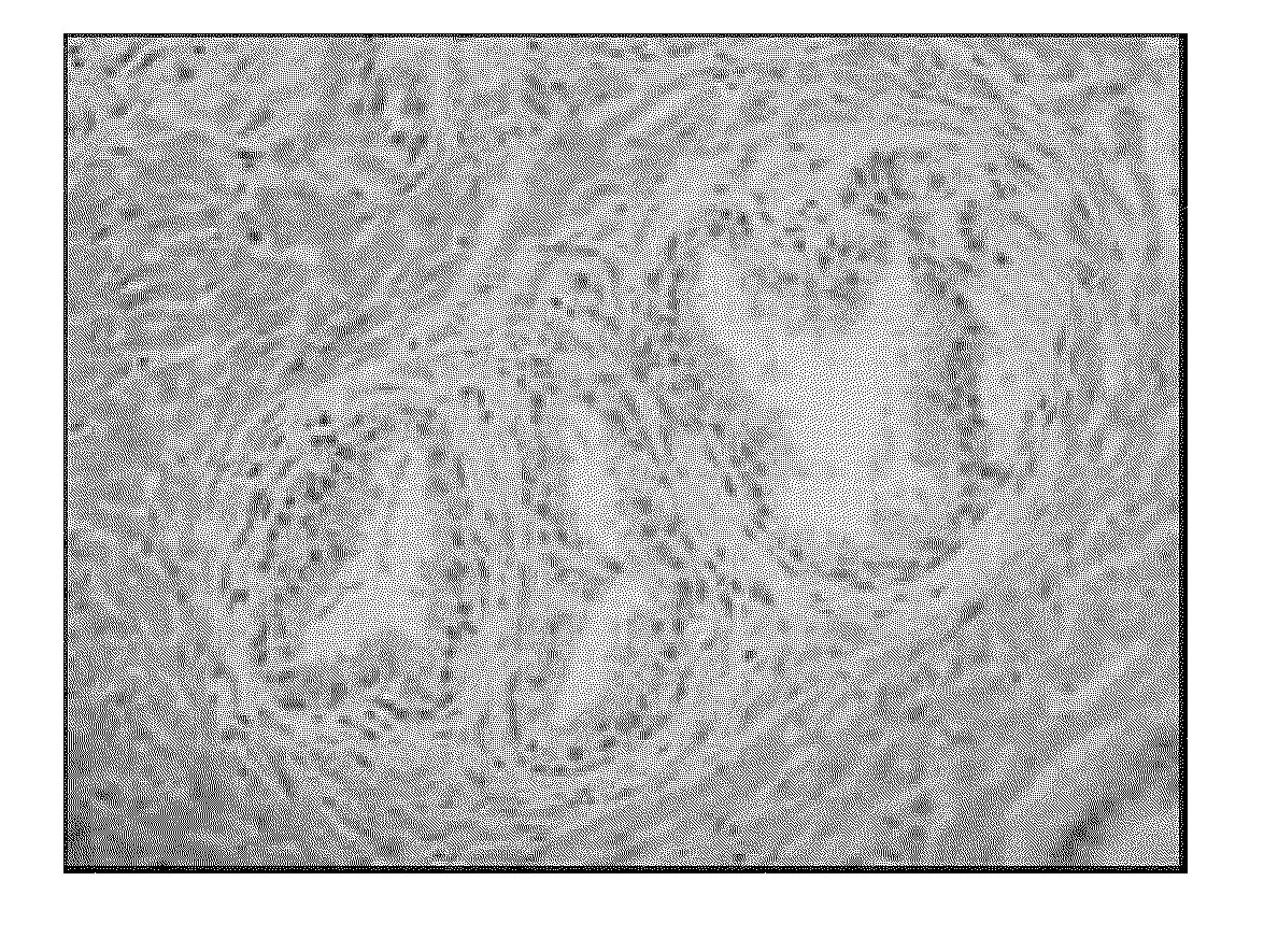
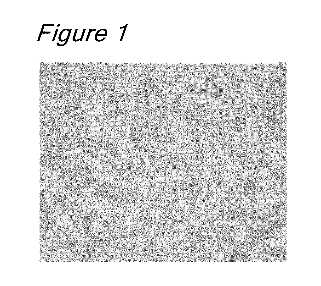
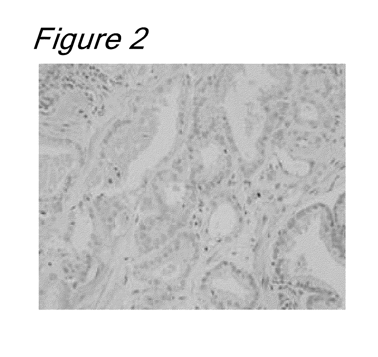
(1) Preparation of Specimens by PLP-Amex-Paraffin Method or PFA-AMeX-Paraffin Method and Observation of Tissue Morphology

[0045]The tissues used were obtained from patients with prostate cancer or benign prostatic hyperplasia who consented to use of their excised tissues for research purposes. The obtained samples were each trimmed to about 5 mm3 size and fixed in PLP fixative (0.01 M periodate, 0.075 M lysine, 0.0375 M phosphate buffer, 4% paraformaldehyde) or in 4% PFA fixative (4% paraformaldehyde / 0.01 M PBS (pH 7.4)) at 4° C. for 16 to 24 hours. The fixed samples were washed with a 0.01 M PBS solution (at 4° C. for 2 hours) and then dehydrated with acetone (at 4° C. overnight and at room temperature for 30 minutes×4), followed by treatment with methyl benzoate (at room temperature for 30 minutes×2) and clearing with xylene (at room temperature for 30 minutes×2). Then, the samples were immersed in a paraffin solution at 60° C. (for 40 minutes×3), embedded i...

example 2

Observation of Tissue Morphology

[0050]For preparation and staining of thin sections from the paraffin blocks prepared by the PFA-AMeX-paraffin method, commercially available standard instruments and reagents were used. More specifically, paraffin sections of 4 μm thickness were prepared in a standard manner from the paraffin blocks, and each section was mounted on a foiled frame for an AS-LMD system (Leica Microsystems) and then dried. After deparaffinization with xylene (at room temperature for 15 seconds×2) and rehydration with acetone (at room temperature for 15 seconds×2), the sections were washed with RNase-free water and immersed in Mayer's hematoxylin solution (at room temperature for 10 seconds). After washing again with RNase-free water, the sections were immersed in an eosin solution (at room temperature for 10 seconds) and then dehydrated with ethanol (100% ethanol, at room temperature for 10 seconds). The sections stained with hematoxylin-eosin (HE) were dried and then o...

example 3

Extraction and Quantification of Total RNA from Specimens Prepared by Four Types of Specimen Preparation Methods, as Well as Comparison of mRNA Quality

[0053]Starting from the same tumor tissue of cultured human prostate cancer cells (LNCaP cells) transplanted into NOG mice, specimens were prepared according to the above four specimen preparation methods (fresh-frozen method, PFA-AMeX-Paraffin method, PFA-Paraffin method, NBF-Paraffin method). Total RNAs were extracted from thin sections of these specimens, followed by comparison of mRNA quality.

(1) Extraction of Total RNA from Thin Sections

[0054]Thin paraffin sections of the specimens prepared by the PFA-AMex-Paraffin method, the PFA-Paraffin method and the NBF-Paraffin method were respectively transferred to separate tubes (0.2 mL). After deparaffinization (xylene, at room temperature for 15 seconds×3) and rehydration (100% ethanol, at room temperature for 15 seconds), the thin sections were dried. Then, 205 μL lysis buffer (20 mM ...


PUM

PropertyMeasurementUnit
Gene expression profileaaaaaaaaaa
Morphologyaaaaaaaaaa

Abstract

The present invention aims to develop a method for specimen preparation, which ensures the maintenance of both tissue morphology and nucleic acid quality (particularly RNA quality). The present invention further aims to prepare a specimen by this method, from which desired cells are then collected by microdissection and analyzed for gene expression. A method for specimen preparation from various frozen or unfrozen organs or tissues (excluding hard tissues) of the whole body, which comprises the following steps: 1) fixing a target organ or tissue with PFA fixative; and 2) embedding the same in paraffin by the AMeX method.

Description

TECHNICAL FIELD[0001]The present invention relates to a method for tissue specimen preparation, which ensures the maintenance of tissue morphology and nucleic acid quality. More specifically, the present invention relates to a method for tissue specimen preparation, which is based on a combination of fixation with PFA fixative and embedding in paraffin by the AMeX method. The present invention also relates to a tissue specimen obtainable by the above method, a method for testing the quality of mRNA in cells within such a tissue specimen, and a method for preparing a sample suitable for DNA microarray gene expression analysis by using such a tissue specimen.BACKGROUND ART[0002]When clinical samples or laboratory animal samples are used in gene expression analysis by Gene Chip (DNA microarray) technology, nucleic acids have usually been extracted from tissue grafts of a certain size and used for analysis. However, tissue grafts contain various cells other than target cells, and hence ...

Claims


Application Information

Patent Timeline
IPC IPC(8): C40B30/00G01N1/36G01N1/30C12N5/07C12Q1/68C40B50/00
CPCC12Q1/6806G01N1/36C12Q2531/113
Inventor WATANABE, TAKESHIYANTING, FANGSUZUKI, MASAMITERASHIMA, HIROMICHIMIZUNO, HIDEAKI
Owner CHUGAI PHARMA CO LTD
Why Eureka
Eureka Blog
Learn More