Method and apparatus for optogenetic treatment of blindness including retinitis pigmentosa

a technology of retinitis pigmentosa and optogenetic treatment, which is applied in the field of optogenetic treatment of blindness including retinitis pigmentosa, can solve the problems of insufficient current generation of one microphotodiode, no therapy that can be restored, and device damage over a period of time, so as to achieve efficient injection into the retina, improve behavioral improvement, and improve the effect of vision

Active Publication Date: 2010-10-21
RGT UNIV OF CALIFORNIA
View PDF3 Cites 11 Cited by
  • Summary
  • Abstract
  • Description
  • Claims
  • Application Information

AI Technical Summary

Benefits of technology

[0028]The method of the illustrated embodiments of the invention aims to target the retinal ganglion cells and eliminates the need of using viral method. Therefore, the systematic approach of the illustrated embodiment of the invention has advantages of (1) in vivo nonviral delivery of the ChR2 gene; (2) targeting of retinal ganglion cells of the adult retina for efficient activation and better behavioral improvement and (3) use of image intensification devices for stimulating the retinal ganglion cells with ambient lighting conditions. The fundamental principle employed by the illustrated embodiment of the invention is that the gene coding ChR2 can be efficiently injected into retina by non-viral (eletcroporation) method and ChR2 can be made to express in retinal ganglion cells of retina by use of a promoter (e.g. Thy 1) and light activation of ChR2 expressed selectively in retinal ganglion cells of blind eye, will cause visually evoked potential in cortex leading to restoration of vision.

Problems solved by technology

Though an experimental ‘rescue’ of retinas by treating eyes with certain stem cells has been shown to preserve visual function in mice that were genetically predisposed to degenerative diseases of the retina, most of the current clinical treatments are primarily focused on slowing down the progression of the disease, as there is no cure that can stop the disease; there is no therapy that can restore any vision lost due to this disease.
The disadvantages with using the subretinal implants also include the fact that the device undergoes damage over a period of time.
Another disadvantage is that one microphotodiode will not be able to produce a current sufficient enough to stimulate adjacent neurons with the use of ambient light.
There are certain disadvantages to using this approach, primarily due to surgically implanting the device.
It is difficult to place the device in the epiretinal space and if damage occurs during the implantation procedure, then cellular proliferation can occur.
Furthermore, a disordered stimulation pattern can result while using epiretinal implants due to the fact that the electrical stimulation can stimulate both the axons and cell bodies of the ganglion cells.
However, the results have not been able to produce any object recognition in individuals.
Though results of the study showed that the visual acuity is improved in some of the patients, critical issues exists in transplantation of a larger number of tissue sheets into the retina.
Besides being highly invasive in nature, all the above methods for restoration of vision are based on very non-specific cellular activation and have low spatial resolution, and hence have not been very successful in restoration of vision.
The recent promoter specific study targeting bipolar cells used in-utero electroporation, but it is not applicable for in-vivo applications.
However, the two reported studies for restoration of vision in blind mice models involve delivery of ChR2 gene either by viral methods or by in-utero electroporation which is not applicable for in vivo applications.
Further, no studies were found describing targeting of retinal ganglion cells specifically and effective behavioral improvement.

Method used


Image

  • Method and apparatus for optogenetic treatment of blindness including retinitis pigmentosa
  • Method and apparatus for optogenetic treatment of blindness including retinitis pigmentosa
  • Method and apparatus for optogenetic treatment of blindness including retinitis pigmentosa

Examples


Embodiment Construction

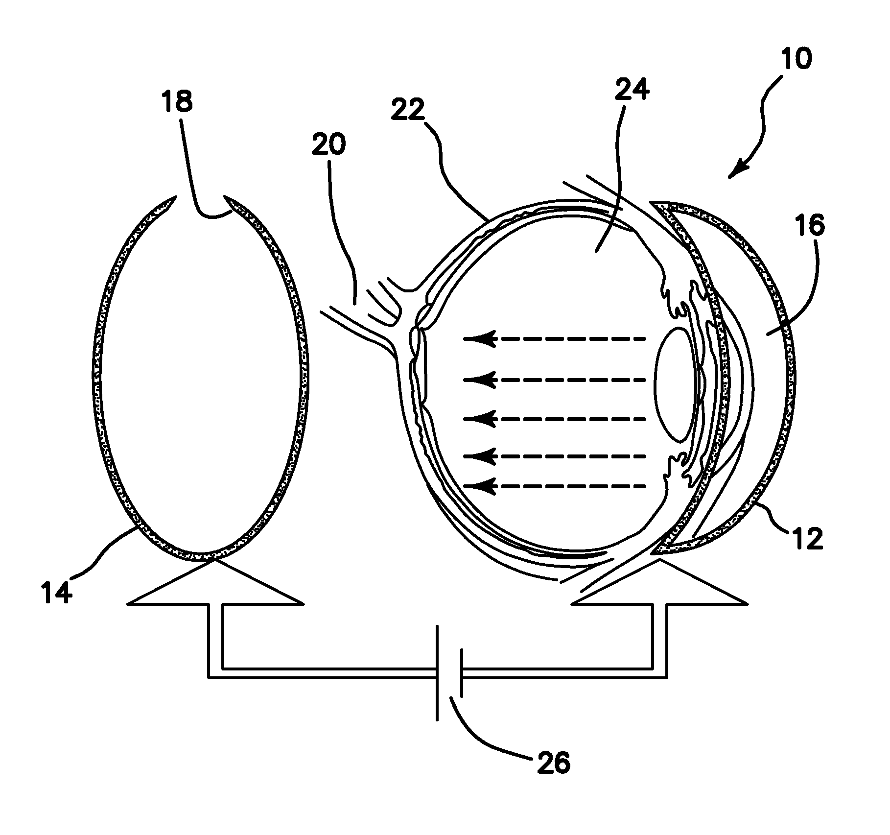
[0039]In diseases such as Retinitis pigmentosa (RP), one of the most common forms of inherited retinal degeneration, progressive loss of photoreceptor cells occurs which eventually leads to blindness. Here, we disclose an in vivo method for efficient delivery of plasmids encoding light-sensitive proteins (ChR2) into the eye of small animal models having retinitis pigmentosa with the aim to restore vision. The illustrated delivery system is comprised of a hemi-spherical cathode to be placed on the cornea and an accommodating anode in back of the eye, driven by optimal electric pulses. Through electroporation of plasmids into a specific layer in the retina (e.g. promoter-specific to retinal ganglion cells), we eliminated the disadvantages of the viral delivery method such as safer administration and avoiding the difficulties in incorporating a large construct of promoter, ChR2, and a fluorescent protein marker into the lentivirus / Adeno associated virus. The specific layer of the retin...


PUM

PropertyMeasurementUnit
electric fieldaaaaaaaaaa
diameteraaaaaaaaaa
diameteraaaaaaaaaa

Abstract

An apparatus for in vivo electroporating a plasmid into a retina of any eye includes a first electrode with a first polarity of voltage placed in contact with a cornea of the eye, a second electrode with an opposite second voltage at least in part behind the retina, and a pulsed voltage source for providing a pulsed DC voltage with an optimized field strength amplitude, frequency, number of pulses, group repetition rate and duration of pulse and group repetition, which are optimized for transfection of the channelrhodospsin-2 (ChR2) gene into the retinal ganglion cells. An in vivo method for treating retinal ganglion cells in an eye without use of viral transfection includes the steps of nonviral in vivo delivering a channelrhodospsin-2 (ChR2) gene to target the specific (retinal ganglion) cells of a retina by intravitreous injection of plasmid DNA, electroporating the plasmid into the retina and use of image intensification device for stimulating the retinal ganglion cells with ambient lighting conditions.

Description

RELATED APPLICATIONS[0001]The present application is related to U.S. Provisional Patent Application, Ser. No. 61 / 170,357, filed on Apr. 17, 2009, which is incorporated herein by reference and to which priority is claimed pursuant to 35 USC 119.BACKGROUND OF THE INVENTION[0002]1. Field of the Invention[0003]The invention relates to the field of apparatus and methods for optogenetic treatment of blindness including retinitis pigmentosa.[0004]2. Description of the Prior Art[0005]Retinitis pigmentosa (RP) refers to disorders characterized by degeneration of photoreceptors in the eye which hinders visual ability by nonfunctional neuronal activation and transmission of signals to the cortex. The prevalence of this disease is at least one million individuals. The disease is most often inherited as an autosomal recessive trait with 50 to 60% of cases having this form of inheritance. However, since not all RP-causing genes have been discovered, if a person chooses to get genetically tested, ...

Claims


Application Information

Patent Timeline
Patent Type & Authority Applications(United States)
IPC IPC(8): A61F9/00A61N1/30
CPCA61F9/0017A61K9/0048A61N1/327A61N1/0412A61N1/0416A61K38/177
Inventor MOHANTY, SAMARENDA K.FICINSKI, MATTHEWWONG, EDWARD K.
Owner RGT UNIV OF CALIFORNIA
Why Eureka
Try Eureka