Myocardial perfusion imaging method
a technology of perfusion imaging and myocardium, applied in the field of myocardial imaging methods, can solve the problems of limited use, limited usefulness of treatment with this compound, and many patients' inability to exercise at the level necessary to provide sufficient blood flow, and achieve the effect of facilitating myocardial imaging
- Summary
- Abstract
- Description
- Claims
- Application Information
AI Technical Summary
Benefits of technology
Problems solved by technology
Method used
Image
Examples
example 1
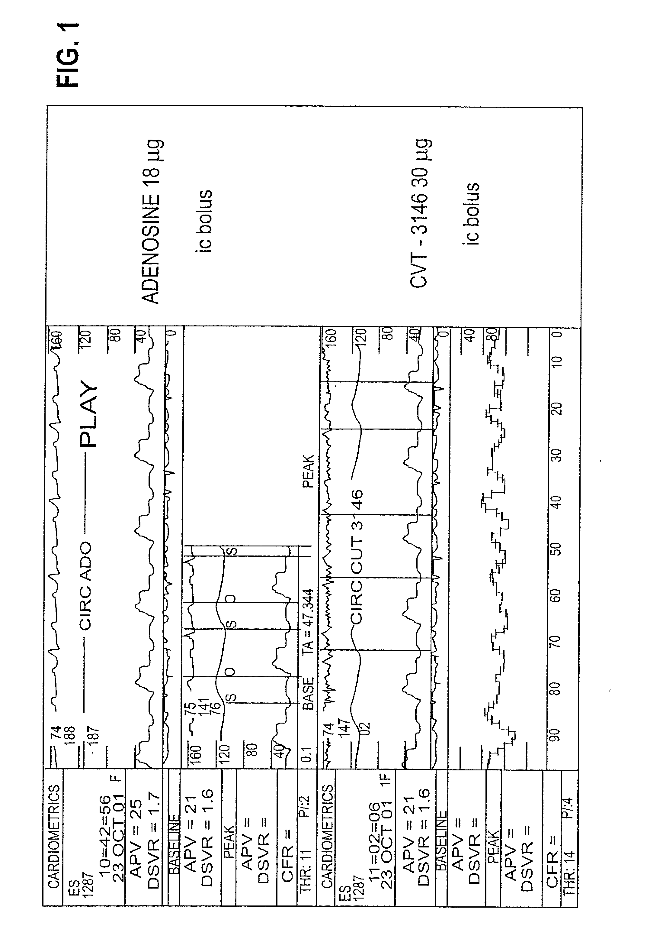
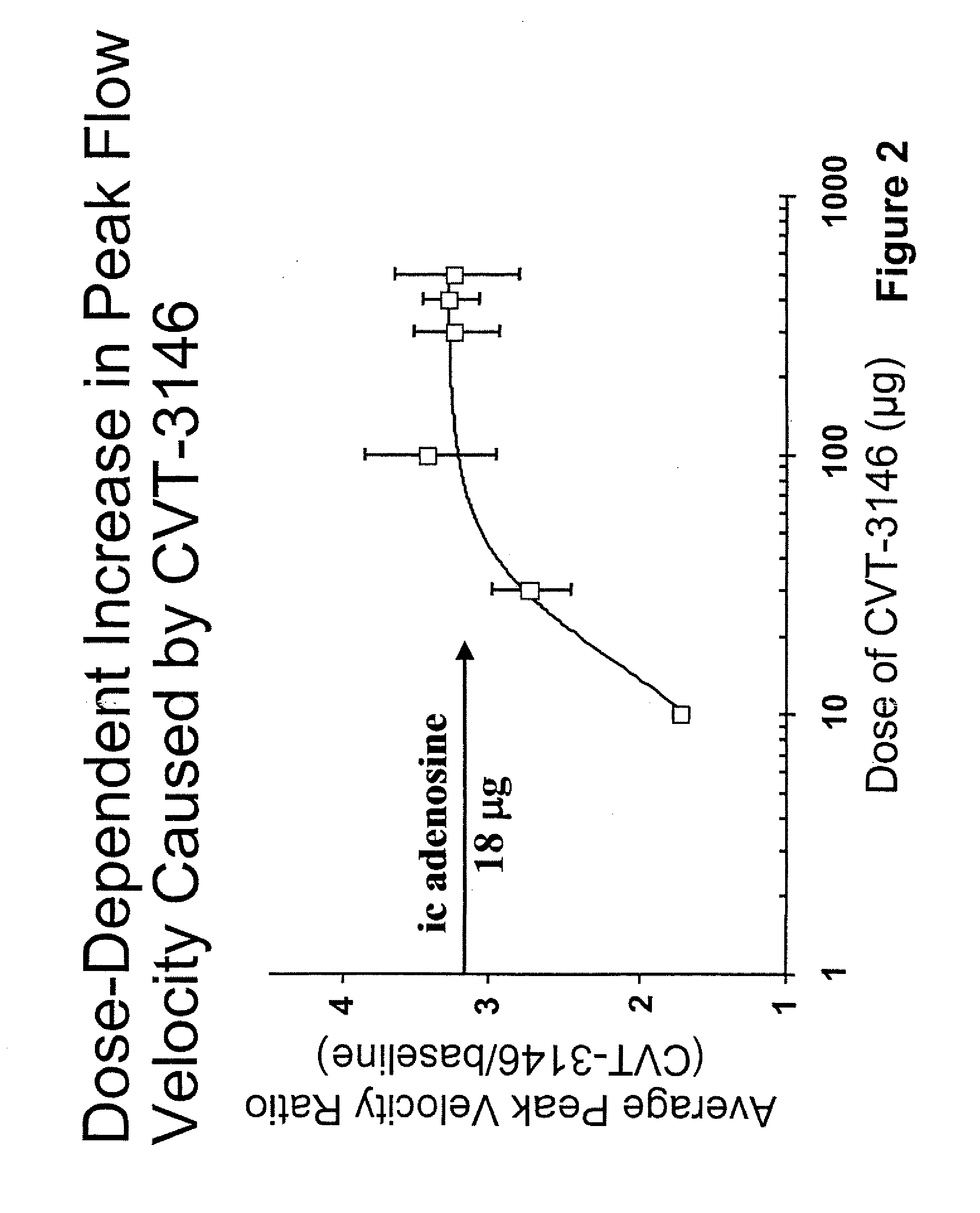
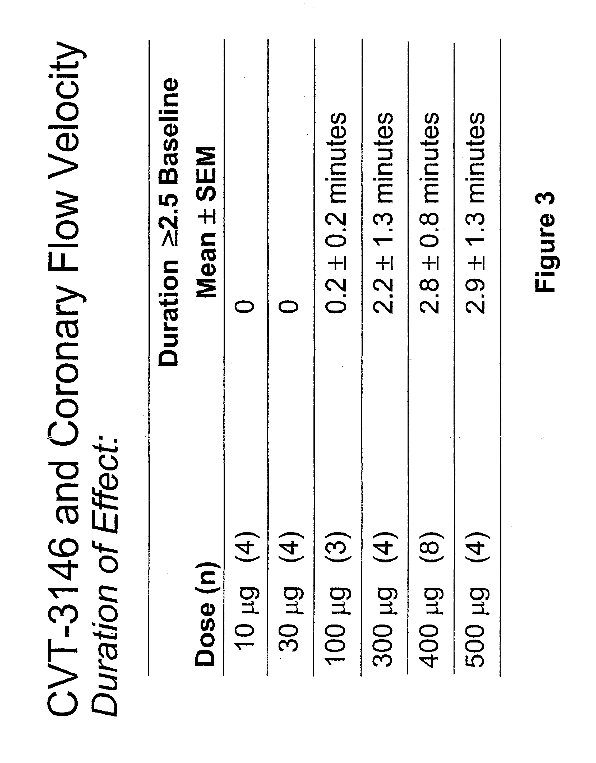
[0147]BACKGROUND: CVT-3146 (CVT), with an initial half-life of 3 minutes with a rapid onset and offset of action, is >100-fold more potent than adenosine (Ado) in increasing coronary blood flow velocity (CBFv) in awake dogs. The purpose of this open label study was to determine the magnitude and duration of effect of CVT-3146 (10-500 μg) on CBFv in humans.
[0148]METHODS: Patients undergoing a clinically indicated coronary catheterization with no more than a 70% stenosis in any coronary artery and no more than a 50% stenosis of the study vessel had CBFv determined by Doppler flow wire. Study subject were selected after measuring baseline and peak CBFv after an intracoronary (IC) injection of 18 μg of Ado. Twenty-three patients, who were identified as meeting the study criteria of having a peak to baseline CBFv ratio of ≧2.5 in response to Adenosine, received a rapid (≦10 sec) peripheral IV bolus of CVT-3146; Doppler signals were stable and interpretable over the time-course of the inc...
example 2
[0151]This example is a study performed to determine the range of dosages over which the selective A2A receptor agonist, CVT-3146 can be administered and be effective as a coronary vasodilator.
[0152]The study included patients undergoing a clinically indicated coronary catheterization with no more than a 70% stenosis in any coronary artery and no more than a 50% stenosis of the study vessel had CBFv determined by Doppler flow wire. Study subject were selected after measuring baseline and peak CBFv after an intracoronary (IC) injection of 18 μg of Ado. 36 subjects were identified as meeting the study criteria of having a peak to baseline CBFv ration of ≧2.5 in response to Adenosine,
[0153]CVT-3146 was administered to the study subjects by IV bolus in less that 10 seconds in amounts ranging from 10 μg to 500 μg.
[0154]The effectiveness of both compounds was measured by monitoring coronary flow velocity. Other coronary parameters that were monitored included heart rate and blood pressure...
example 3
[0165]This Example is a study performed to evaluate (1) the maximum tolerated dose of CVT-3146 and (2) the pharmacokinetic profile of CVT-3146 in healthy volunteers, after a single IV bolus dose.
Methods
[0166]The study was performed using thirty-six healthy, non-smoking male subjects between the ages of 18 and 59 and within 15% of ideal body weight.
[0167]Study Design
[0168]The study was performed in phase 1, single center, double-blind, randomized, placebo-controlled, crossover, ascending dose study. Randomization was to CVT-3146 or placebo, in both supine and standing positions.
[0169]CVT-3146 was administered as an IV bolus (20 seconds) in ascending doses of 0.1, 0.3, 1.3, 10, 20 and 30 μg / kg.
[0170]Subjects received either CVT-3146 of placebo on Day 1 supine, then crossover treatment on Day 2 supine. On Day 3, subjects received CVT-3146 or placebo standing, then crossover treatment on Day 4 standing.
Assessments
[0171]Patient safety was monitored by ECG, laboratory assessments, and col...
PUM
| Property | Measurement | Unit |
|---|---|---|
| weight | aaaaa | aaaaa |
| weight | aaaaa | aaaaa |
| time | aaaaa | aaaaa |
Abstract
Description
Claims
Application Information
 - R&D
- Intellectual Property
- Life Sciences
- Materials
- Tech Scout
- Unparalleled Data Quality
- Higher Quality Content
- 60% Fewer Hallucinations
Browse by: Latest US Patents, China's latest patents, Technical Efficacy Thesaurus, Application Domain, Technology Topic, Popular Technical Reports.
© 2025 PatSnap. All rights reserved.Legal|Privacy policy|Modern Slavery Act Transparency Statement|Sitemap|About US| Contact US: help@patsnap.com


