Urine markers for detection of bladder cancer

Active Publication Date: 2010-10-28
PACIFIC EDGE
View PDF6 Cites 12 Cited by
  • Summary
  • Abstract
  • Description
  • Claims
  • Application Information

AI Technical Summary

Benefits of technology

[0011]Using a combination of microarray analysis and quantitative polymerase chain reaction (qPCR), we have been able to identify specific genetic markers that are selective for bladder cancer. In some embodiments, we have found markers that can be used to differentiate the stage of a bladder tumor, and in other embodiments, we have identified markers that can distinguish types of tumors. In other embodiments, we have unexpectedly found that combinations of two or more markers can provide for a highly reliable and sensitive detection of bladder cancer. In still further embodiments, we have identified markers that are highly expressed in bladder cancer cells and not in blood cells. Thus, in many embodiments, tests for bladder cancer are unexpectedly better than prior art tests.
[0017]We have unexpectedly found that certain urine markers, in particular those not found in blood at high levels, when used in combination or alone can provide highly reliable, sensitive and specific diagnosis of bladder cancer.

Problems solved by technology

However, for a number of major cancers, the available markers suffer from insufficient sensitivity and specificity.
However, this technique is time consuming, invasive and its sensitivity is only approximately 90%, meaning that about 10 percent of cancers are not detected using these methods.
Although cytology has a specificity of about 95%, it has poor sensitivity (9-25%) for low-grade lesions, is extremely dependent on sample quality and suffers from high inter-observer variability.
However, array technology is relatively non-quantitative and is highly variable.
Tests for these markers offer better sensitivity than cytology, but tend to suffer from sub-optimal specificity because elevated levels of these markers are also commonly observed in patients with non-malignant diseases including inflammation, urolithiasis and benign prostatic hyperplasia.
The high variability of NMP22 means that it is not ideal for rapid, easy detection of bladder cancer.
Such artifacts include differences in the number of ligand oligonucleotides placed on an array dot, uneven and unpredictable binding of dyes to hybridized oligonucleotides on an array spot, uneven washing of non-specific materials from array spots and other problems.
At present, however, there are no methods to easily and reliably distinguish between superficial and invasive bladder cancer classes without performing cystoscopy.
Thus, in conditions characterized by reduced urine production (e.g., reduced urine volume), the concentration of a marker may be increased, yet may not reflect bladder cancer.
On the other hand, increased urine production may dilute a tumor marker, and thereby tend to mask the presence of bladder cancer.

Method used


Image

  • Urine markers for detection of bladder cancer
  • Urine markers for detection of bladder cancer
  • Urine markers for detection of bladder cancer

Examples


example 1

Identification of Markers for Superficial and Invasive Malignancy of the Bladder

[0153]Hierarchal clustering of microarray data from the gene expression patterns of invasive and superficial bladder cancer showed large numbers of significant differences. As a result, these cancer types were treated separately in the following analyses. Nevertheless, a high proportion of genes are over-expressed in both cancer types. FIG. 3 depicts a table that shows results of microarray studies for markers for invasive bladder malignancy. Thirty-one of the 199 invasive markers meet the above-stated criteria for serum markers (Denoted by “S” in figure). FIG. 4 depicts a table that shows results of microarray studies for superficial bladder malignancy. Thirty-four of the 170 superficial markers meet the above criteria for serum markers. FIGS. 3 and 4 include the HUGO symbol for the gene (“symbol”), the MWG Biotech oligonucleotide number, the NCBI mRNA reference sequence number, the protein reference se...

example 2

qPCR Analysis

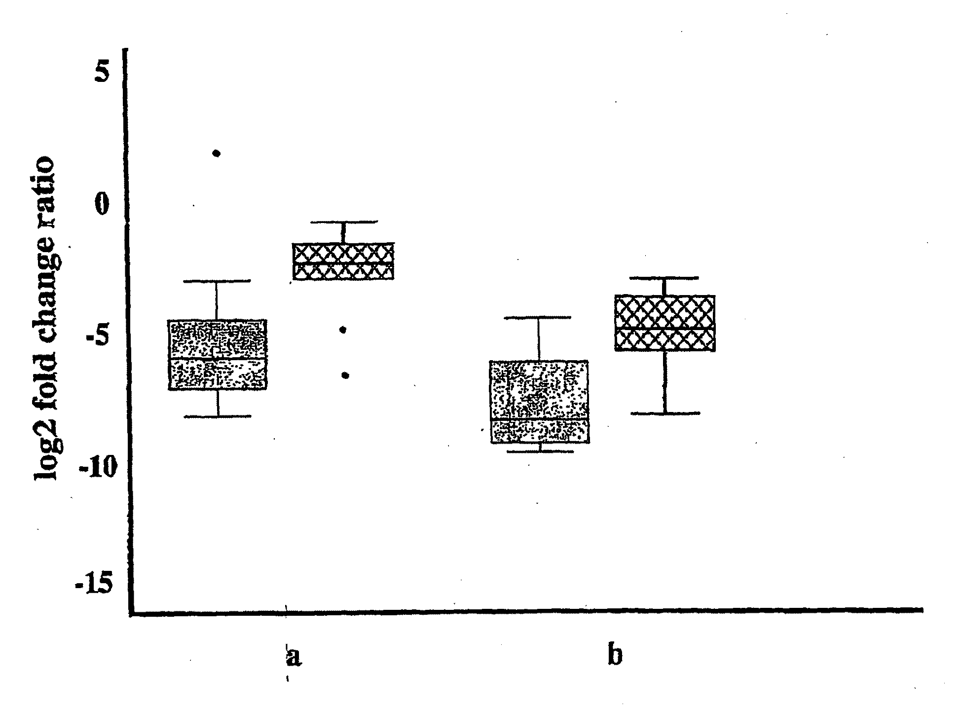
[0155]More sensitive and accurate quantitation of gene expression was obtained for a subset of the genes shown in FIGS. 3 & 4 using qPCR. Messenger RNA from up to 30 invasive bladder tumors, 25 superficial bladder tumors, and 18 samples of normal urothelium were analyzed for 18 genes identified by the microarray analysis (FIGS. 3 & 4), with the results shown in FIG. 5. Data for both invasive and superficial type bladder cancer is shown for markers SPAG5, TOP2a, CDC2, ENG, NRP1, EGFL6, SEM2, CHGA, UBE2C, HOXA13, MDK, THY1, BIRC5 and SMC4L1. Markers SEMA3F, IGFBP5, and NOV were only over-expressed compared to normal urothelium in the superficial type alone, and MGP was only over-expressed in the invasive type alone; these markers maintained similar expression to normal urothelium in the tumor samples that were not over-expressed. FIG. 5 includes the gene name, gene aliases, the gene symbol, the median fold change between tumor (T) and non-malignant (N) tissue, the maximum...

example 3

Use of Multiple Markers in Detection of Bladder Cancer

[0167]FIGS. 12a-12b depict histograms of the number of genes exhibiting a significantly increased expression (“over-expression”) in individual tumor samples compared to normal samples. The histograms were based on qPCR data obtained from the first twelve markers shown in FIG. 5. Of the 30 invasive tumors in the PCR analysis, 27 (90%) over-expressed at least four genes greater than the 95th percentile (FIG. 12a). Of the 25 superficial tumors in the analysis, 23 (92%) over-expressed at least four genes greater than the 95th percentile (FIG. 12b). These findings indicates that, in situations in which multiple genes are over-expressed relative to normal tissue, the reliability of cancer detection can be very high, making diagnosis of cancer more certain. However, in some cases, elevation of expression of a single marker gene is sufficient to lead to the diagnosis of cancer.

[0168]The reliability of successful discrimination of tumor a...


PUM

PropertyMeasurementUnit
Volumeaaaaaaaaaa
Volumeaaaaaaaaaa
Fractionaaaaaaaaaa

Abstract

Early detection of tumors is a major determinant of survival of patients suffering from tumors, including bladder tumors. Members of the BTM or UBTM family can be highly and consistently accumulated in bladder tumor tissue and other tumor tissue, and / or can be accumulated in urine of patients, and thus are markers for bladder and other types of cancer. In certain embodiments, BTMs or UBTMs can accumulate in the urine, and detection of UBTM family members can be an effective diagnostic approach. In some embodiments, quantitative PCR methods have advantages over microarray methods. In other embodiments, detection and quantification of a plurality of BTMs or UBTMs can increase the sensitivity and specificity of detection of bladder cancer, and therefore provides methods for determining the stage and type of bladder cancer. Kits provide easy, convenient ways for carrying out the methods of this invention.

Description

CLAIM OF PRIORITY[0001]This application claims priority to New Zealand Provisional Patent Application No: 534,289 filed Jul. 23, 2004 titled “Markers for Detection of Bladder Cancer,” Applicant: Pacific Edge Biotechnology Ltd., to New Zealand Provisional Patent Application No: 539,219 filed Apr. 4, 2005 titled “Markers for Detection of Bladder Cancer,” Applicant: Pacific Edge Biotechnology Ltd., and to and U.S. Provisional Patent Application No. 60 / 692,619 filed Jun. 20, 2005 titled “Urine Markers for Detection of Bladder Cancer,” Inventors: Parry John Guilford, Natalie Jane Kerr and Robert Pollock. Each of the above applications is incorporated herein fully by reference.FIELD OF THE INVENTION[0002]This invention relates to detection of cancer. Specifically, this invention relates to the use of markers for the detection of bladder cancer. More specifically, this invention relates to use of urine markers for the detection of bladder cancer. Yet more specifically, this invention relat...

Claims


Application Information

Patent Timeline
IPC IPC(8): C12Q1/68G01N33/68C12Q1/26C40B30/00G01N33/566G01N33/573C12M1/00
CPCC07K14/47C07K14/4748C12Q1/6886C12Q2600/112G01N33/57407G01N33/57488G01N2800/52C12Q2600/158G01N2333/47
Inventor GUILFORD, PARRY JOHNKERR, NATALIE JANEPOLLOCK, ROBERT CRAIG
Owner PACIFIC EDGE
Why Eureka
Try Eureka