Isolation, Characterization and Propagation of Germline Stem Cells

a germline stem cell and cell technology, applied in the field of germline stem cell isolation, characterization and propagation, can solve the problems of affecting the repair system, limited types, and accumulation of damage to resident stem cells

Inactive Publication Date: 2010-11-11
PRIMEGEN BIOTECH LLC
View PDF4 Cites 10 Cited by
  • Summary
  • Abstract
  • Description
  • Claims
  • Application Information

AI Technical Summary

Benefits of technology

[0023]In another embodiment, the higher level of telomerase activity is at least 20% of the telomerase activity of embryonic stem cells. In another embodiment, the higher level of telomerase activity is at least 50% the telomerase activity of embryonic stem cells.

Problems solved by technology

However, before ES cell applications can be realized, at least ethical issues must be resolved, and teratomas formation after transplantation must also be overcome.
At the next stage, cells become multipotent, meaning they can give rise to several other cell types, but those types are limited in number.
Unfortunately, virtually every somatic cell in the adult animal's body, including stem cells, possess a genome ravaged by time and repeated cell division.
As an organism ages, it is possible that damage to resident stem cells accumulates and that the repair systems are compromised.
It has been postulated that similar types of stem cells may exist in the female germline, but those cells have proven difficult to identify and isolate.
Whether precursor stem cells exist for SSC, i.e., germline precursor stem cells, has been difficult to ascertain.
Presumably present in very small numbers, their sheltered location has complicated isolation and purification.
Unfortunately, the location and identifying characteristics of these cells within the seminiferous tubule have been difficult to discern.
Generation of embryonic stem cell lines had been thought to provide a renewable source of embryonic stem cells for both research and therapy but reports now indicate that existing cell lines are less robust than originally believe and many have also been contaminated during culture with immunogenic animal molecules.
However, embryonic stem cells may not be appropriate for direct transplant as they form teratomas after transplantion.
Additionally there are moral and ethical issues associated with the isolation of embryonic stem cells from human embryos.

Method used


Image

  • Isolation, Characterization and Propagation of Germline Stem Cells
  • Isolation, Characterization and Propagation of Germline Stem Cells
  • Isolation, Characterization and Propagation of Germline Stem Cells

Examples


example 1

Isolation of Human Fetal Gonocytes

[0159]Aborted fetuses from 11-22 weeks gestation were donated by approved clinics and maintained under sterile conditions. Gonads were decapsulated and one gonad was placed in 2.5 mL of phosphate buffered saline (PBS) and minced with sterile scissors. The minced gonadal tissue was incubated with 2.5 mL of a 2× collagenase solution (2 mg collagenase and 20 unites of DNase I per milliliter of DPBS(−)) for 30 minutes at 37° C. in a shaking water bath. During the dissociation period the sample is triturated several times to promote dissociation of the tissue. After dissociation, 1 mL of fetal bovine serum is added to deactivate the collagenase and the sample is triturated once more. The sample is then centrifuged at 400×g for 5 minutes and the cells resuspended at 5−7.5×10⁵ per mL in PM-10™ medium plus growth factors as disclosed in co-pending U.S. patent application Ser. No. 11 / 694,687, filed on Mar. 30, 2007 which is incorporated by reference herein f...

example 2

Long Term Culture of Human Fetal Gonocytes

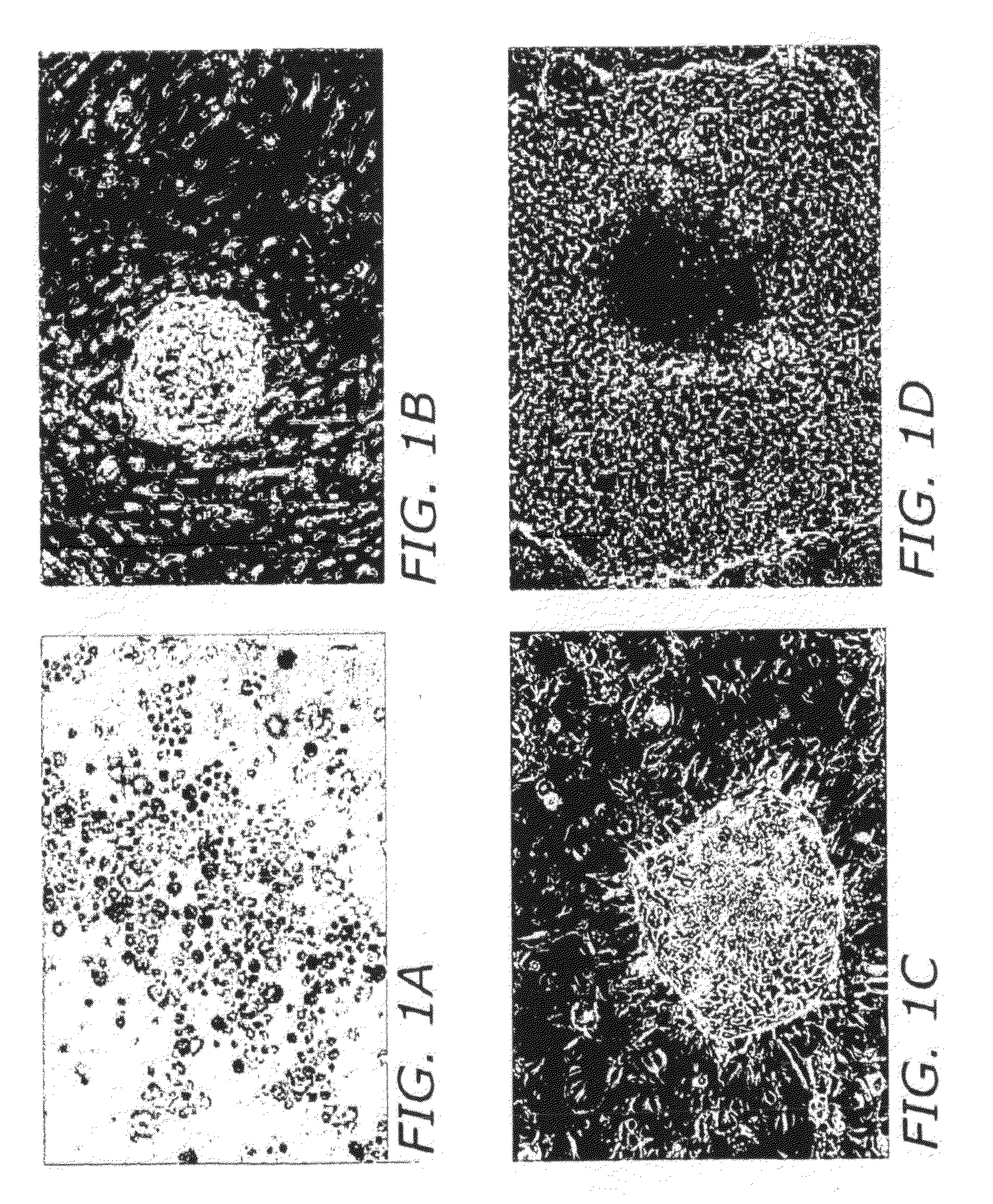
[0160]The isolated fetal gonocytes showed a high level of alkaline phosphatase staining at the onset of culture (FIG. 1A). Gonocytes were then cultured on STO / c (feeder cells) plates or under feeder-free conditions for colony formation. Alternatively, gonocytes can be cultured in PM-10™ medium plus growth factors on gelatin-coated plates prior to culturing on STO feeder cells.

[0161]After two weeks in culture, the human fetal gonocytes formed ‘mushroom’ colonies (FIG. 1B). At this time the mushroom colonies were transferred to fresh STO plates. After 1-3 passages, the mushroom colonies formed outgrowths indicating that the colonies are expanding (FIG. 1C). These outgrowths continued to grow on the STO feeders and form flat colonies resembling human embryonic stem cell colonies (FIG. 1D).

[0162]The cultured gonocytes were passaged onto fresh STO cells every two weeks in serum-free culture medium (PM-10™+ growth factors) for further expansion.

example 3

Expression of Pluripotent Markers by Cultured Human Fetal Gonocytes

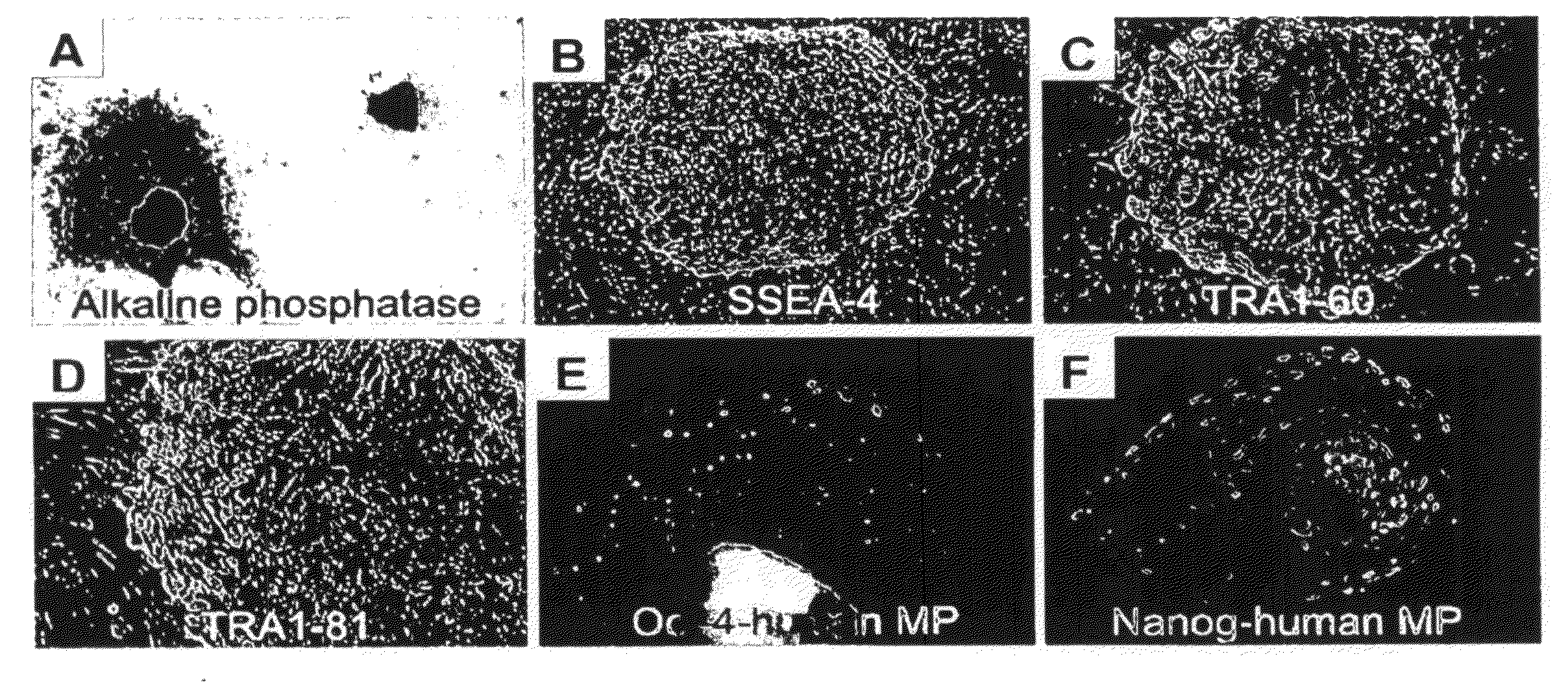
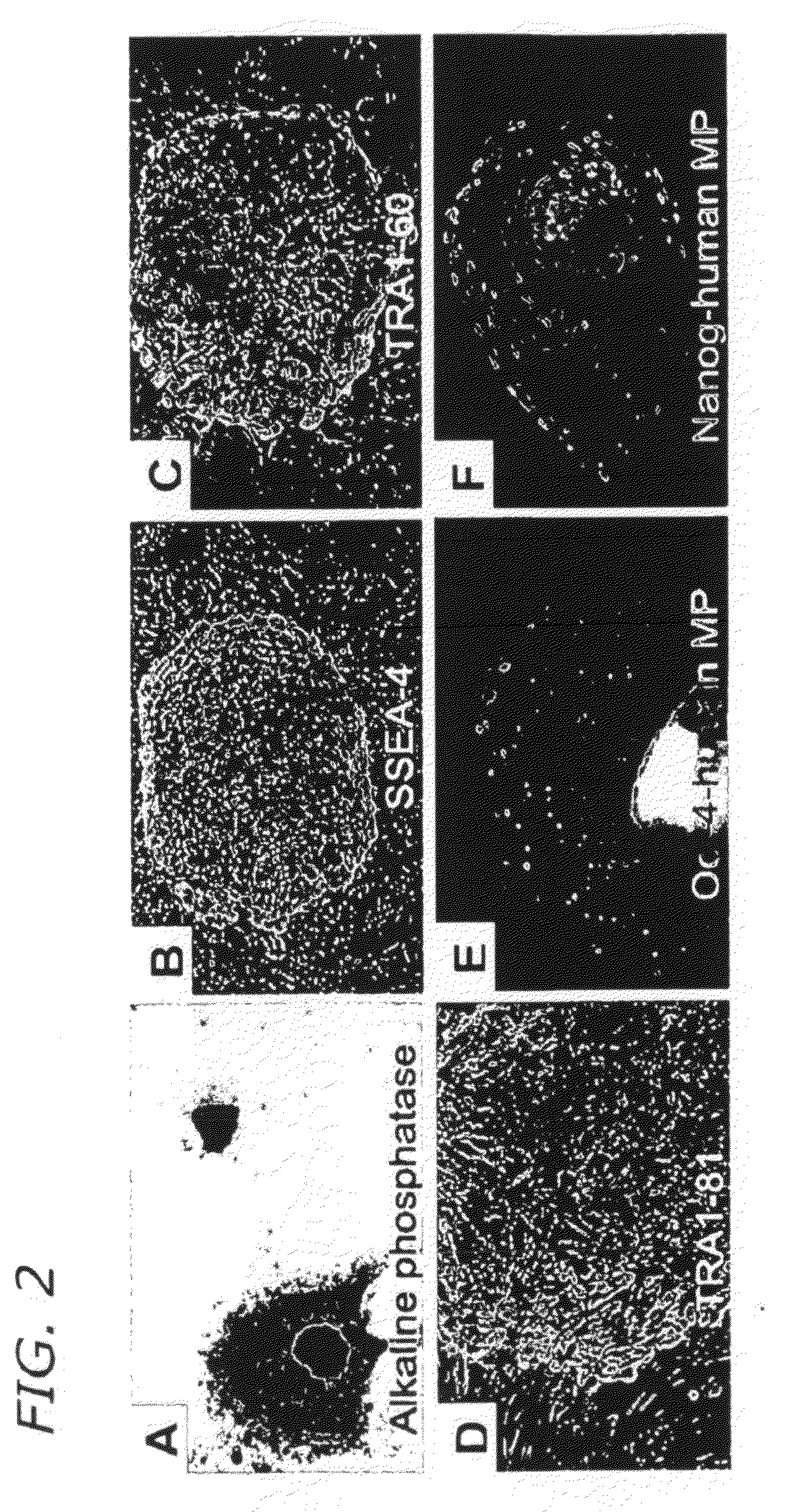
[0163]Long-term culture in the presence of growth factors changes the gene expression profile of the gonocytes, therefore cultured gonocytes express markers that normally would be expressed in pluripotent stem cells (FIGS. 2 and 4) including alkaline phosphatase (FIG. 2A), SSEA-4 (FIG. 2B), TRA1-60 (FIG. 2C), TRA1-81 (FIG. 2D), Oct-4 and human mitochondrial protein (MP, FIG. 2E), and Nanog and human MP (FIG. 2F). Human MP is used as a human-specific marker.

[0164]FIG. 3 depicts two colonies of long-term cultured gononocyes (FIG. 3A: HF-89-p2 colony; FIG. 3B: HF-22-p7 colony) that have been cultured under serum and feeder-free conditions such that they are not exposed to animal products in the reprogramming or culture process and are not contaminated with immunogenic non-human molecules.

[0165]FIG. 4 depicts expression of a wide range of pluripotency-associated markers such as Oct-4. Nanog, Sox2 and Rex1 in expanded gon...


PUM

PropertyMeasurementUnit
cell cycle doubling timeaaaaaaaaaa
cytofluorimetric size analysisaaaaaaaaaa

Abstract

Methods are provided for the isolation, characterization and propogation of germline cells stem cells from fetal and adult mammals. Additionally, isolated populations of germline cells having different phenotypes are disclosed wherein the subpopulations are capable of forming long-term cultures of multipotent or pluripotent cells or are capable of differentiating into mature germline cells and repopulating a sterile reproductive organ. The multipotent or pluripotent germline cells are also suitable for differentiation into tissue-specific somatic cells for therapeutic purposes.

Description

CROSS REFERENCE TO RELATED APPLICATIONS[0001]The present application claims priority under 35 U.S.C. §119(e) to U.S. Provisional Patent Application Nos. 60 / 954,496 filed Aug. 7, 2007 and 61 / 060,356 filed Jun. 10, 2008, the entire contents of which are incorporated herein by reference in their entirety.FIELD OF THE INVENTION[0002]The present invention relates to the field of male and female germline stem cells, isolated during fetal and post-natal development and the use of these cells for therapeutic purposes. Specifically, the present invention relates to identification, isolation, and differentiation of distinct germline stem cell populations, and cell lines generated therefrom, with different potential uses in cell replacement therapy.BACKGROUND OF THE INVENTION[0003]Generation of pluripotent cell lines which can be safely used in regenerative medicine has a great potential impact in cell replacement therapy. In this regard, embryonic stem (ES) cells have been considered as poten...

Claims


Application Information

Patent Timeline
Patent Type & Authority Applications(United States)
IPC IPC(8): C12N5/07C12N5/074
CPCC12N5/0609C12N2502/13C12N2501/13C12N5/0611C12N5/0608
Inventor IZADYAR, FARIBORZPACHIAROTTI, JASONMAKI, CHADRAMOS, THOMAS V.
Owner PRIMEGEN BIOTECH LLC
Why Eureka
Try Eureka