Activated protease indicator

a protease indicator and activation technology, applied in the field of activation protease indicators, can solve the problems of insufficient quantification, limited number of cells that can be analyzed, and methods that require complex assay procedures, and achieve the effect of restoring luminescent function

Inactive Publication Date: 2010-11-25
THE UNIV OF TOKYO +1
View PDF1 Cites 16 Cited by
  • Summary
  • Abstract
  • Description
  • Claims
  • Application Information

AI Technical Summary

Benefits of technology

[0011]Luc-C and Luc-N are molecules prepared by splitting a single luciferase into two fragments. In this case, the “single luciferase” may include a full-length luciferase or a part of a luciferase required for the luminescent function. Additionally, these “half fragments” are a state that the luciferase is split at a site where the C-terminal amino acid residue of the Luc-N and the N-terminal residue of the Luc-C can be ligated together with a peptide bond so that the resulting product can restore the luminescent function upon the protein splicing with an intein as described below.

Problems solved by technology

These methods therefore require complex assay procedures like preparation of cell lysates and elimination of sediments upon analysis of intracellular protease.
However, the results obtained are not quantitative but rather qualitative, since the changes in the fluorescence signals are very small and the number of cells that can be analyzed is limited.
In addition, it is difficult to apply the indicators to living animals because the light for excitation of the fluorophores is mostly absorbed by their tissues [Weissleder, R.
However, the molecular size of the indicator is quite large and the intensity of the luminescence signals is relatively low because of insufficient digestion of the DEVD sequence during apoptosis.

Method used


Image

  • Activated protease indicator
  • Activated protease indicator
  • Activated protease indicator

Examples


example 1

1. Method

1-1. Construction of Plasmid

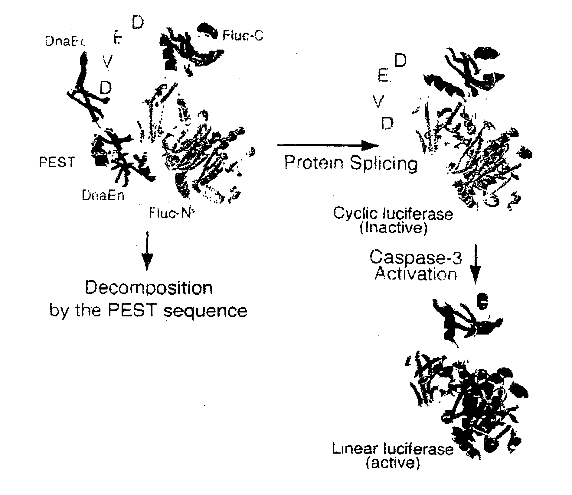
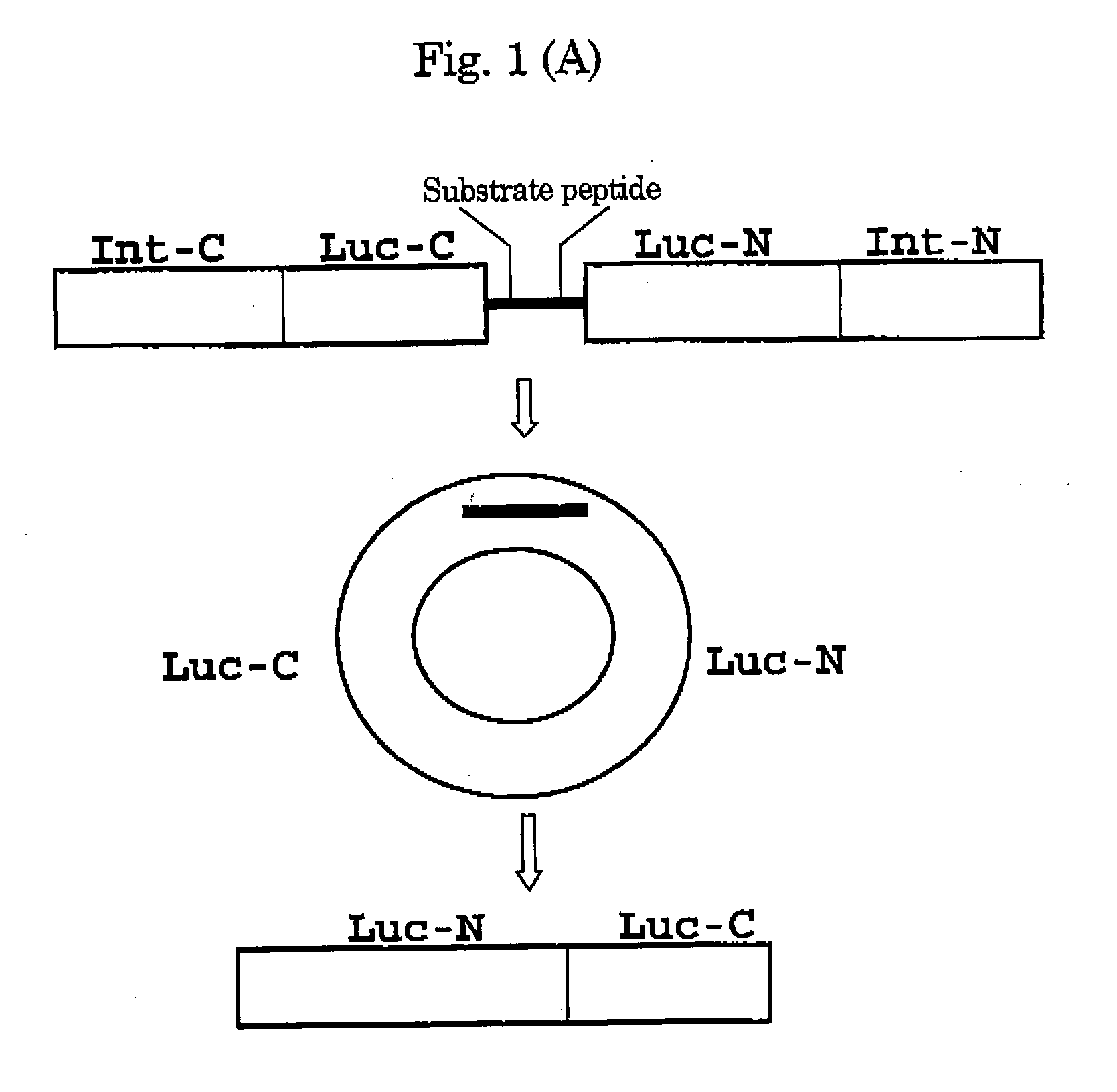
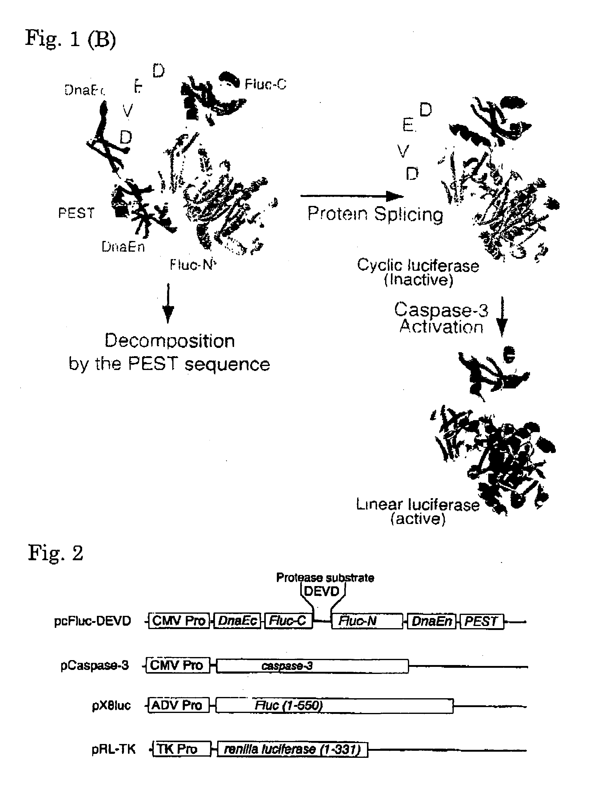
[0070]Using the Escherichia coli strain DH5-α as a host, all plasmids were constructed. By PCR, an initiation codon and enzyme sites BamH I and Mun I were individually introduced in cDNA of the C-terminal fragment of DnaE. Using the Sal I site, cDNA of the C-terminal fragment (399-550 aa) of Fluc was conjugated to cDNA of the N-terminal fragment (2-416 aa) of Fluc, and subsequently, the Mun I and Hind III sites were independently introduced in the 5′ and 3′ termini, respectively of the resulting chimera DNA. By PCR, the PEST sequence and enzyme sites Hind III and Xho I were introduced in cDNA of the N-terminal fragment of DnaE. The resulting PCR products were sequenced with a DNA sequencer ABI prism 310 (Applied Biosystems, Tokyo, Japan), to determine the fidelity. These fragments were conjugated together, for subcloning into the BamH I site and Xho I site of an expression vector pcDNA 3.1 (+) (Invitrogen, Carlsbad, Calif.). Caspase-3 cDNA was ki...


PUM

PropertyMeasurementUnit
pHaaaaaaaaaa
timeaaaaaaaaaa
body weightaaaaaaaaaa

Abstract

It is an object of the invention to provide novel approaches capable of detecting activated protease and also detecting protease activation on real time at a high sensitivity in a noninvasive manner. By the method for detecting activated protease of the invention, an indicator in a circular form comprising the C-half fragment of luciferase (Luc-C) and the N-half fragment of luciferase (Luc-N) linked together through a substrate peptide for a protease is introduced in an in vitro assay system or in cells. Upon digestion of the substrate peptide by the protease, Luc-N and Luc-C together reconstructs active luciferase, so that the activated protease can be detected by assaying the luminescence signal from the luciferase.

Description

TECHNICAL FIELD[0001]The present invention relates to an activated protease indicator, and an indicator of protease activation capable of noninvasive real-time detection of protease activation at a high sensitivity.BACKGROUND ART[0002]Eukaryotic cells contain many proteases involved in cell growth, division, differentiation, migration, and intra- or extracellular signaling. One of the complicated proteolytic signaling processes is used to decide programmed cell death or apoptosis. Improper apoptosis causes many diseases including Alzheimer disease, Huntington disease, ischemia, autoimmune disorder and immortality of cancer cells [Nijhawan, D. et al. (2000) Annu. Rev. Neurosci. 23, 73-87; Rideout, H. J. & Stefanis, L. (2001) Histol. Histopathol. 16, 895-908]. For example, as cytosolic caspases play a central role in mediating the initiation and propagation of apoptosis, chemical compounds that can either inhibit or accelerate caspase activity are a major concern. Also, an understandi...

Claims


Application Information

Patent Timeline
Patent Type & Authority Applications(United States)
IPC IPC(8): C12Q1/68C12N9/02C12N15/63C12Q1/66
CPCC07K2319/50C12N9/0069G01N2500/04C12Q1/66C12Q1/37
Inventor UMEZAWA, YOSHIOOZAWA, TAKEAKIKANNO, AKIRA
Owner THE UNIV OF TOKYO
Why Eureka
Try Eureka