Magnetic cells for localizing delivery and tissue repair

Active Publication Date: 2011-01-06
EMMETROPE
View PDF6 Cites 25 Cited by
  • Summary
  • Abstract
  • Description
  • Claims
  • Application Information

AI Technical Summary

Benefits of technology

[0011]In general, it is expected that the magnetic nanoparticles will have a mean diameter of between 5 and 500 nm, more particularly between 40 and 400 nm, most particularly between 40 and 100 nm, with a standard deviation of 50% or lower, more usually 20% or lower. Difficulties in using nanoparticles over micron-scale particles include particle aggregation, particle tracking and observation, and ability to mobilize the particles by external magnetic fields, all of which are considerably easier when using micron-scale or larger particles. For this reason, previous efforts likely focused on micron-scale particles and ignored the possible advantages of nanoparticles. We have overcome a number of these difficulties, and have now discovered a number of advantages of using nanoparticles over micron-scale particles. Advantages include ability to bind to cell surfaces without stimulating endocytosis; ability to be shed from cell surfaces or, when internalized, excreted from cells; and ability to be excreted from the eye or the body when shed from cells.

Problems solved by technology

However, transplantation procedures are invasive, have varying rates of success, and are not yet even available for many types of diseases or disorders, in particular for a number of eye diseases, for example, including diseases of the cornea (including but not limited to endothelial or stromal dystrophies), diseases of retinal ganglion cells and the optic nerve (including but not limited to glaucoma, ischemic optic neuropathies, other optic neuropathies), and diseases of retinal photoreceptors and retinal pigment epithelium (including but not limited to Leber's congenital amaurosis, retinitis pigmentosa and age-related macular degeneration)
Although in many cases it would seem desirable to administer new “healthy” cells, e.g. by injection or infusion, simply injecting such cells generally does not work as they do not remain localized and stick to or become incorporated into the patients' tissue.
For example, healthy corneal endothelial cells are inefficiently incorporated into a patient's diseased cornea when injected into the anterior chamber of the eye (e.g. Mimura et al, Invest. Oplrthalmol. Vis. Sci. 2005, 46(10):3637-44), and healthy retinal ganglion cells are not incorporated into the retina when injected into the vitreous cavity of the eye.
Most current technology depends on whole tissue transplants, or in the case of stem cell clinical trials, there are no techniques for controlling the cells' localization in vivo.
A stem cell transplantation clinical trial for retinitis pigmentosa, for example, uses subretinal injection of hematopoetic stem cells, but does nothing to control their localization there, or keep them from floating or migrating away after surgical implantation.

Method used


Image

  • Magnetic cells for localizing delivery and tissue repair
  • Magnetic cells for localizing delivery and tissue repair
  • Magnetic cells for localizing delivery and tissue repair

Examples


example 1

Coating Magnetic Nanoparticles for Surface Attachment

[0029]For coating various magnetic nanoparticles for surface attachment to neurons, a procedure analogous to that effective for coating 1 μm particles activated with carboxylic acid (Dynal Biotech, Oslo, NORWAY) or for coating 50 nm particles (e.g. Miltenyi Biotech) with anti-TrkB (BD Bioscience, San Jose, Calif., USA) can be used. The coating procedure is performed according to the manufacturer's standard protocols. Briefly, particles are washed twice with 25 mM MES at approximate pH6 buffer for approximately 10 min each time. Approximately 150 μg of anti trkB in MES buffer is used for functionalizing particles, and slow tilt rotated for approximately 30 min. Then, 0.3 mg of EDC in MES buffer is added, and incubated overnight at 4° C. with tilt rotation. Finally, particles are washed in PBS for four times and PBS is added to a final 1 mg / ml. We found that 1 μm magnetic particles coated in this manner can strongly bind to RGCs (FI...

example 2

Optimal Functionalization (Surface Coating) of Commercially Available Superparamagnetic Nanoparticles to Maximize Binding to Retinal Ganglion Cells

[0030]Commercially available surface activated superparamagnetic nanoparticles as small as 25 nm (MicroMod Partikeltechnologie GmbH, GERMANY) can be coated according to manufacturers' protocols with functional molecules selected for their ability to strongly and specifically bind neurons. Briefly, tosyl-activated or carboxyl-activated magnetic nanoparticles can be used for attaching antibodies, proteins and other biomolecules that contain primary amino or sulphydryl groups. We will use manufacturers' suggested protocols for nanoparticle and protein / antibody concentrations as a starting point to covalently attach the following proteins: antibodies to the trkB receptor, antibodies to the surface adhesion molecule L1, antibodies to surface integrin receptors, and cholera toxin subunit B, which binds to the GM1 ganglioside on the surfaces of ...

example 3

Measurement of Binding Specificity of Magnetic Nanoparticles in Purified and Mixed Cultures

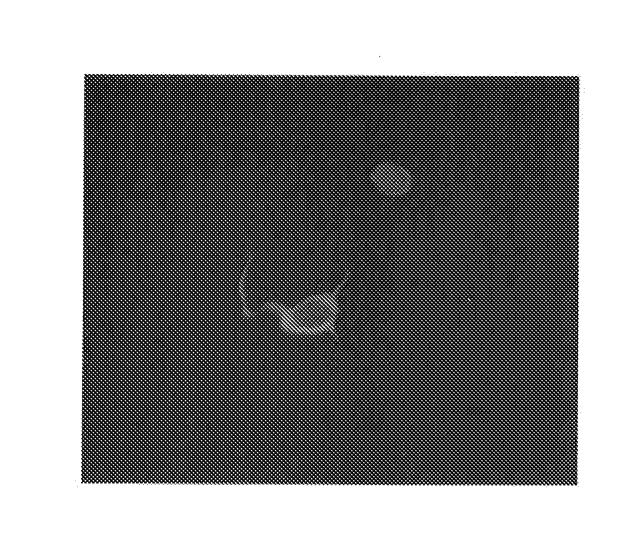
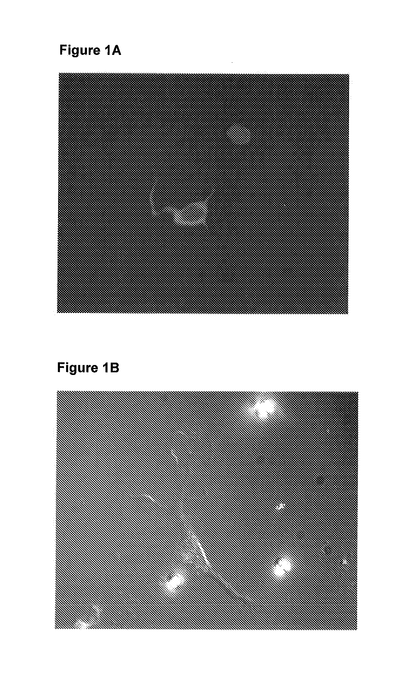
[0031]To assay for nanoparticle binding by neurons, retinal ganglion cells (RGCs) can be cultured according to standard protocols (Meyer-Franke et al., 1995; Goldberg et al., 2002b). We will add functionalized nanoparticles generated as described above to the RGCs 2 hours after plating, leave them for an additional 1 hour at 37° C., and then exchange the media to remove excess unbound nanoparticles. We will leave the neurons in culture for 1 hour to 3 days, to examine whether the nanoparticles remain attached with time. At the end of the culture period we will use three techniques to confirm nanoparticle binding: (1) direct visualization using high-magnification microscopy available in the lab; (2) commercially available iron staining kits (Sigma) in the case of nanoparticles with exposed iron surfaces; and (3) standard immunohistochemistry with fluorescent secondary antibodies directed agains...


PUM

PropertyMeasurementUnit
Diameteraaaaaaaaaa
Diameteraaaaaaaaaa
Magnetismaaaaaaaaaa

Abstract

Normal or genetically modified cell(s) having magnetic nanoparticle(s) bound (affixed) to their surfaces and methods of delivery to target tissues, e.g. for treatment of disease and or injury.

Description

[0001]The invention described herein was developed in part using funds from a grant from the Government of the United States of America (NEI / NIH grant no. R21-017971). The United States Government has certain rights in the invention.FIELD OF THE INVENTION[0002]The invention relates to magnetic cell(s), and methods of use. The cells are either naturally occurring or genetically altered animal somatic or stem cell(s), generated either from a patient / individual or from a living or deceased donor. Magnetic particles are affixed to the outer surface of the cell. The magnetic cell(s) can then be administered to a subject, e.g. by injection or implantation, and their localization controlled by application of an external magnetic field, e.g. a magnet positioned outside the body. This allows the cells to be magnetically directed to specific sites in the body, wherein the cells themselves, or various contents thereof such as enzymes, hormones, transmitters, and the like, will be useful for in...

Claims


Application Information

Patent Timeline
IPC IPC(8): A61K9/00A61K35/12C12N5/079C12N5/071A61P27/02A61K35/44
CPCA61K35/12A61K35/44A61K41/00A61K47/48776A61K9/5094C12N5/0006A61K9/0034A61K9/5115A61K47/48884A61N2/002A61K47/6901A61K47/6929A61P27/02A61K47/68A61K47/6923A61K47/6803A61K9/0048A61K35/28A61K35/30A61K47/6931A61K47/6829
Inventor GOLDBERG, JEFFREY L.HALPERN, ALAN
Owner EMMETROPE
Why Eureka
Try Eureka