
Methods and Algorithms for Cell Enumeration in a Low-Cost Cytometer

a low-cost, cell-enumeration technology, applied in the direction of character and pattern recognition, material analysis, packaging goods type, etc., can solve the problems of considerable laborious experimentation and optimization in the development of image acquisition and data reduction algorithms, and achieve the effect of reducing purchase and operating costs and functional simplicity in design

Inactive Publication Date: 2011-02-24
UNIVERSITY OF TWENTE
View PDF41 Cites 35 Cited by
  • Summary
  • Abstract
  • Description
  • Claims
  • Application Information

AI Technical Summary

Benefits of technology

[0025]One particularly advantageous aspect of the present invention is its utility both as a research tool in cell biology and as a clinical tool in the diagnosis of diverse cellular, fungal and viral pathologies including but not limited to HIV and cancer. Further advantages provided by this invention are the functional simplicity in design, ruggedness, compactness, AC or DC power options, and substantially lower purchase and operating costs relative to conventional commercial devices with comparable performance characteristics. The features and improvements of the devices of this invention, exemplified as compact clinical cell cytometers, make them particularly useful for operation in primitive laboratories or under field conditions prevalent in resource-poor countries.

Problems solved by technology

The development of the algorithms for image acquisition and data reduction required considerable laborious experimentation and optimization.

Method used


Image

  • Methods and Algorithms for Cell Enumeration in a Low-Cost Cytometer
  • Methods and Algorithms for Cell Enumeration in a Low-Cost Cytometer
  • Methods and Algorithms for Cell Enumeration in a Low-Cost Cytometer

Examples


example 1

System Characterization

[0145]To evaluate the theoretical predictions, the following experiment was done. A photodiode was placed in front of the objective, at the sample plane. A diaphragm restricted the illuminated area on the photodiode to a disk with a diameter of 1.6 mm. The LED and the condenser lens were placed in different configurations, so that an image of the diode chip was created at the back focal plane of the objective, and the size of this image was varied from almost a pin-point source to 25 mm.

[0146]The result of the experiment is presented in FIG. 20 where the curve obtained from the ray-tracing algorithm is shown together with the experimental data. As was predicted by the algorithm, an optimal value was found for the size of the image at the back focal plane of the objective. The shape of the curve resembles the situation predicted by the algorithm, except for large values of BB′, where the experiment shows an efficiency of almost zero. This may be the result of t...

example 2

Total White Blood Cell Counting

[0160]Isolated white blood cells were spiked into a leukocyte-depleted red cell concentrate at known leukocyte concentrations, which ranged from 5 to 30,000 cells / μl. The samples were then processed according to the following total leukocyte selection protocol. To 100 μl of EDTA anti-coagulated whole blood in a 12×75 mm glass tube, 20 μl 100 μ / μl biotinylated CD45 monoclonal antibodies were added. After 30 minutes of incubation at room temperature, 10 μl of 0.4 mg / ml streptavidin-ferrofluid was added. Then, the sample was placed in and out of a HGMS magnetic quadrupole (QMS13, Immunicon® Corp., PA) three times (10 seconds each time). After standing for another 30 minutes, 5 μl of 3 mg / ml acridine orange was added and the sample was diluted to a final volume of 2 ml with Cell Buffer (Immunicon Corp, comprised of mainly phosphate buffered saline or PBS) and a 320 μl aliquot of the sample was then inserted into the sample chamber. The chamber was capped a...

example 3

CD4+Cell Counting

[0167]The number of CD4+ lymphocytes in 95% of all normal individuals fall between 355 to 1298 cells / μl. In AIDS patients, a CD4 count of 500 cells / μl is often used to initiate antiretroviral therapy, a count of 200 CD4 / μl is used to start prophylactic anti-microbial treatment, a count of 100 CD4 / μl is often associated with an increase in opportunistic infections and a count below 50 CD4 / μl has a high occurrence of HIV related death. It is therefore important to accurately determine the number of lymphocytes expressing CD4.

Linearity

[0168]CD4 counts were measured in whole blood samples from ten donors by Becton Dickinson's TruCount® flow cytometer and the method of the invention outlined below. Whole blood (200 μl) was added to 12 mm×75 mm polystyrene test tubes and mixed with 20 uL 0.1 mg / ml 10× biotinylated-anti-CD4 Mab (2 μg added Mab) and 8.5 μL 0.47 mg / ml Streptavidin ferrofluid (4 μg added iron). The sample was mixed and incubated for 10 minutes in a QMS13. Aft...


PUM

PropertyMeasurementUnit
central wavelengthaaaaaaaaaa
average cell velocitiesaaaaaaaaaa
areaaaaaaaaaaa

Abstract

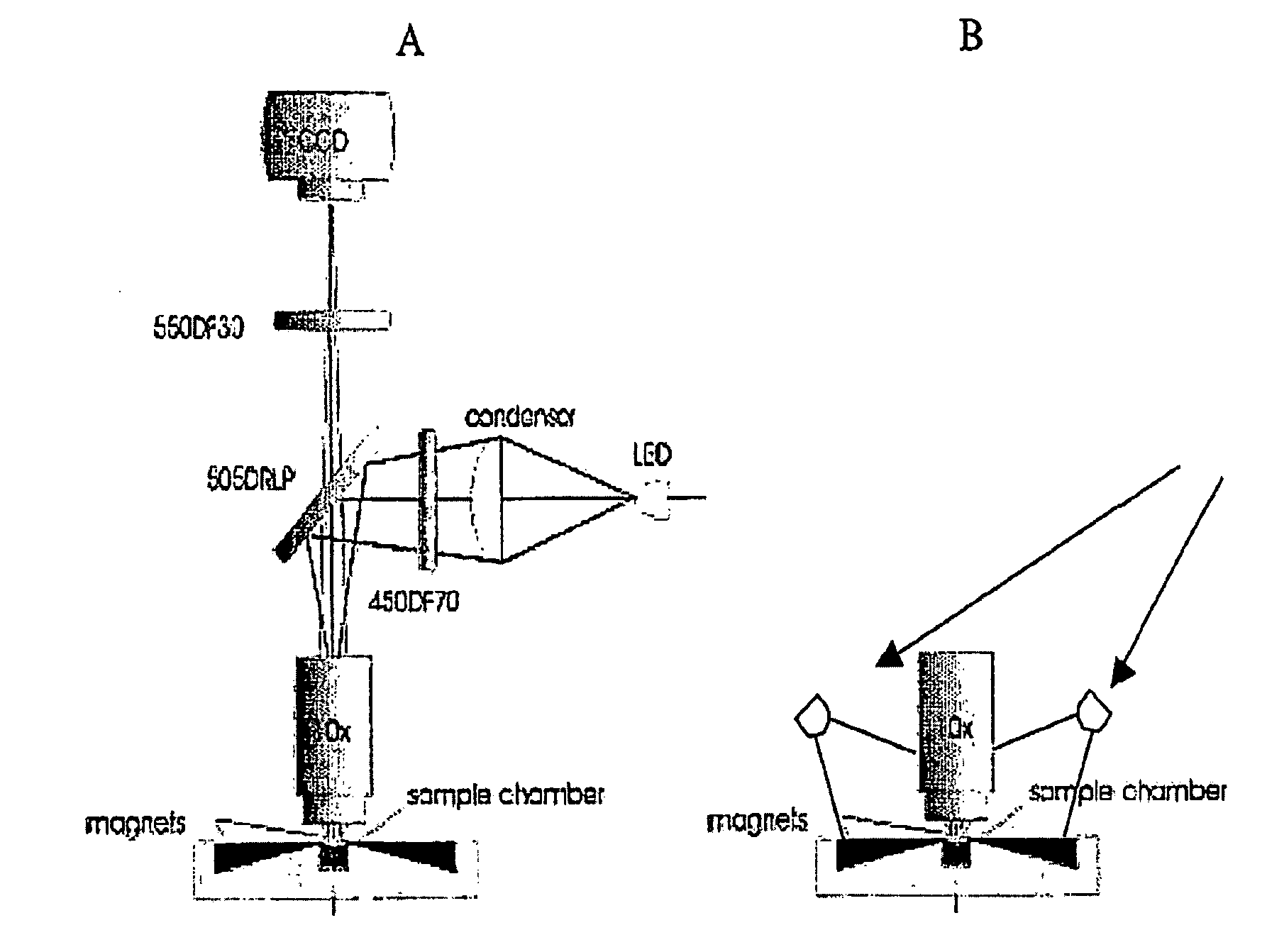
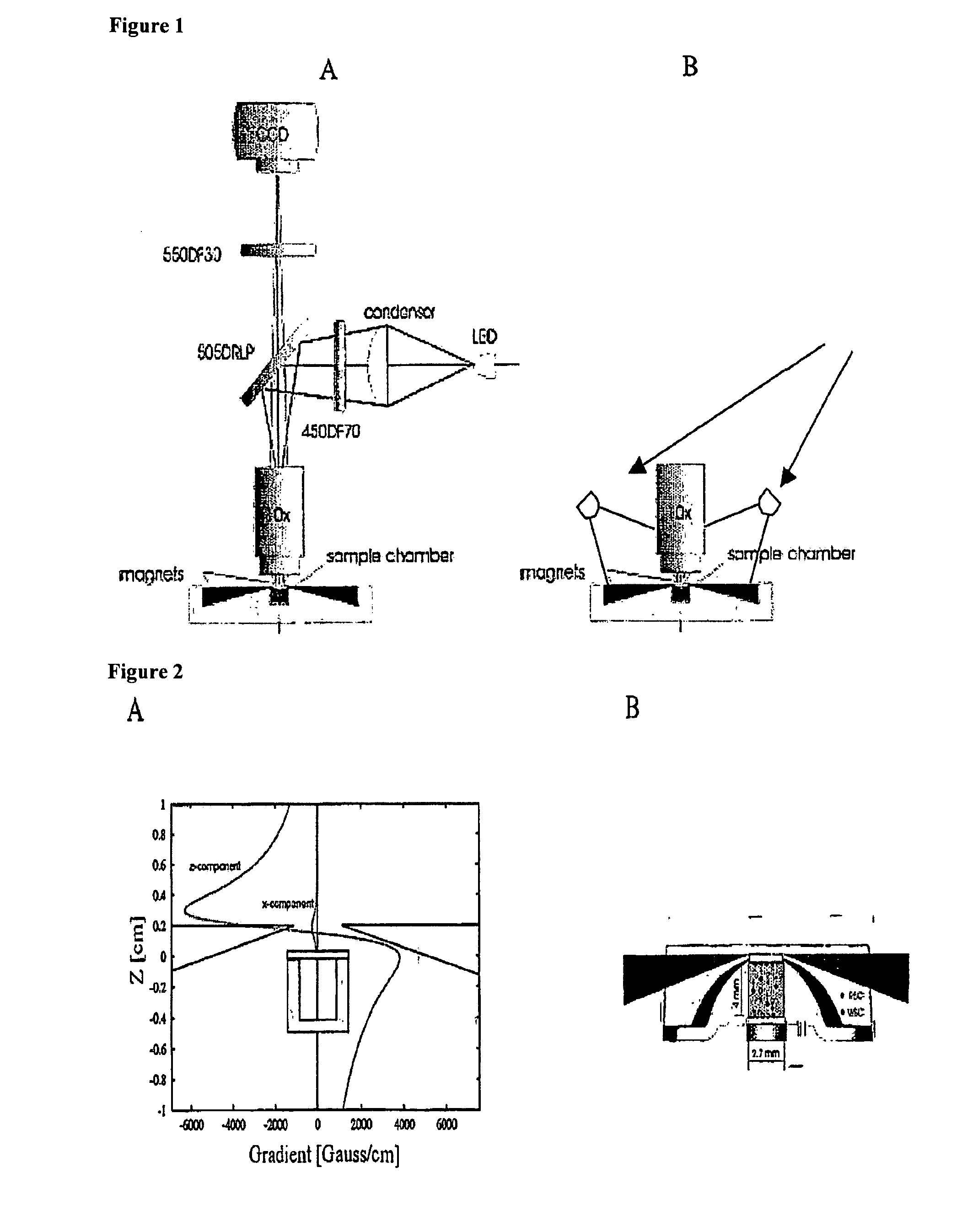
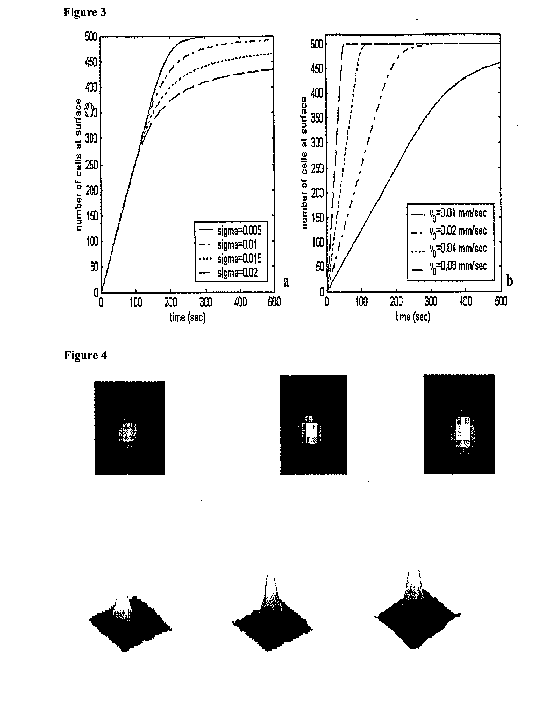
The enumeration of cells in fluids by flow cytometry is widely used across many disciplines such as assessment of leukocyte subsets in different bodily fluids or of bacterial contamination in environmental samples, food products and bodily fluids. For many applications the cost, size and complexity of the instruments prevents wider use, for example, CD4 analysis in HIV monitoring in resource-poor countries. The novel device, methods and algorithms disclosed herein largely overcome these limitations. Briefly, all cells in a biological sample are fluorescently labeled, but only the target cells are also magnetically labeled. The labeled sample, in a chamber or cuvet, is placed between two wedge-shaped magnets to selectively move the magnetically labeled cells to the observation surface of the cuvet. An LED illuminates the cells and a CCD camera captures the images of the fluorescent light emitted by the target cells. Image analysis performed with a novel algorithm provides a count of the cells on the surface that can be related to the target cell concentration of the original sample. The compact cytometer system provides a rugged, affordable and easy-to-use technique, which can be used in remote locations.

Description

CROSS REFERENCE TO RELATED APPLICATIONS[0001]This application is a divisional application of U.S. Ser. No. 10 / 903,798, filed 30 Jul. 2004, which is the U.S. national stage of PCT / US03 / 04468, filed 14 Feb. 2003, which claims the benefit of claim priority under 35 U.S.C. 365(c) to U.S. Provisional Application No. 60 / 357, 170, filed 14 Feb. 2002, all incorporated by reference herein.FIELD OF THE INVENTION[0002]This invention relates generally to simple and low cost electronic optical devices, methods and algorithms for enumeration of microscopic particles distributed in a two-dimensional plane. The novel counting techniques are particularly applicable to enumerating is magnetically selected fluorescent cells in complex biological specimens such as blood by means of an inexpensive cell cytometer.BACKGROUND OF THE INVENTION[0003]The enumeration of absolute levels of cells and their subsets in body fluids is of primary importance in determining the state of health of human beings and mamm...

Claims


Application Information

Patent Timeline
Patent Type & Authority Applications(United States)
IPC IPC(8): G06K9/00G01N21/64C12M1/34C12Q1/06G01N15/00G01N15/14G01N21/78G01N33/53G01N33/543G01N33/545
CPCG01N15/1463G01N15/1475G01N33/5094Y10T436/25375G01N2015/008G01N2015/1486Y10T436/101666G01N33/54333G01N33/48G01N37/00C12Q1/00G06F18/00
Inventor TIBBE, ARJANGREVE, JANGOHEL, DHANESHDROOG, ERIKTERSTAPPEN, LEON W.M.M.
Owner UNIVERSITY OF TWENTE
Learn More