Methods for Assaying MC1R Variants and Mitochondrial Markers in Skin Samples

a technology of mitochondrial markers and variants, which is applied in the field of methods for predicting, diagnosing and monitoring skin states and skin diseases, can solve the problems of increasing the chances of infection, not necessarily effective detection methods based on visible observations, and not providing an enriched sample of cells on the surface of skin, so as to achieve the effect of high risk

Inactive Publication Date: 2011-02-24
MITOMICS
View PDF6 Cites 5 Cited by
  • Summary
  • Abstract
  • Description
  • Claims
  • Application Information

AI Technical Summary

Problems solved by technology

Skin disease represents a major health care challenge in today's world.
Detection methods that rely on visible observations, however, are not necessarily effective for diagnosing skin states or diseases, and do not detect risk or disease until after clinical manifestation.
Furthermore, invasive methods such as biopsies, are not only traumatic for a subject being tested, they also increase the chances of infection.
These methods must also be performed by a medical practitioner in order to be safely conducted, and typically do not provide an enriched sample of cells on the surface of skin, which are the cells generally involved in a reaction.
Ultraviolet Radiation (UVR) Damage
Unknown or poorly quantifiable factors often contribute to skin damage as a result of ultraviolet radiation (UVR).
Behavioural factors play a large role in the severity and level of damage to skin from UVR but are extremely difficult to assess clinically as they are dependent upon the accuracy of self-reporting and often a flawed awareness of a patient's sun lifestyle habits.
Many individuals who use sunscreen regularly also use it improperly, failing to apply it in sufficient quantity or to reapply at the recommended intervals, creating a false sense of protection that can lead to increased exposure to UVR.
Human skin tissue is highly complex and comprises numerous cell types.
This approach is limited because non-melanoma skin cancer (NMSC) is predominantly formed on body sites which are “usually” exposed to the sun when outdoors as opposed to sites that are “occasionally” exposed to the sun (Armstrong, 2004).
In addition, these samples were obtained by painful methods of skin collection previously known in the art.
Evaluation of phenotypic characteristics associated with sun sensitivity alone would not be able to elucidate this increased risk.
Current methods for the collection of skin samples for use in the diagnosis or characterization of diseases, such as skin cancer, include invasive or painful methods that can cause substantial discomfort to the individual being tested.
Such invasive techniques have other disadvantages including risk of infection, inconvenience of sample collection, and the possibility that collected samples can be lost or misidentified.
Further, the time and cost associated with these invasive test methods make it difficult to rapidly genotype and assess DNA damage for large populations of individuals.

Method used


Image

  • Methods for Assaying MC1R Variants and Mitochondrial Markers in Skin Samples
  • Methods for Assaying MC1R Variants and Mitochondrial Markers in Skin Samples
  • Methods for Assaying MC1R Variants and Mitochondrial Markers in Skin Samples

Examples


example 1

Analysis of 3895 by Human mtDNA Deletion

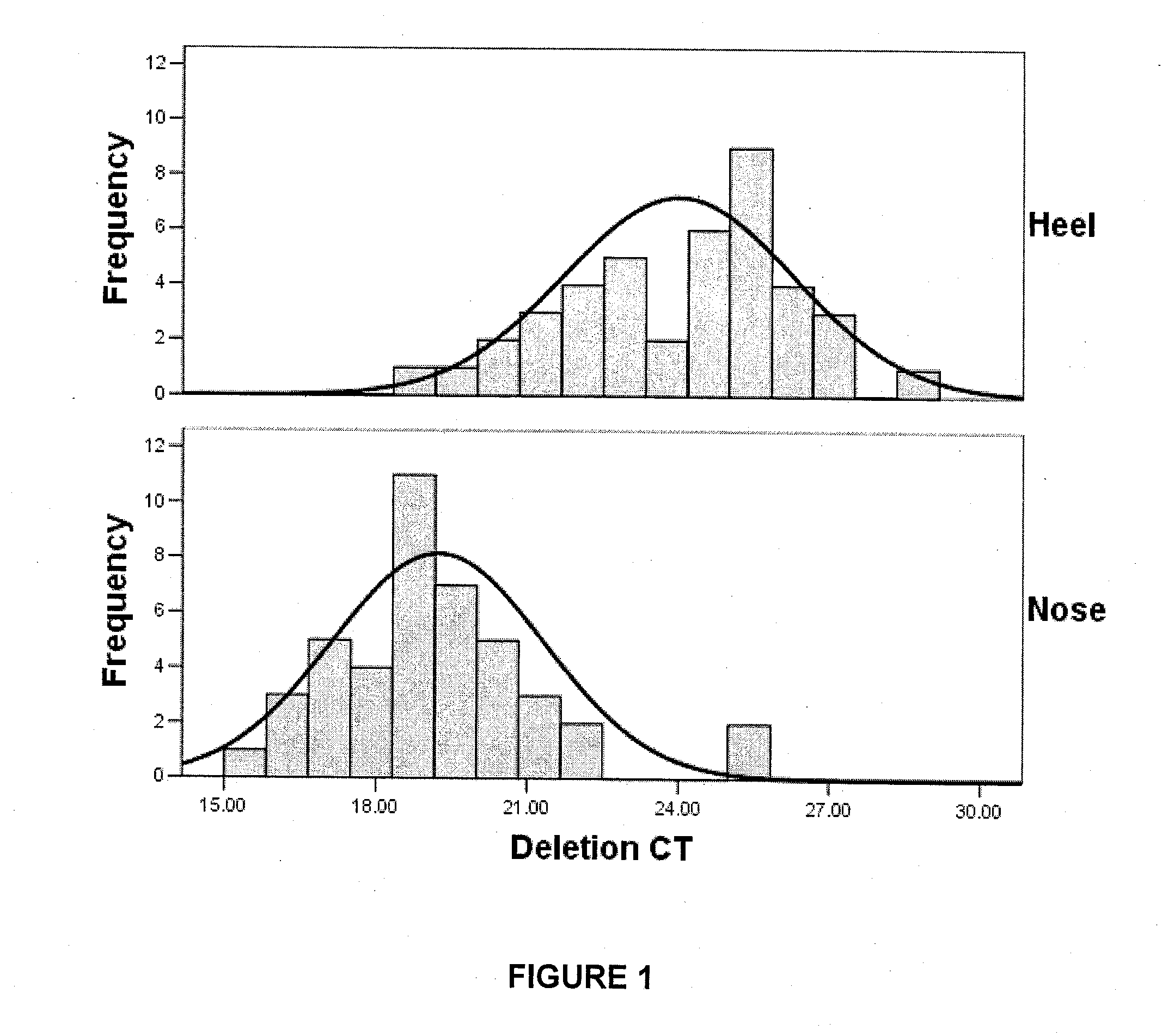
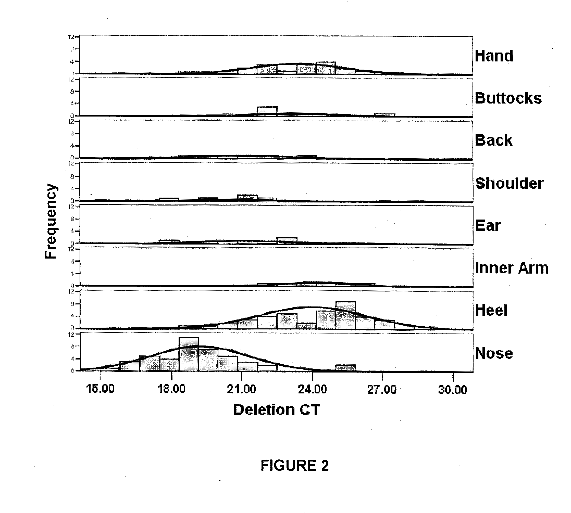
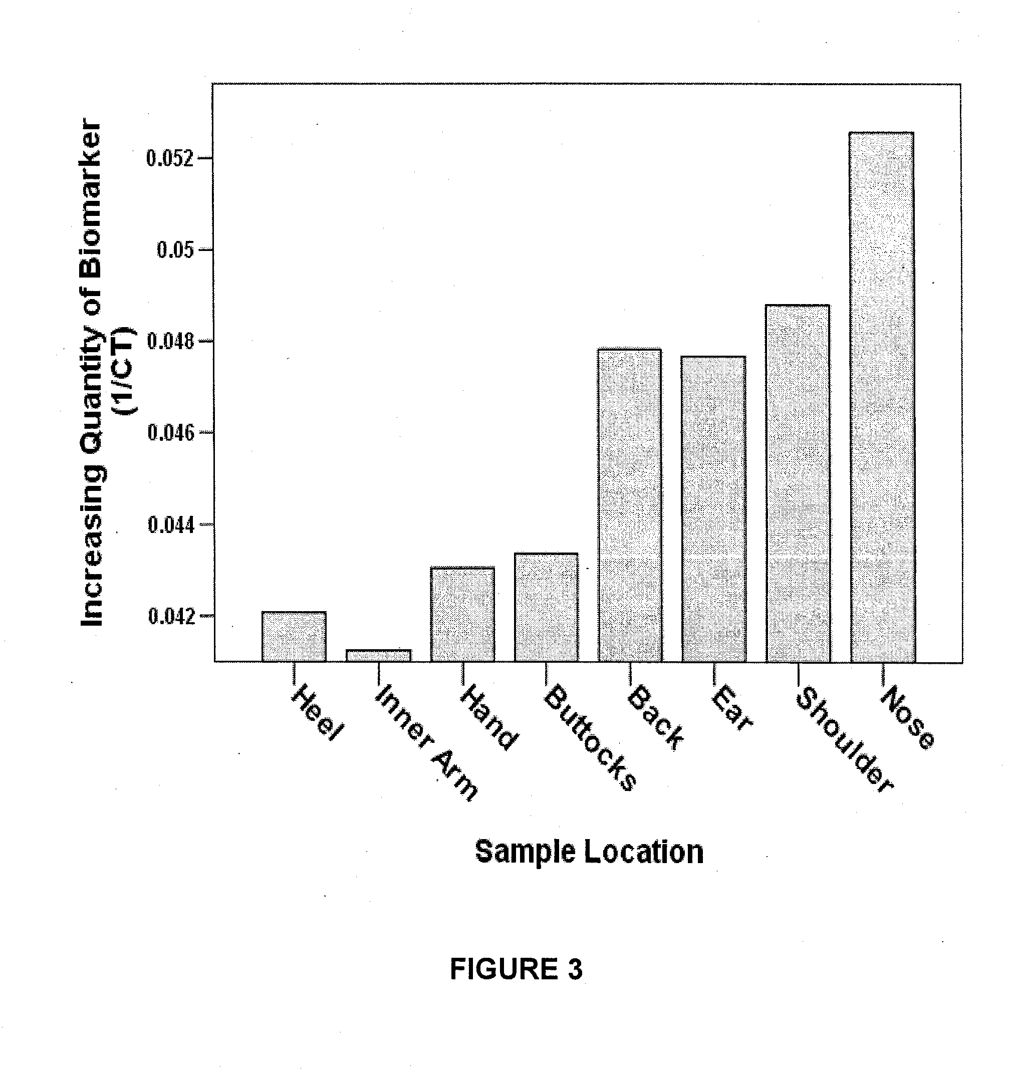
[0137]The method of the present invention was used to analyze the 3895 by mtDNA deletion identified in PCT application no. WO / 06 / 111029. Collection and extraction of the mtDNA was conducted as provided below.

[0138]1. Skin samples were collected by swabbing a skin site approximately 15 times with a sterile swab. Skin samples were collected from heel (n=41), nose (n=43), inner arm (n=20), ear (n=5), shoulder (n=5), buttock (n=5), and back (n=5).

[0139]2. mtDNA was extracted using a commercially available kit (QiaAMP™ DNA Micro Kit, product no. 56304, Qiagen, Maryland USA) according to the manufacturer's protocol.

[0140]3. Double stranded DNA was quantified using the HS-DNA Quant-it™ dsDNA HS Assay Kit (product no. Q32851, Invitrogen, California USA) on the Qubit™ Fluorometer (product no. Q32857), Invitrogen, California USA).

[0141]4. The level of the 3895 by deletion was then quantified by real-time PCR (rt-PCR) using the iQ Sybr Green Supermix™ (p...

example 2

Comparison of Skin Collection Methods

[0162]Five different non-invasive skin collection methodologies were tested in order to identify which, if any, would yield sufficient quantity and quality of nucleic acids for molecular analyses such as quantitative real-time PCR. The five methods tested were:[0163]Tapelift using surgical tape;[0164]Biore® adhesive strip;[0165]Sterile swab wetted with 8% mandelic acid;[0166]Sterile swab wetted with distilled water; and[0167]Wax strip.

[0168]The tapelift, Biore strip and wax strip were applied to the surface of the skin following the application of 70% isopropanol to sterilize the area. Firm pressure was applied and then the tape or strip was removed quickly. The swabs were first deposited in a sterile solution of either 8% mandelic acid, or distilled water and then rubbed firmly on the skin site of interest after the skin had been cleaned with 70% isopropanol.

[0169]Following the collection of skin cells from 3 individuals each collection medium w...

example 3

Comparison of Additional Skin Collection Methods

[0216]In this example five additional methods for the non-invasive collection of skin samples were tested in order to identify which, if any, would yield sufficient quantity and quality of nucleic acids for molecular analyses such as quantitative real-time PCR.

[0217]From a single individual, skin samples were collected twice using the following methods:[0218]scraping of skin using a sterile surgical blade[0219]scraping of skin using a wooden scraper[0220]sticky surface of an adhesive pad (CapSure™ Clean-up Pad, Arcturus)[0221]film from LCM MacroCap™ (Arcturus)[0222]heated film from LCM MacroCap™ (Arcturus)

[0223]The skin was first prepared by cleansing with a 70% isopropanol wipe. The wooden scraper and the surgical blade were passed firmly over the skin surface to remove skin cells and then deposited into a centrifuge tube. The adhesive pad and films were pressed firmly against the skin without rubbing to collect skin cells.

[0224]The m...


PUM

PropertyMeasurementUnit
timeaaaaaaaaaa
skin colouraaaaaaaaaa
frequency ofaaaaaaaaaa

Abstract

The present invention relates to methods for predicting, diagnosing and monitoring skin states and skin diseases. The methods combine the use of non-invasive skin collecting techniques with one or more assays for determining mitochondrial DNA (mtDNA) aberrations and Melanocortin 1 Receptor (MC1R) variants, thereby providing a comprehensive tool for identifying, predicting and / or monitoring photoageing, ultraviolet radiation (UVR) damage or skin disease. The methods of the invention may also be effective in screening for new therapeutic agents, skin care products and treatment regimes, and may also be useful for monitoring the response of a subject to a preventative or therapeutic treatment.

Description

CROSS-REFERENCE TO RELATED APPLICATIONS[0001]This application claims priority from PCT Application No. PCT / CA2007 / 001790, filed Oct. 11, 2007, and U.S. Application No. 60 / 999,074, filed Oct. 15, 2007, the entire contents of which are incorporated herein by reference.FIELD OF THE INVENTION[0002]The present invention relates to methods for predicting, diagnosing and monitoring skin states and skin diseases. In particular, the present invention pertains to methods coupling non-invasive skin sampling techniques with assays for determining mitochondrial mutations and Melanocortin 1 Receptor (MC1R) variants for use as a comprehensive tool for predicting and monitoring disease, photoageing and ultraviolet radiation (UVR) damage. The methods are also useful for assessing the effectiveness of and response to therapeutic agents, skin care products and treatment regimes.BACKGROUND OF THE INVENTION[0003]Skin disease represents a major health care challenge in today's world. With more than one m...

Claims


Application Information

Patent Timeline
Patent Type & Authority Applications(United States)
IPC IPC(8): C12Q1/68
CPCC12Q1/6883C12Q2600/156C12Q2600/136G01N33/15G01N33/48
Inventor PARR, RYANBIRCH-MACHIN, MARKHARBOTTLE, ANDREWTHAYER, ROBERTCREED, JENNIFERMAGGRAH, ANDREAROBINSON, KERRYDAKUBO, GABRIELREGULY, BRIANMAKI, KATRINA
Owner MITOMICS
Why Eureka
Try Eureka