Minimally invasive endoscopic systems for placing intramedullary nails and methods therefor

a technology of intramedullary nail and minimally invasive, applied in the field of systems, devices and methods for treating fractures, can solve the problems of reducing the risk of hardware failure, reducing the risk of post-operative wound drainage, osteomyelitis, sepsis, and death associated with wound drainage, and reducing the risk of post-operative wound infection. , the effect of reducing the risk of bleeding

Inactive Publication Date: 2011-03-03
BRANDON MARK LEONARD
View PDF47 Cites 14 Cited by
  • Summary
  • Abstract
  • Description
  • Claims
  • Application Information

AI Technical Summary

Benefits of technology

[0018]The present desirably provides an endoscopic system for placing intramedullary nails in long bones. The intramedullary nailing system disclosed herein preferably results in less trauma to the soft tissues caused by blunt dissection. In one embodiment, the present invention uses a series of tissue dilators so as to provide for gentle spreading of soft tissues as opposed to sharp soft tissue dissection, which may injury nerves, tendons and blood vessels in soft tissue between the skin and the long bone. The intramedullary nailing system of the present invention also preferably results in less bleeding and shorter surgical procedures than occur when using sharp soft dissection because the present invention obviates the need for suture repair in the soft tissue deep to the dermis. The present invention potentially also preferably lowers the risk of deep vein thromboses, lowers the risk of intra-operative myocardial infarction (heart attack) and lowers mortality risk due to the shortened surgical procedure time. In one embodiment, the present invention also improves healing of the soft tissue deep in the dermis due to the decreased trauma to the soft tissue, improves positioning of the guide wires and the intramedullary nail and all corresponding hardware devices due to the ability to directly visualize and select the optimal long bone cortical entry site of the guides wires and the intramedullary nail and all corresponding devices. The present invention also decreases the likelihood of hardware failure due to improved hardware positioning in the affected long bone, potentially decreases the risk of post-operative wound drainage due to decreased trauma to the soft tissues and decreased risk of traumatizing blood vessels in the soft tissue, and potentially decreases risk of wound infection, osteomyelitis, sepsis, septic shock and death associated with wound drainage.

Problems solved by technology

The present invention also decreases the likelihood of hardware failure due to improved hardware positioning in the affected long bone, potentially decreases the risk of post-operative wound drainage due to decreased trauma to the soft tissues and decreased risk of traumatizing blood vessels in the soft tissue, and potentially decreases risk of wound infection, osteomyelitis, sepsis, septic shock and death associated with wound drainage.

Method used


Image

  • Minimally invasive endoscopic systems for placing intramedullary nails and methods therefor
  • Minimally invasive endoscopic systems for placing intramedullary nails and methods therefor
  • Minimally invasive endoscopic systems for placing intramedullary nails and methods therefor

Examples


Embodiment Construction

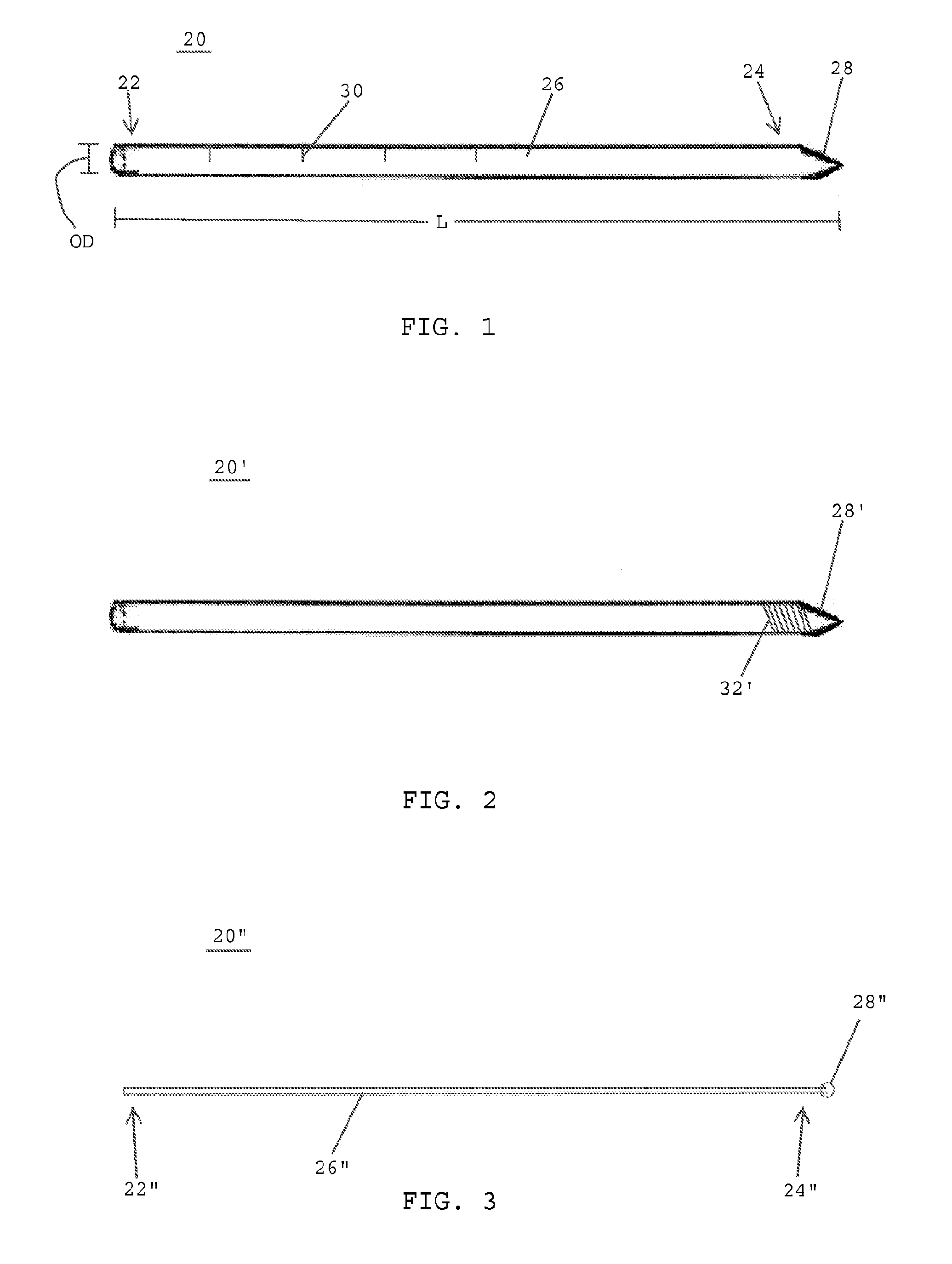
[0044]Referring to FIG. 1, in one embodiment, an intramedullary nail placement system includes a guide wire 20 having a proximal end 22, a distal end 24, and an elongated shaft 26 that desirably extends between the proximal and distal ends. The elongated shaft 26 of the guide wire 20 is preferably smooth. The distal end 24 of the guide wire 20 desirably has a pointed tip 28 for passing through skin, soft tissue and bone. The pointed tip 28 may also function to hold the guide wire 20 in place after the distal end 24 has been inserted into bone.

[0045]In one embodiment, the guide wire 20 is preferably made of stainless steel. In one embodiment, the guide wire is made of a durable, biocompatible material. In one embodiment, the guide wire 20 has an outer diameter OD of approximately 3.0 mm and a length L of between about 32-150 cm. In one embodiment, the guide wire 20 preferably includes visual indicia 30 provided along the outer surface of the elongated shaft 26 to provide an indicatio...


PUM


Abstract

A system for percutaneously placing intramedullary nails includes a guide wire (20) percutaneously insertable into tissue (T) for forming an opening in the tissue and engaging a cortex of a bone (FB), at least one tissue dilator (40A) slideable over an outer surface of the guide wire (20) for widening the tissue opening formed by said guide wire, and a cannula (60) slideable over an outer surface of said at least one tissue dilator (40A) for further widening the tissue opening and providing access to the cortex of the bone. The cannula (60) has an outer wall (66) extending between proximal and distal ends thereof, the outer wall having a smaller portion including a door (76) that extends between the proximal end distal ends of the cannula and a larger portion that bounds the door. The door (76) is selectively detachable from the larger portion of the outer wall (66) for opening an elongated slot (74) in the outer wall that extends from the proximal end to the distal end of the cannula.

Description

CROSS-REFERENCE TO RELATED APPLICATIONS[0001]The present application claims the benefit of U.S. Provisional Application Ser. No. 61 / 141,346, filed Dec. 30, 2008, entitled “Endoscopic Minimally Invasive System for Placement of Intramedullary Nails,” the disclosure of which is hereby incorporated by reference herein.BACKGROUND OF THE INVENTION[0002]1. Field of the Invention[0003]The present invention generally relates to systems, devices and methods for treating bone fractures and more specifically relates to systems, devices and methods for treating fractures in long bones using intramedullary nails.[0004]2. Description of the Related Art[0005]Vertebrates maintain their upright posture and resist the earth's gravitational pull with the aid of their bony structure. Vertebrates' bones are formed of a hard outer cortex that surrounds a soft, spongy cancellous portion. The cortical portion of the bone is stiffer than cancellous portion. In a bony structure, the long bones are those bones...

Claims


Application Information

Patent Timeline
Patent Type & Authority Applications(United States)
IPC IPC(8): A61B17/56A61F2/958
CPCA61B17/1725A61B17/175A61B17/3421A61B17/72A61B2019/521A61B17/8897A61B19/5202A61B2017/0046A61B2017/3443A61B17/8872A61B90/30A61B2090/309
Inventor BRANDON, MARK LEONARD
Owner BRANDON MARK LEONARD
Why Eureka
Try Eureka