Cell detection, monitoring and isolation method

a cell detection and monitoring technology, applied in the field of cell detection, monitoring and isolation methods, can solve the problems of impeded non-invasive tests for determining fetal genetic abnormalities, invasive approaches, etc., and achieve the effect of facilitating the method of treatmen

Inactive Publication Date: 2011-04-14
MURDOCH CHILDRENS RES INST
View PDF7 Cites 4 Cited by
  • Summary
  • Abstract
  • Description
  • Claims
  • Application Information

AI Technical Summary

Benefits of technology

[0025]Furthermore, the ability to detect rare cell types facilitates methods of treatment involving rare cells. For example, autologous or heterologous stem cells may be identified and the population expanded ex vivo before being re-introduced to the subject or another subject. In addition, using the CNV signature of the present invention, the fate and / or distribution of these stem cells can be followed and monitored.

Problems solved by technology

However, this approach can be invasive, particularly in the prenatal setting.
Non invasive tests for determining fetal genetic abnormalities are impeded by the high proportion of maternal cells to fetal cells.

Method used


Image

  • Cell detection, monitoring and isolation method
  • Cell detection, monitoring and isolation method
  • Cell detection, monitoring and isolation method

Examples


example 1

Detection of Donor Cells in Bone Marrow Transplant Patients

[0099]A copy number deletion present in the transplant patient or recipient's DNA which is not present in the donor's DNA is detected to distinguish the donor cells in the recipient.

[0100]By using two color FISH test using a probe within and an adjacent probe outside the copy number deletion, the recipient non-deleted locus is one color such as yellow (red+green) and the copy number deletion is red. If the copy number deletion is not present, the donor cells show a signal which is two yellow signals (red+green).

[0101]The test is also done with only one color (e.g. red) using a probe within the recipient copy number deletion. Recipient cells show one red signal and the donor cells show two red signals. As false positive cells showing two signals are unlikely, i.e. Recipient cells masquerading as donor cells, this single color test is applicable here.

[0102]This test is not restricted to using host / recipient specific copy numbe...

example 2

Detection of Down Syndrome

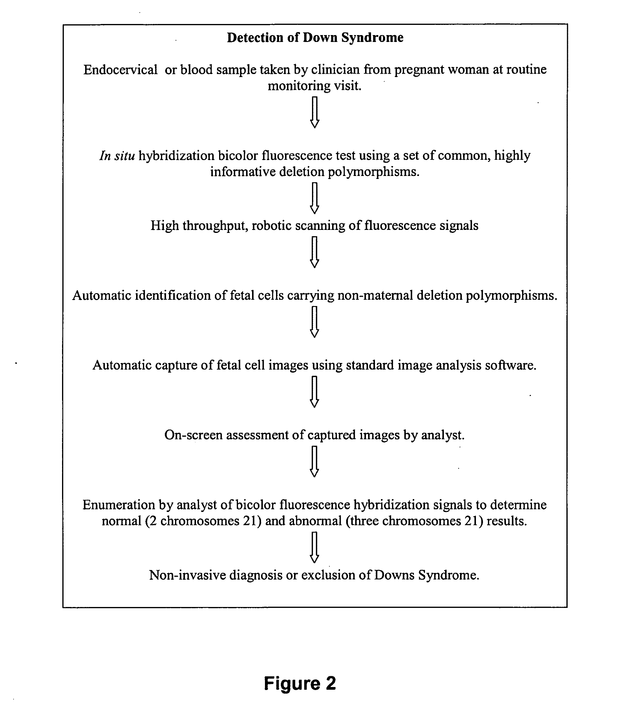
[0105]A schematic protocol for detecting Down Syndrome is shown in FIG. 2.

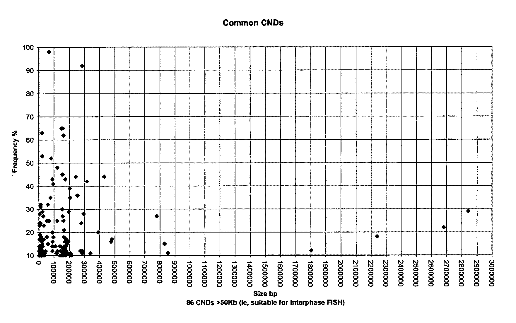
[0106]Individuals with Down Syndrome have trisomy 21, i.e. have three instead of two chromosomes 21. The assay is a non-invasive pre-natal test for trisomy 21, which is performed in situ on the microscope slide, thus avoiding the need to manually isolate fetal cells. The test is based on the analysis of a class of DNA polymorphism called Copy Number Variation (CNV). Fetal cells are distinguished from maternal cells through detection of non-maternal copy number variations. Specifically, deletion polymorphisms are used that have been inherited by the fetus from the father and are not carried by the mother. As only fetal cells carry these non-maternal polymorphisms, they can be distinguished from maternal cells. Deletion polymorphisms are located throughout the human genome. By using deletion polymorphisms located on chromosome 21 with the in situ assay, not only are fetal cells identifi...

example 3

Detection of Cells from a Fetus with Down Syndrome Method 1

[0110]An endocervical sample is taken by a clinician from a pregnant woman at routine monitoring visit at approximately seven weeks gestation. Such a sample contains mainly maternal endothelial cells with small numbers of cells of fetal origin. An aliquot of the cell preparation is pipetted onto a standard glass microscopy slide and air dried at ambient temperature.

[0111]The Fluorescence in situ hybridization (FISH) test discriminates maternally and paternally derived DNA loci within the cells. Only fetal cells contain paternally derived loci and this is the basis on which fetal cells are distinguished from maternal cells. Any paternally derived locus is suitable for identification of fetal cells but those on chromosome 21 are most suitable for simultaneous assay of the number of chromosome 21s present for diagnosis of Down syndrome (see FIG. 3).

[0112]A locus is selected either by:

(1) prior knowledge of its presence through ...


PUM

PropertyMeasurementUnit
temperaturesaaaaaaaaaa

Abstract

The present invention relates generally to a method for identifying or distinguishing one type of cell from other cells within a population of cells. The present invention further provides the detection, monitoring and isolation of sub-populations cell types within a population of cells including a biological entity comprising such cell types. Kits, diagnostic agents and panels of nucleic acid probes for identifying and distinguishing cell types also form part of the present invention. The detection of particular cell types is based upon the use of a labelled probe that hybridizes to chromosomal DNA at the flanking sequences of a deletion. Alternatively two probes with distinguishable reporter molecules are used wherein only one probe is capable of hybridizing to chromosomal DNA in one cell type whereas both are capable of hybridizing to chromosomal DNA in another cell type. The methods are useful in the identification of cells with copy number variations, chromosomal deletions, additions or aberrations.

Description

RELATED APPLICATION DATA[0001]This application is associated with and claims priority from Australian Patent Application No. 2007905324, filed on 28 Sep. 2007, the content of which is incorporated herein in its entirety by reference.FIELD[0002]The present invention relates generally to a method for identifying or distinguishing one type of cell from other cells within a population of cells. The present invention further provides the detection, monitoring and isolation of sub-populations cell types within a population of cells including a biological entity comprising such cell types. Kits, diagnostic agents and panels of nucleic acid probes for identifying and distinguishing cell types or for nucleic acid probes useful for same also form part of the present invention.BACKGROUND[0003]Reference to any prior art in this specification is not, and should not be taken as, an acknowledgment or any form of suggestion that this prior art forms part of the common general knowledge in any count...

Claims


Application Information

Patent Timeline
Patent Type & Authority Applications(United States)
IPC IPC(8): C12Q1/68
CPCC12Q1/6881C12Q1/6841C12Q2600/156
Inventor BRUNO, DAMIEN LUISSLATER, HOWARD ROBERT
Owner MURDOCH CHILDRENS RES INST
Why Eureka
Try Eureka