System and method for imaging with enhanced depth of field

a technology of depth of field and enhanced image, applied in the field of imaging, can solve the problems of imperfect focus in the acquired image, unfavorable sample collection, and further exacerbated problems, and achieve the effect of enhancing the depth of field

Inactive Publication Date: 2011-04-21
GENERAL ELECTRIC CO
View PDF21 Cites 23 Cited by
  • Summary
  • Abstract
  • Description
  • Claims
  • Application Information

AI Technical Summary

Benefits of technology

[0009]In accordance with aspects of the present technique, a method for imaging is presented. The method includes acquiring a plurality of images corresponding to at least one field of view at a plurality of sample distances. Furthermore, the method includes determining a figure of merit corresponding to each pixel in each of the plurality of acquired images. The method also includes for each pixel in each of the plurality of acquired images identifying an image in the plurality of images that yields a best figure of merit for that pixel. Moreover, the method includes generating an array for each image in the plurality of images. In addition, the method includes populating the arrays based u...

Problems solved by technology

Additionally, even within a single field of view of the microscope, it may not be possible to bring an entire sample into focus at one time merely by adjusting the optics.
Moreover, this problem is further exacerbated in the case of a scanning microscope, where the image to be acquired is synthesized from multiple fields of view.
The mechanism for translating the slide in a plane normal to the optical axis of the microscope may also introduce imperfections in image quality while raising, lowering and tiling the slide, thereby leading to imperfect focus in the acquired image.
Additionally, the problem of imperfect focus is further aggravated in an event that a sample disposed on a slide is not substantially flat within a single field of view of the microscope.
Specifically, these samples disposed on the slide may have significant amounts of material that is out of a plane of the slide.
However, use of these techniques results in inadequate focus when the depth of the ...

Method used


Image

  • System and method for imaging with enhanced depth of field
  • System and method for imaging with enhanced depth of field
  • System and method for imaging with enhanced depth of field

Examples


Embodiment Construction

[0019]As will be described in detail hereinafter, a method and system for imaging a sample, such as a sample that has significant material out of a plane of a slide, while enhancing image quality and optimizing scanning speed are presented. By employing the method and device described hereinafter, enhanced image quality and substantially increased scanning speed may be obtained, while simplifying the clinical workflow of sample scanning

[0020]Although, the exemplary embodiments illustrated hereinafter are described in the context of a digital microscope, it will be appreciated that use of the imaging device in other applications, such as, but not limited to, a telescope, a camera, or a medical scanner such as an X-ray computed tomography (CT) imaging system, are also contemplated in conjunction with the present technique.

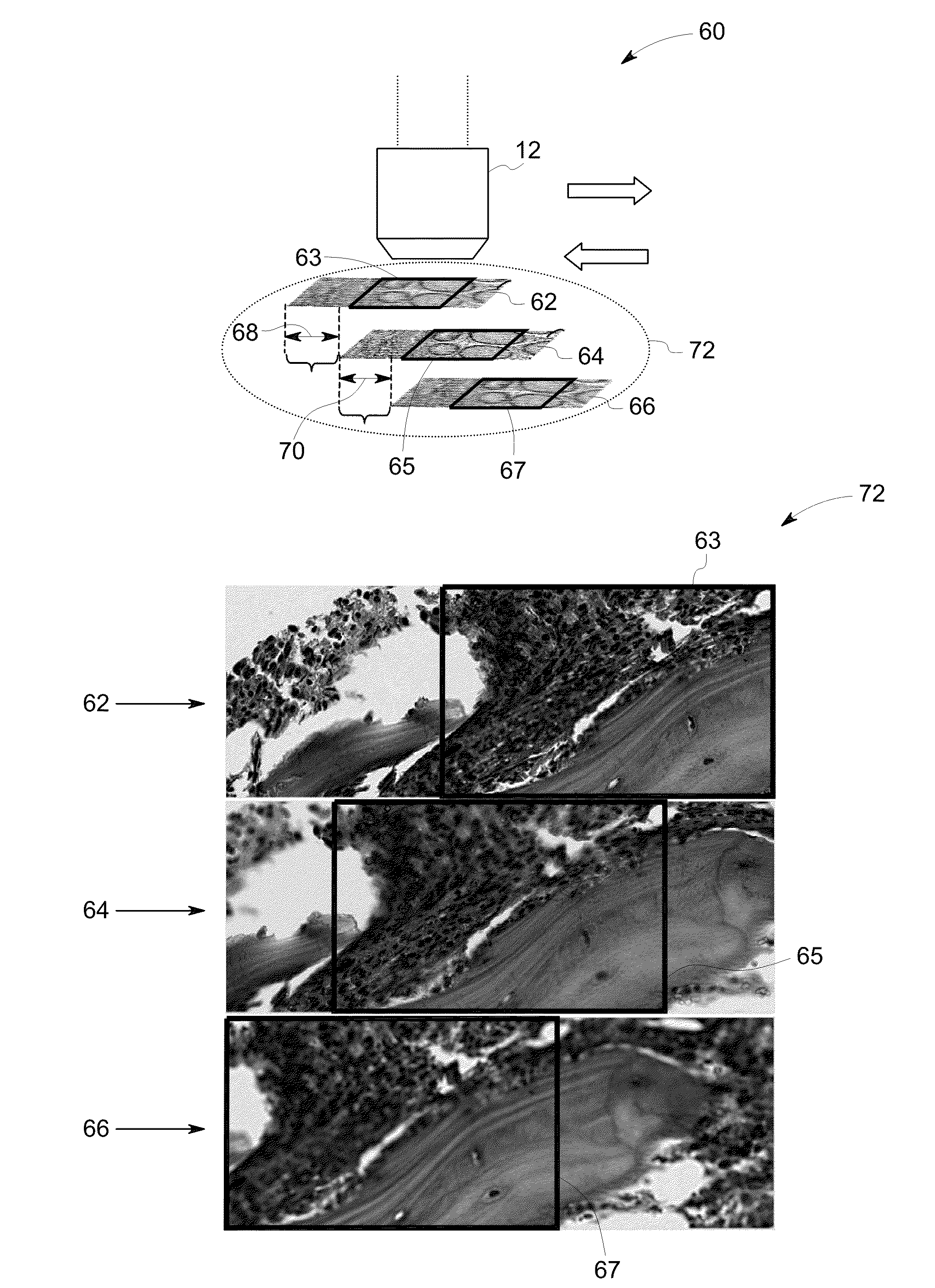
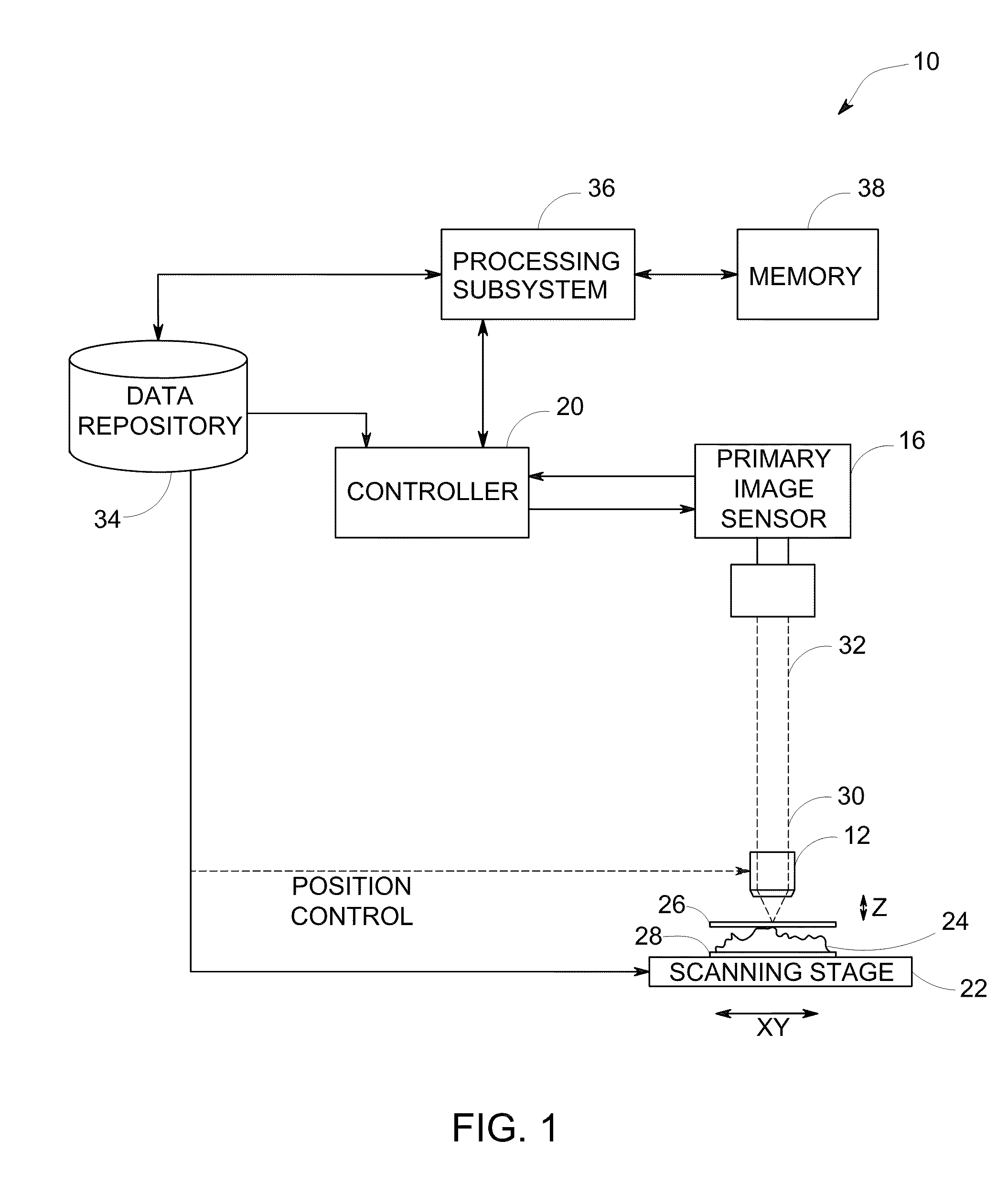
[0021]FIG. 1 illustrates one embodiment of an imaging device 10, such as a digital optical microscope, that incorporates aspects of the present invention. The imagin...


PUM


Abstract

A method for imaging is presented. The method includes acquiring a plurality of images corresponding to at least one field of view at a plurality of sample distances. Furthermore, the method includes determining a figure of merit corresponding to each pixel in each of the plurality of acquired images. The method also includes for each pixel in each of the plurality of acquired images identifying an image in the plurality of images that yields a best figure of merit for that pixel. Moreover, the method includes generating an array for each image in the plurality of images. In addition, the method includes populating the arrays based upon the determined best figures of merit to generate a set of populated arrays. Also, the method includes processing each populated array in the set of populated arrays using a bit mask to generate bit masked filtered arrays. Additionally, the method includes selecting pixels from each image in the plurality of images based upon the bit masked filtered arrays. The method also includes processing the bit masked arrays using a bicubic filter to generate a filtered output. Further, the method includes blending the selected pixels as a weighted average of corresponding pixels across the plurality of images based upon the filtered output to generate the composite image having an enhanced depth of field.

Description

BACKGROUND[0001]Embodiments of the present invention relate to imaging, and more particularly to construction of an image with an enhanced depth of field.[0002]Prevention, monitoring and treatment of physiological conditions such as cancer, infectious diseases and other disorders call for the timely diagnosis of these physiological conditions. Generally, a biological specimen from a patient is used for the analysis and identification of the disease. Microscopic analysis is a widely used technique in the analysis and evaluation of these samples. More specifically, the samples may be studied to detect presence of abnormal numbers or types of cells and / or organisms that may be indicative of a disease state. Automated microscopic analysis systems have been developed to facilitate speedy analysis of these samples and have the advantage of accuracy over manual analysis in which technicians may experience fatigue over time leading to inaccurate reading of the sample. Typically, samples on ...

Claims


Application Information

Patent Timeline
IPC IPC(8): G06K9/40G02B21/00
CPCG02B21/367G06T2200/21G06T11/00
Inventor KENNY, KEVIN BERNARDHENDERSON, DAVID LAVAN
Owner GENERAL ELECTRIC CO
Why Eureka
Try Eureka