Computer-Based Method And System For Imaging-Based Dynamic Function Evaluation Of An Organ

a dynamic function and computer-based technology, applied in the field of imaging-based organ function assessment, can solve the problems of inability to detect deterioration, significant delay between impairment in hepatic function and detectable change in serum levels of analytes, cumbersome clearance tests, etc., to facilitate synergetic determination of a physiological function of these organs and facilitate inter-relationship of their function.

Inactive Publication Date: 2011-04-28
BAYER PHARMA AG
View PDF0 Cites 14 Cited by
  • Summary
  • Abstract
  • Description
  • Claims
  • Application Information

AI Technical Summary

Benefits of technology

[0023]Some embodiments provide for simultaneous determination of a function of more than one organ at the same time, for instance of a liver and kidneys. In this way a synergetic determination of a physiological function of these organs and an inter-relationship of their function is facilitated. For instance waste products from the liver are carried by the blood to the kidneys. The kidneys filter out these waste products and expel them from the body in urine. Diagnosis of dysfunctions in this delicate organ interaction are thus provided.
[0024]Some embodiments provide for identification of segments or sub-segments of organs that are in dysfunction. This in turn provides for facilitating virtual planning of surgical procedures to treat the dysfunction.

Problems solved by technology

Furthermore there is often a significant delay between impairment in hepatic function and a detectable change in serum levels of analytes.
Clearance tests and analyte measurements are indicators of global liver function, and cannot detect deterioration in hepatocyte function or bile excretion on a segmental or regional level.
Clearance tests are cumbersome and are generally seldom used in clinical practice.
However, this application has not gained a widespread clinical use due to implementation reasons and patient dosage limitations, amongst others.
However, scintigraphic methods are hampered by a number of drawbacks, such as the low resolution and limited anatomic detail in the images obtained.
In the liver, regional differences in hepatocyte function may therefore be hard or impossible to detect.

Method used


Image

  • Computer-Based Method And System For Imaging-Based Dynamic Function Evaluation Of An Organ
  • Computer-Based Method And System For Imaging-Based Dynamic Function Evaluation Of An Organ
  • Computer-Based Method And System For Imaging-Based Dynamic Function Evaluation Of An Organ

Examples


example 1

Subjects

[0177]T1-weighted Gd-EOB-DTPA-enhanced DHCE-MRI was performed on 20 healthy volunteers, 10 men and 10 women, ages ranging from 22 to 45 years. Routine serum liver function tests were performed at inclusion in the study. Test subjects had no history of hepato-biliary disease, previous hepato-biliary surgery or alcohol abuse.

[0178]Protocol

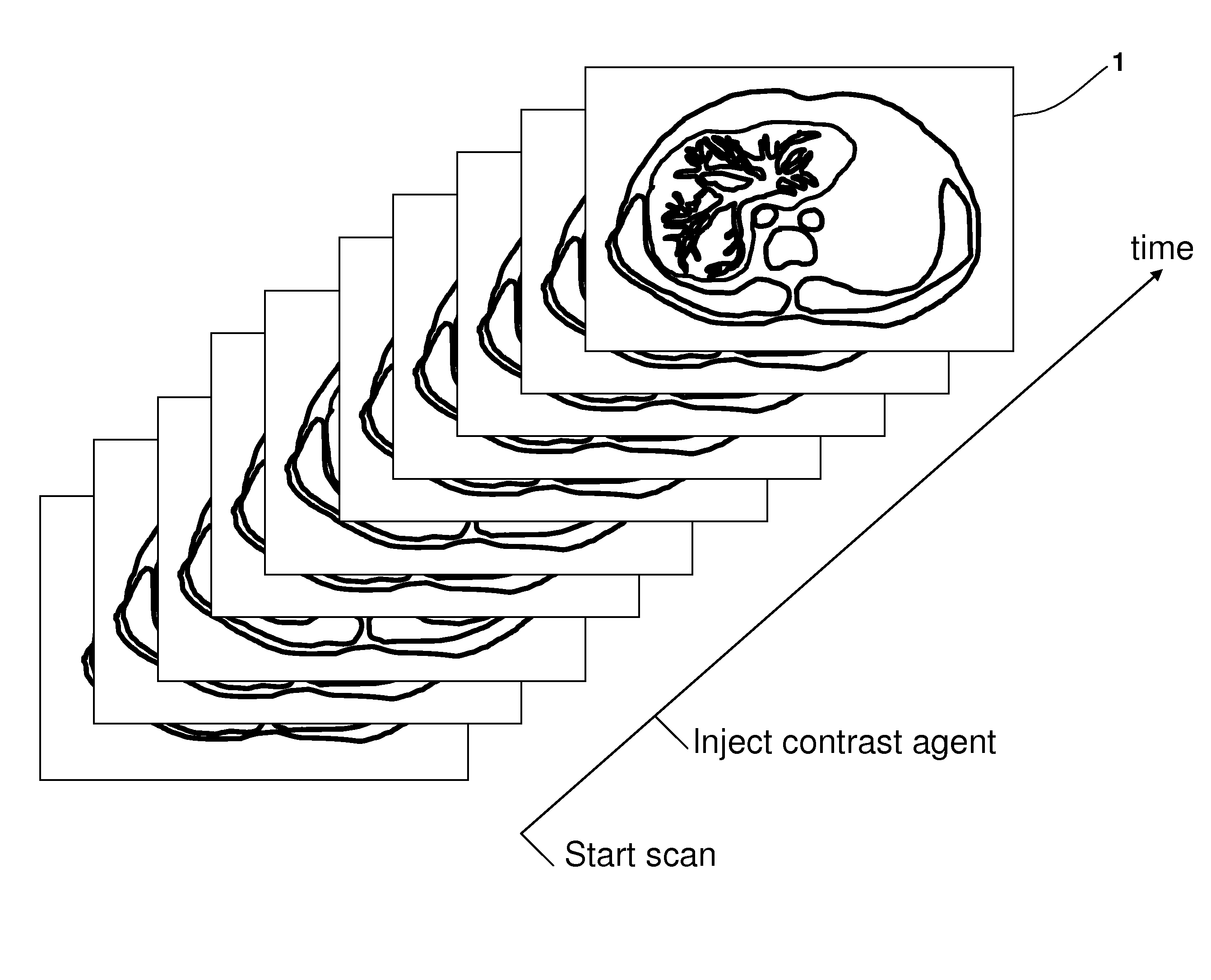
[0179]Data was collected using a Philips Intera 1.5T scanner (Best, Holland), with a Philips four-channel SENSE body coil. A T1-weighted 3D spoiled-gradient-echo pulse sequence (Repetition Time / Echo Time / Flip Angle 4.1 ms / 2.0 ms / 10 deg, Field Of View=415 mm, matrix resolution 256×192, 40 slices, slice thickness 10 mm and SENSE factor R=2) was used. The volume was imaged in a single breath hold at 41 different time points (12 seconds scan time per acquired volume). Three volumes were acquired pre-contrast for baseline calculations, followed by 38 volumes with step-wise increase in sampling intervals. The sampling density was chosen with respec...

example 2

Investigation of Patients with Primary Biliary Cirrhosis (PBC)

[0192]Subjects

[0193]T1-weighted Gd-EOB-DTPA-enhanced DHCE-MRI was performed on 20 healthy volunteers, 10 men and 10 women, and on patients with an established diagnosis of PBC. Routine serum liver function tests were performed at inclusion in the study on the healthy volunteers, and for the patients it was recorded from the most recent visit documented in their clinical charts. The healthy volunteers had no history of hepato-biliary disease, previous hepato-biliary surgery or alcohol abuse. All subjects were asked to be fasting for at least four hours prior to the examination. For each patient, relevant clinical data was documented and together with the results from the liver function tests they were used to calculate the CPS, Mayo risk score and MELD score.

[0194]MR Procedure

[0195]T1-weighted Gd-EOB-DTPA-enhanced DHCE-MRI was performed using a Philips Intera 1.5T scanner (Best, Holland), with a Philips four-channel SENSE ...

example 3

Investigation of Patients with Primary Sclerosing Cholangitis (PSC)

[0201]Subjects

[0202]T1-weighted Gd-EOB-DTPA-enhanced MRI was performed on 20 healthy volunteers, 10 men and 10 women, and on patients with an established diagnosis of PSC. Routine serum liver function tests were performed at inclusion in the study on the healthy volunteers, and for the patients it was recorded from the most recent visit documented in their clinical charts. The healthy volunteers had no history of hepato-biliary disease, previous hepato-biliary surgery or alcohol abuse. All subjects were asked to be fasting for at least four hours prior to the examination. For each patient, relevant clinical data was documented and together with the results from the liver function tests they were used to calculate the CPS, Mayo risk score and MELD score.

[0203]MR Procedure

[0204]T1-weighted Gd-EOB-DTPA-enhanced MRI was performed using a Philips Intera 1.5T scanner (Best, Holland), with a Philips four-channel SENSE body ...


PUM


Abstract

A computer-based method of determining a functional assessment of at least one organ having secretional or excretional functions, such as a liver or kidneys, of a human is disclosed. The method comprises processing a four-dimensional (4D) image data set of said human comprising data for an assessment of said organ function, wherein said 4D image data is acquired by an image modality; and wherein said processing said 4D image data comprises performing a deconvolutional analysis (DA) comprising a matrix inversion using singular value decomposition (SVD) based on said 4D image data.

Description

FIELD OF THE INVENTION[0001]This invention pertains in general to the field of imaging-based organ function assessment. More particularly the invention relates to a method and system for imaging-based dynamic function evaluation of at least one organ having secretional or excretional functions, such as a liver and / or kidneys of a human, and related methods and use thereof. Even more particularly some embodiments of the invention pertain to Magnetic resonance imaging (MRI)-based dynamic functional assessment of at least one organ having secretional or excretional functions, in particular hepatic and renal function assessment taking advantage of organ specific contrast enhancement agents, such as Gd-EOB-DTPA.BACKGROUND OF THE INVENTION[0002]The assessment of liver function currently relies mostly on serum analyte measurements, scoring models such as Child-Pugh and MELD and to some extent, clearance tests. The simplicity and low cost of analyte measurement accounts for their frequent u...

Claims


Application Information

Patent Timeline
Patent Type & Authority Applications(United States)
IPC IPC(8): A61B5/055G06F19/00
CPCA61B5/42G01R33/485G06F19/345G01R33/56308G01R33/56366G01R33/5601G16H50/20A61B5/055G16H30/40G01R33/5608A61B5/0033
Inventor BLOMQVIST, LENNARTNORDELL, ANDERSJONAS, EDUARDNILSSON, HENRIK
Owner BAYER PHARMA AG
Why Eureka
Try Eureka