
Central nervous system tissue-labeling composition, method for labeling central nervous system tissue, and screening method using central nervous system tissue-labeling composition

a central nervous system and composition technology, applied in the direction of luminescence/biological staining preparation, x/gamma/cosmic radiation measurement, instruments, etc., can solve the problems of not being able to migrate into the brain of many of the compounds that can migrate into a normal tissue, and not being suitable for morphological imaging of a specific site in the brain, etc., to achieve novel, effective tools, and simple and highly precise assessment and analysis

Inactive Publication Date: 2011-08-04
CANON KK
View PDF18 Cites 29 Cited by
  • Summary
  • Abstract
  • Description
  • Claims
  • Application Information

AI Technical Summary

Benefits of technology

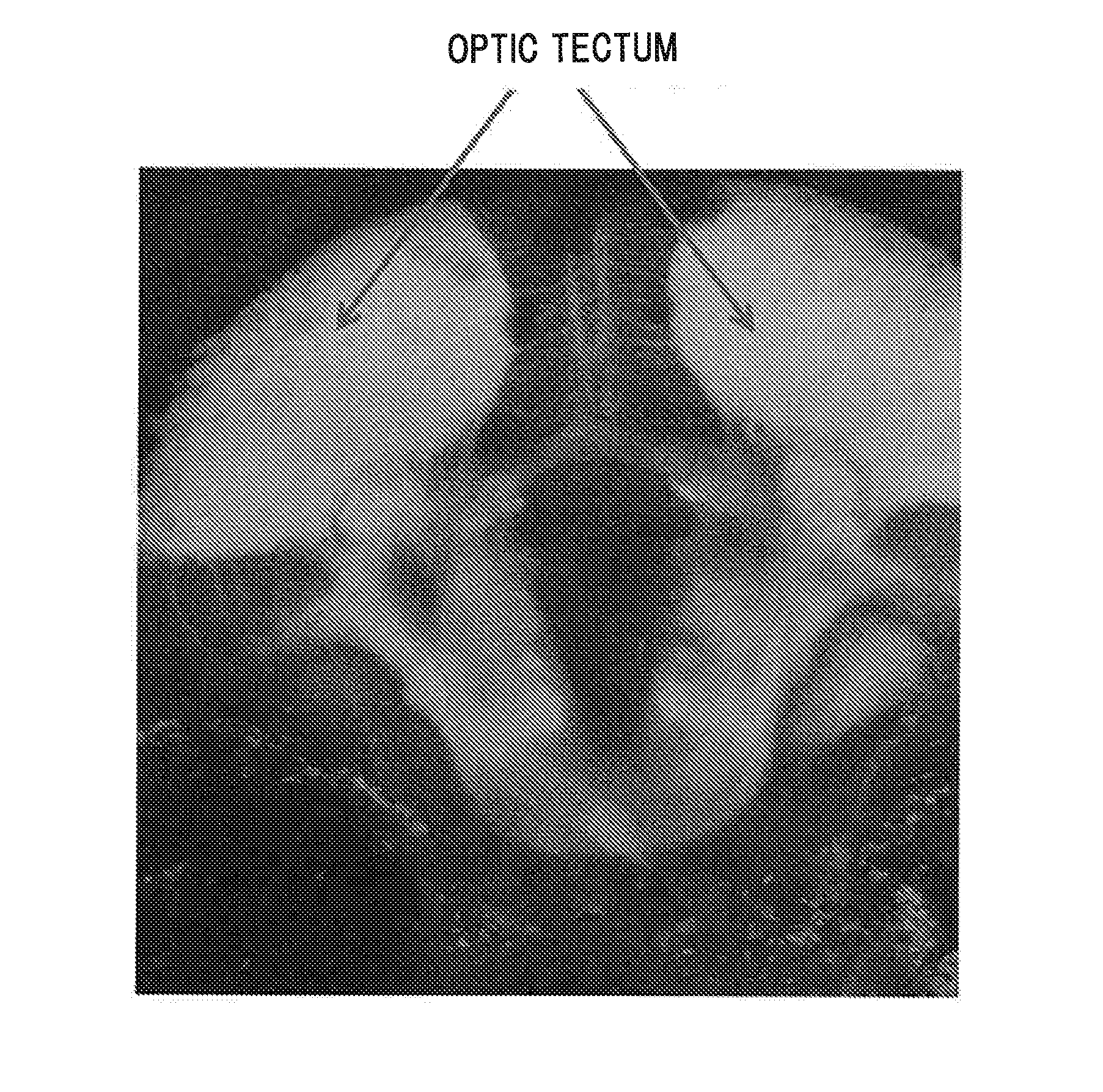
[0017]The present inventors conducted an intensive study to solve the aforementioned problems pertaining to the conventional technology. As a result, they have found that dye compounds represented by the following general formulas (1) and (7) are capable of labeling the central nervous system tissue of the living body alive. That is, they have found that the compounds label at least any one of the tissues including optic nerve, optic tract, superior colliculus (optic tectum), pituitary gland, tectospinal (tectobulbar) tract, and reticular formation with high sensitivity, serving as a novel central nervous system tissue-labeling compound enabling highly accurate diagnosis and screening of a drug, thereby completing the present invention. Also, the present inventors have established a method for labeling the central nervous system tissue of the living body. Further, the present inventors have developed a screening method using a labeling composition of the present invention, thereby completing the present invention.
[0019]Provision of the central nervous system tissue-labeling composition of the present invention has enabled selective labeling of a brain tissue such as optic nerve, optic tract, superior colliculus (optic tectum), pituitary gland, tectospinal (tectobulbar) tract, and reticular formation, which has been conventionally difficult. This enables simple and highly precise assessment and analysis of the morphology and the state of cells of a specific site in central nervous system tissues. Further, a screening method using the central nervous system tissue-labeling composition of the present invention can be a novel, effective tool for the research and the discovery of a drug for the central nervous system.

Problems solved by technology

The conventionally-employed probes utilize their properties such as accumulating in a site where a large amount of glucose is taken up, or of specifically accumulating to β-amyloid; therefore, they are not suitable for morphological imaging of a specific site in the brain in diseases not associated with such a specific site.
Also, there is a problem with the aforementioned glial cell-staining compound because the compound requires such a highly invasive treatment that involves direct administration to the brain.
In the first place, the transferability of a compound to the brain is regulated by the blood-brain barrier (BBB) and the blood-cerebrospinal fluid barrier (BCSFB), and many of the compounds that can migrate into a normal tissue may not be able to migrate into the brain.

Method used


Image

  • Central nervous system tissue-labeling composition, method for labeling central nervous system tissue, and screening method using central nervous system tissue-labeling composition
  • Central nervous system tissue-labeling composition, method for labeling central nervous system tissue, and screening method using central nervous system tissue-labeling composition
  • Central nervous system tissue-labeling composition, method for labeling central nervous system tissue, and screening method using central nervous system tissue-labeling composition

Examples


first embodiment

[0043]The central nervous system tissue-labeling composition according to a first embodiment of the present invention is characterized by containing, as an active ingredient, at least one of compounds represented by the general formula (1) or (7).

[0044]In the general formula (1), R1 to R2 each independently represent a hydrogen atom, an alkyl group, an aralkyl group, or an aryl group. Also, an aromatic ring A represents a skeletal structure represented by the following general formulas (2) to (4), or an aromatic ring A represents, through binding to R2 via N, a skeletal structure represented by the following general formulas (5) to (6):

[0045]In the general formula (2), R3 represents a hydrogen atom, an alkyl group, an aralkyl group, or an aryl group. In the general formula (3), R4 represents an oxygen atom, a sulfur atom, or N(R6), R5 represents a hydrogen atom, an alkyl group, an alkoxy group, or a sulfonic acid group, and R6 represents a hydrogen atom, an alkyl group, or an aryl g...

example 1

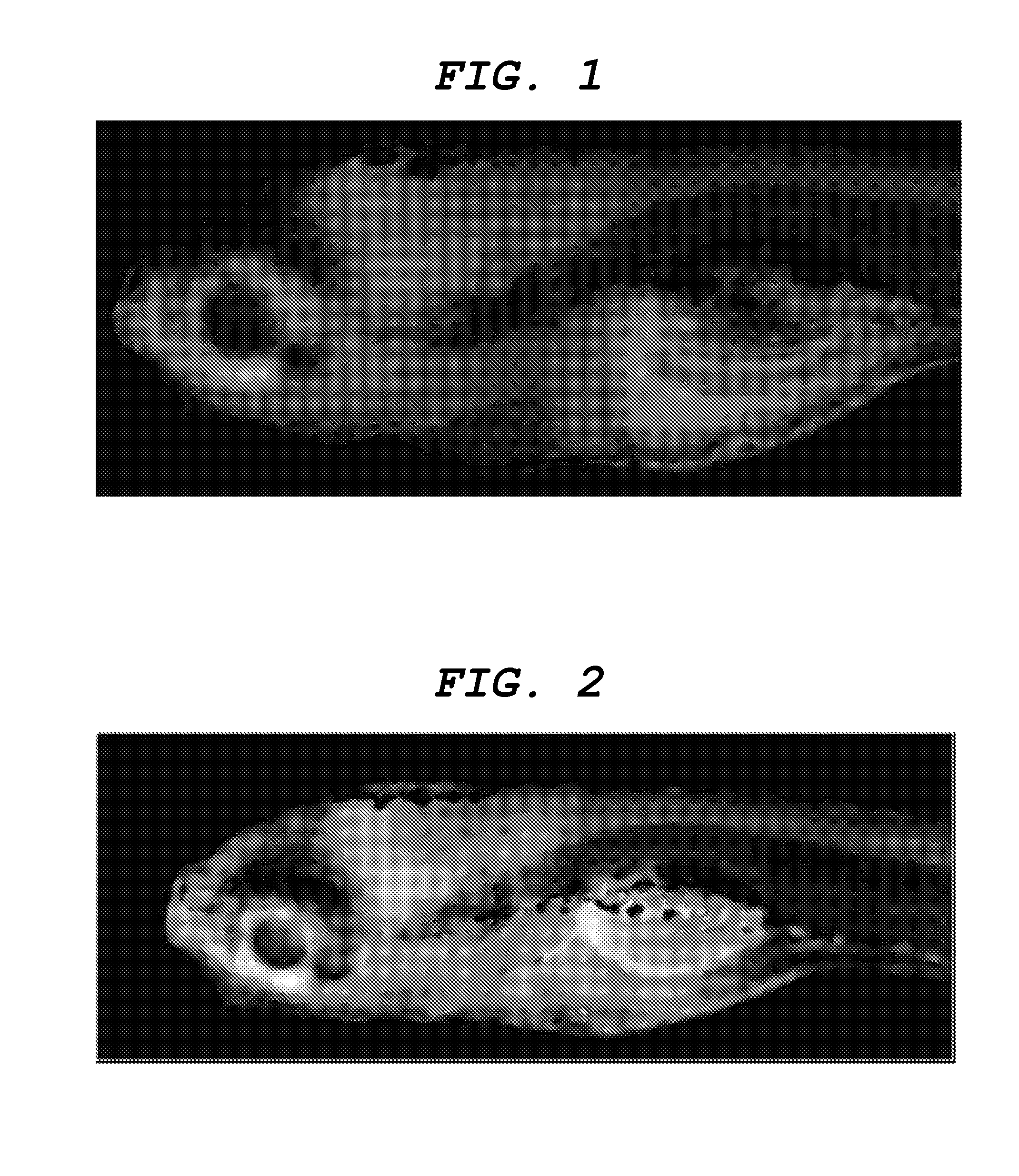
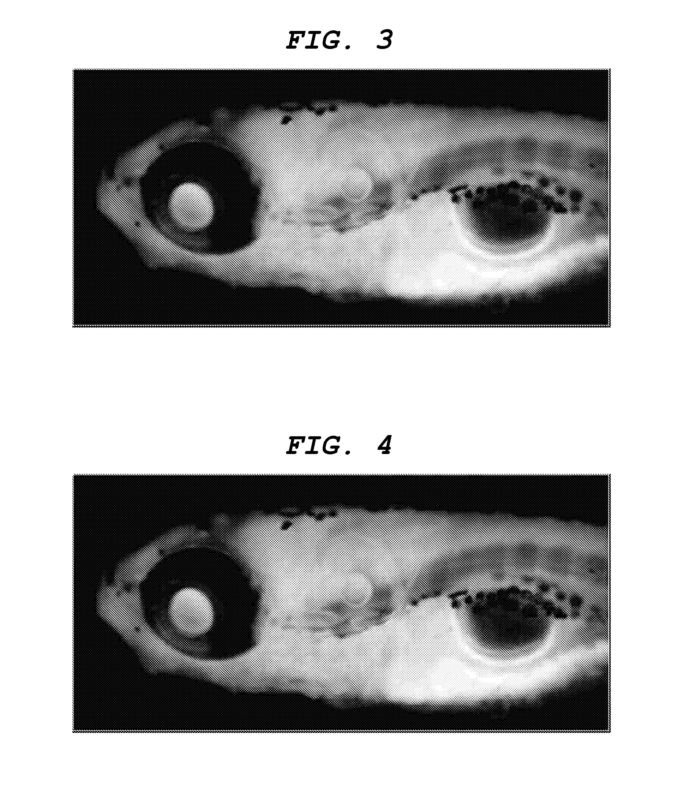
[0109]Labeling the central nervous system tissue with the central nervous system tissue-labeling composition Distilled water was added to a 1 mg / mL solution of the aforementioned compound (8) in DMSO to prepare a labeling solution 1 having a concentration of the aforementioned compound (8) of 1 μg / mL. Into an arbitrary well of a 24-well multiplate (manufacture by IWAKI), 1 mL of the labeling solution 1 and a day-7 embryo (7 dpf) of juvenile zebrafish were placed, and the plate was left to stand for one hour. Subsequently, the labeling solution 1 in the well was removed and replaced by 1 mL of distilled water. This operation was repeated three times. Then, juvenile zebrafish was removed from the well and embedded in 5% low melting point agarose gel on a slide glass so as to restrict movement, and the state of labeling in the central nervous system tissues were observed from the lateral side of zebrafish by a fluorescence stereomicroscope (manufactured by Leica Microsystems, MZ16FA). ...

examples 2 to 7

[0110]Zebrafish was labeled and observed by similar operations to Example 1 except for changing the dye compound (8) of Example 1 to the dye compounds (9) to (14) listed in Table 1 and for using labeling solutions 2 to 7.


PUM

PropertyMeasurementUnit
excitation wavelengthaaaaaaaaaa

Abstract

To provide a central nervous system tissue-labeling composition labeling the central nervous tissue system. Also, another object of the present invention is to provide a method for non-invasively labeling the central nervous tissue system. Further, another object of the present invention is to provide a screening method using the above central nervous system tissue-labeling composition. A central nervous system tissue-labeling composition containing, as an active ingredient, at least one of compounds represented by the general formula (1) or (7).

Description

CROSS-REFERENCE TO RELATED APPLICATIONS[0001]This application is a continuation of International Application No. PCT / JP2010 / 007519, filed Dec. 24, 2010, which claims the benefit of Japanese Patent Application No. 2009-296270, filed Dec. 25, 2009.BACKGROUND OF THE INVENTION[0002]1. Field of the Invention[0003]The present invention relates to a labeling composition capable of clearly labeling a central nervous system tissue, a method for labeling a central nervous system tissue using the central nervous system tissue-labeling composition, and a screening method using the central nervous system tissue-labeling composition.[0004]2. Description of the Related Art[0005]Recently, a number of patients with central nervous system diseases has been on the increase along with the aging of society. Representative examples of the diseases include Parkinson's disease, Alzheimer's disease, epilepsy, migraine, spinocerebellar degeneration, brain tumor, cerebral hemorrhage, and cerebral infarction. ...

Claims


Application Information

Patent Timeline
Patent Type & Authority Applications(United States)
IPC IPC(8): A61K49/00C07D221/18C07D491/147C07D221/14C07D417/10C07D417/06
CPCC07D417/06A61K49/00A61K51/00G01N33/58G01N33/15G01N33/50G01T1/161A61K49/0039A61K49/0032A61K49/006
Inventor WATANABE, KOHEISHINTOU, TAICHINOMOTO, TSUYOSHIMIYAZAKI, TAKESHIOKANO, MIETANAKA, TOSHIONISHIMURA, YUHEISHIMADA, YASUHITO
Owner CANON KK
Learn More