
Protein Biomarkers and Methods for Diagnosing Kawasaki Disease

a kawasaki disease and protein biomarker technology, applied in the field of kawasaki disease diagnosis, can solve the problems of increased risk of coronary aneurysm, heart damage and death, and significant risk of coronary artery disease in kd patients, and achieve the effect of increasing the accuracy of diagnosis

Inactive Publication Date: 2011-08-04
RGT UNIV OF CALIFORNIA
View PDF0 Cites 29 Cited by
  • Summary
  • Abstract
  • Description
  • Claims
  • Application Information

AI Technical Summary

Benefits of technology

The present invention provides a method for diagnosing Kawasaki Disease using a combination of cardiomyocyte and inflammatory biomarkers. The method involves detecting the levels of these biomarkers in a biological sample from the patient and comparing them to reference values established for healthy subjects. The invention also provides a diagnostic biomarker panel that includes at least two biomarkers, such as N-terminal pro-B-type natriuretic peptide (NT-proBNP) and interferon type-I biomarker, to differentiate between Kawasaki Disease and other non-specified viral infections. The invention can be used in diagnosing Kawasaki Disease in patients with a biological sample, such as whole blood or plasma.

Problems solved by technology

If not detected and treated immediately, it can result in heart damage and death.
Although KD is self-limiting, failure to administer timely treatment with IVIG results in a significant risk that the KD patient will develop coronary artery disease.
Moreover, there is an increased risk of coronary aneurysm for patients who have had prolonged KD or are afflicted at less than one year of age.
However, not only is the cause of KD not known, there is no diagnostic test available to this date.
Awaiting the observation of clinical signs can result in missing the critical window for IVIG treatment, thereby making the patient susceptible to coronary artery damage.
Attempts to identify a single biomarker for KD have not been successful, as the markers identified lacked sensitivity and / or lacked specificity for KD.

Method used


Image

  • Protein Biomarkers and Methods for Diagnosing Kawasaki Disease
  • Protein Biomarkers and Methods for Diagnosing Kawasaki Disease
  • Protein Biomarkers and Methods for Diagnosing Kawasaki Disease

Examples


example 1

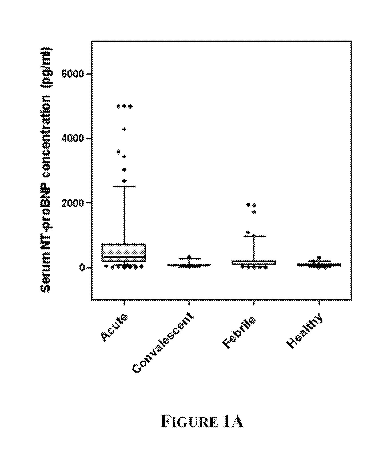
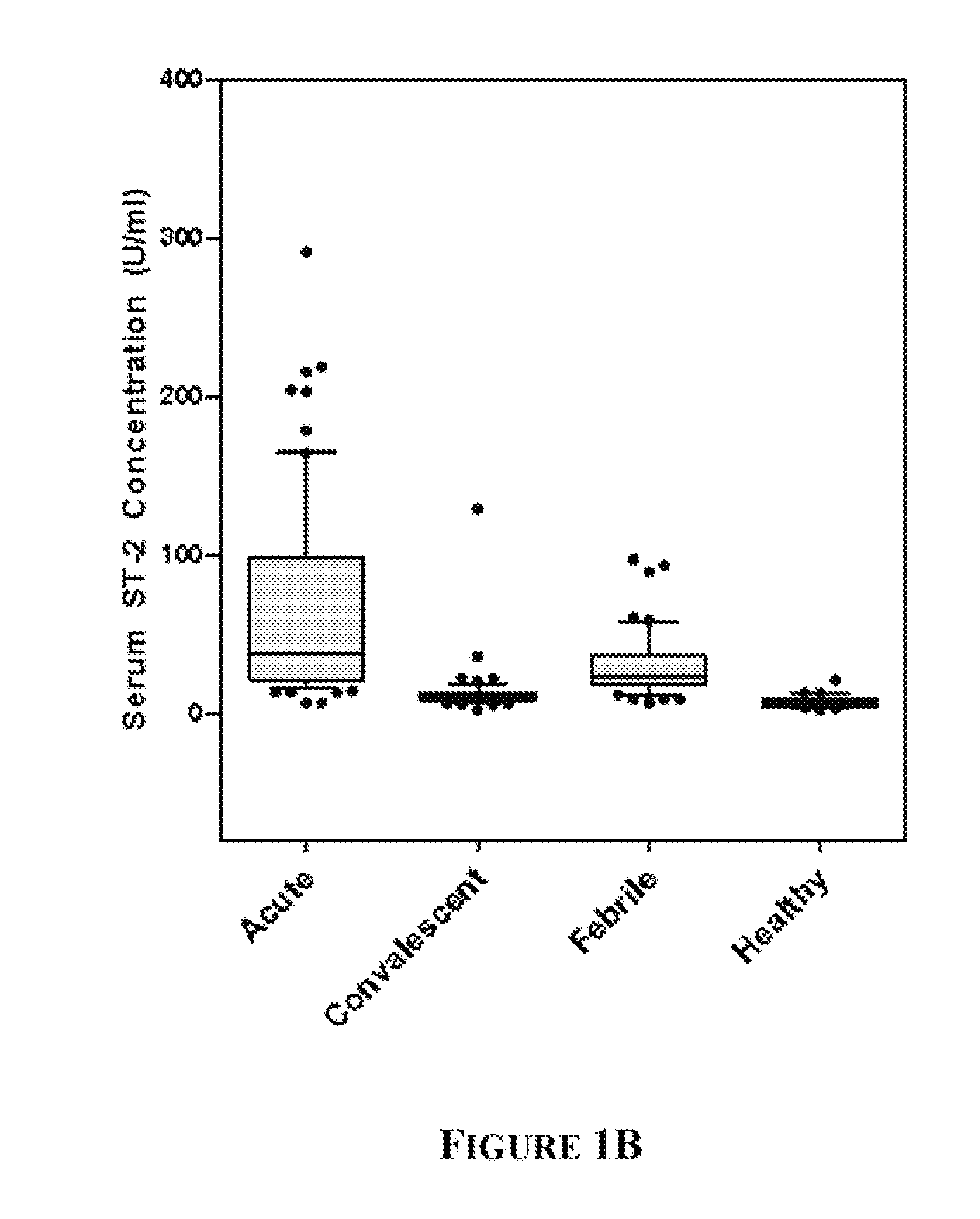
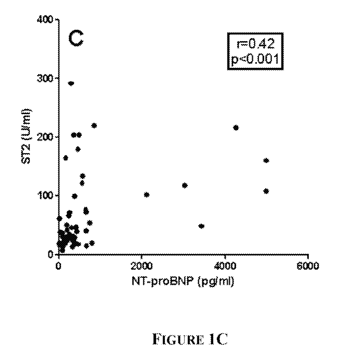
NT-proBNP and ST2 as Indicators of Myocardial Strain in Acute Kawasaki Disease (KD)

Study Population and Sample Collection

[0087]KD patients (n=96), febrile controls (n=93), and healthy pediatric controls (n=50) were enrolled at Rady Children's Hospital, San Diego, Calif. after parental informed consent. The human subjects protocol was reviewed and approved by the Institutional Review Board at UCSD. All KD patients had fever and ≧4 of the 5 principal clinical criteria for KD (rash, conjunctival injection, cervical lymphadenopathy, changes in the oral mucosa, and changes in the extremities) or 3 criteria plus coronary artery abnormalities documented by echocardiography (Liu et al (2006) Curr Opin Microbiol. 9:312-9). All febrile control patients had nasopharyngeal and stool viral cultures. Controls classified as having acute adenoviral infection (n=14) had fever for at least 3 days, a negative throat culture for Group A β-hemolytic streptococcus (GABHS), and a positive nasopharyngeal (...

example 2

Transcript Abundance Profiles Distinguish Kawasaki Disease from Other Rash / Fever Illnesses

[0096]In this study, patterns of whole blood gene expression in acute KD patients, and patients with illnesses of similar clinical presentation and well-defined etiology were compared. Patterns of gene expression and corresponding biological programs that differed between KD and the other illnesses were identified, which were able to distinguish between KD and adenovirus infection on the basis of gene expression patterns. The results from this study can be exploited to devise a diagnostic test for KD that may lead to more accurate and timely diagnosis of these patients.

[0097]To identify unique features of KD, gene expression patterns in the blood of 23 children with acute KD and 18 age-matched febrile controls were compared. Genes associated with platelet and neutrophil activation were expressed at higher levels in KD patients than patients with adenovirus infections or systemic toxic drug reac...

example 3

Inflammatory Biomarkers as Indicators in Acute Kawasaki Disease (KD)

[0117]Plasma samples from twenty-eight (28) acute Kawasaki Disease subjects and twenty-eight (28) age- and sex-matched febrile control children were analyzed using the Luminex bead system (RulesBasedMedicine, Inc.) for eighty-nine (89) analytes in pathways related to inflammation. Of the eighty-nine (89) analytes, thirteen (13) analytes showed significantly different levels between acute Kawasaki Disease and febrile control subjects. The median and 95% CI for the two groups are shown for each analyte in these graphs. (FIGS. 7-19).

[0118]The thirteen analytes showing significantly different levels in acute Kawasaki Disease are alpha-1 antitrypsin, calcitonin, CD40, C reactive protein, EN-RAGE, erythropoietin, fibrinogen, ICAM-1, IL-6, MIP-1 alpha, myeloperoxidase, TIMP-1, and VEGF.


PUM

PropertyMeasurementUnit
swellingaaaaaaaaaa
concentrationsaaaaaaaaaa
concentrationaaaaaaaaaa

Abstract

A method, kit and device for diagnosing Kawasaki Disease are provided. The invention provides detecting an expression level of at least two Kawasaki Disease diagnostic biomarkers in a biological sample from a patient with a capture agent and diagnosing the patient as having Kawasaki Disease when the expression levels of the at least two diagnostic biomarkers in the patient biological sample are higher than the normal expression levels of the same biomarkers in a biological sample from a control subject. The first Kawasaki Disease diagnostic biomarker disclosed in the present invention is a cardiomyocyte biomarker, and the second Kawasaki Disease diagnostic biomarker is an inflammatory biomarker. The invention further provides detecting an expression level of a third biomarker, interferon type-I biomarker, in the patient biological sample with a capture agent and diagnosing the patient as having Kawasaki Disease when the expression level of interferon type-I biomarker is lower than the expression level in a control subject.

Description

CROSS-REFERENCE TO RELATED APPLICATIONS[0001]This patent application is a continuation of PCT Application No. PCT / US09 / 055,140, filed Aug. 28, 2009, which claims priority benefit of U.S. Provisional Application No. 61 / 092,451 filed Aug. 28, 2008, each of which is incorporated herein by reference in their entireties.STATEMENT OF GOVERNMENT INTEREST[0002]This invention was made with government support under NHLBI R01-HL69413 awarded by National Institutes of Health. The government has certain rights in the invention.FIELD OF THE INVENTION[0003]The invention relates generally to the field of diagnosis of Kawasaki Disease (KD).BACKGROUND OF THE INVENTION[0004]Various publications, including patents, published applications, technical articles and scholarly articles are cited throughout the specification. Each of these cited publications is incorporated by reference herein, in its entirety.[0005]Kawasaki Disease (KD) is a self-limited acute vasculitis, the cause of which remains unknown. ...

Claims


Application Information

Patent Timeline
Patent Type & Authority Applications(United States)
IPC IPC(8): G01N33/68G01N33/74G01N33/573G01N30/00
CPCG01N33/6893G01N2800/60G01N2800/328
Inventor BURNS, JANE C.
Owner RGT UNIV OF CALIFORNIA
Learn More