

Device for quantification and monitoring of cardiovascular function during induced stress or physical activity and at rest
a technology of cardiovascular function and measurement device, which is applied in the field of quantification and monitoring of cardiovascular function during induced stress or physical activity and at rest, can solve the problems of not providing all the necessary information, high morbidity, mortality, and cost, and achieves the effect of easy calculation of pulmonary pressure values and easy differentiation of vibrations within the aorta and pulmonary arteries
- Summary
- Abstract
- Description
- Claims
- Application Information
AI Technical Summary
Benefits of technology
Problems solved by technology
Method used
Image

Examples
example 2
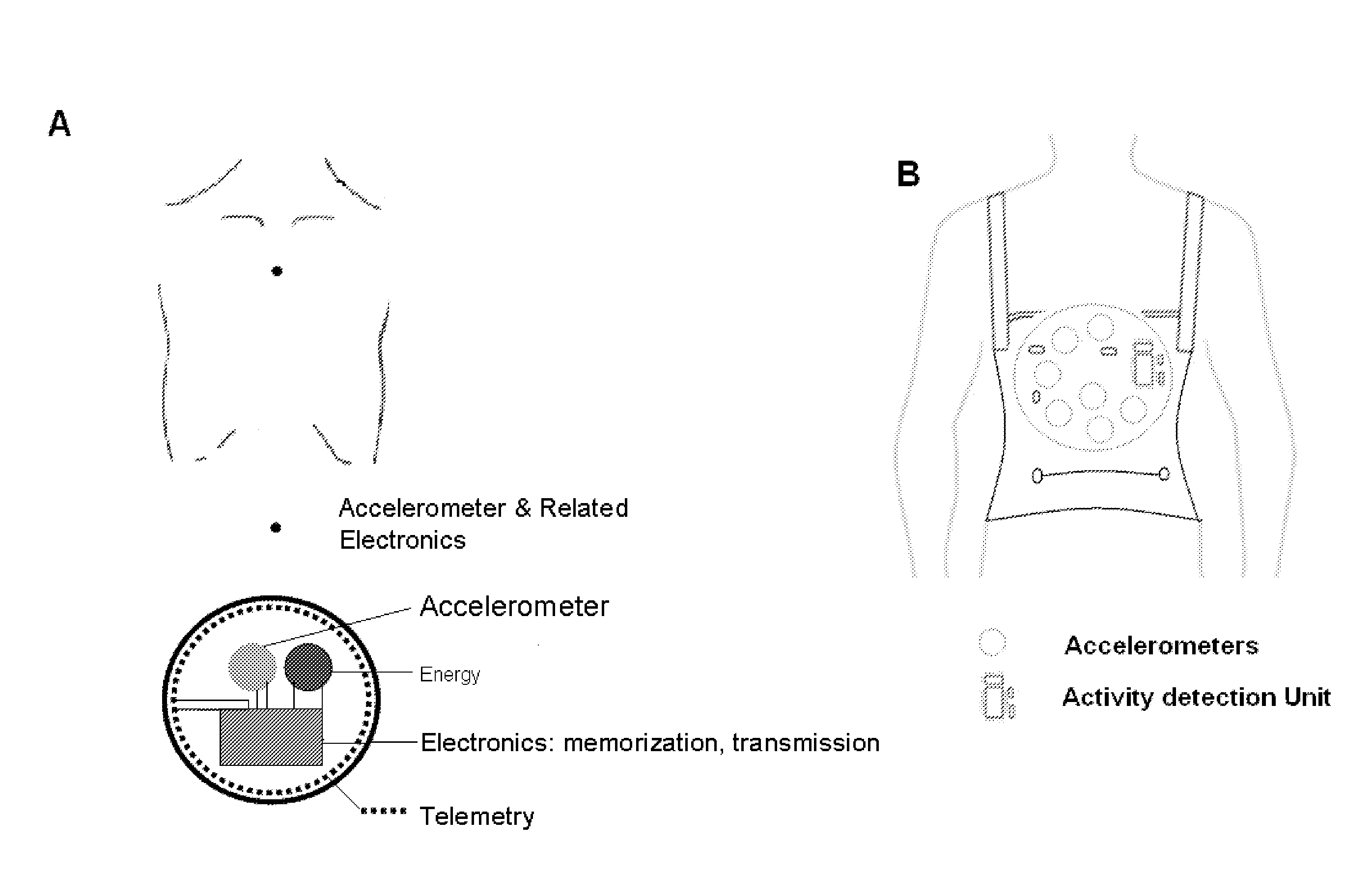
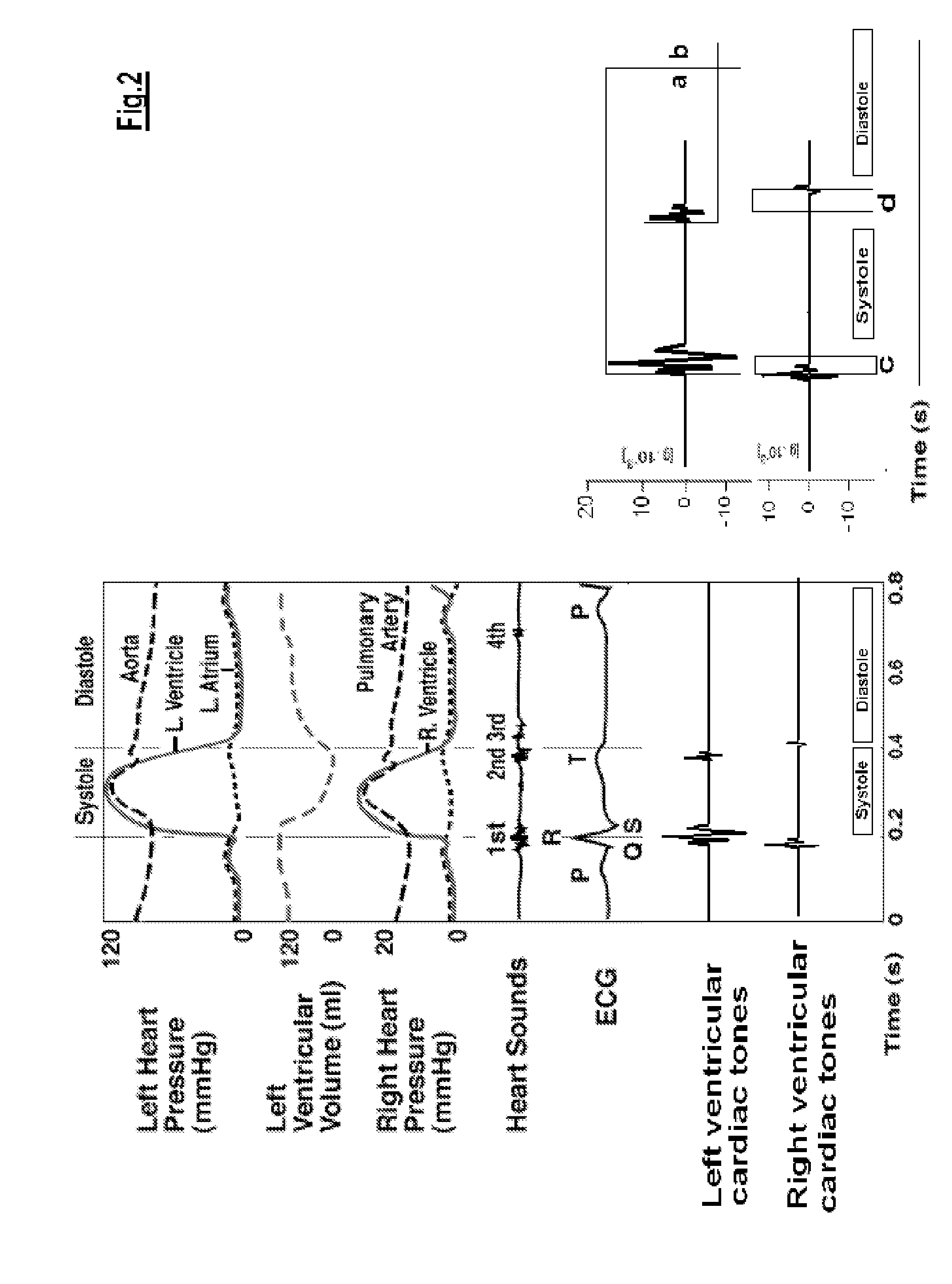
Shown in FIG. 2
[0074]Cardiac tones accelerations signals and cardiac cycle hemodynamic events Left: the different phases of the cardiac cycle in which the sensors quantify the amplitude, the spectral characteristics and timing of myocardial and vessels vibrations (left ventricular cardiac tones and right ventricular cardiac tones).
[0075]Right: the amplitude of the vibration signals (a=first cardiac tone vibration amplitude; b=second cardiac tone vibration amplitude) the times between vibrations as time markers (c and d=time gaps from right and left ventricular vibrations related mechanical events) are acquired as instantaneous values at baseline and during activity / stress. For each cardiovascular function-derived parameter, the curve of the cardiovascular function variation as a function of heart rate is finally computed. The data can be also read remotely by a telemetric connection.
example 3
Shown in FIG. 3
The Sensor for Measuring the Force-Frequency Relation
[0076]Upper panel. X-axis: time (sec); Y-axis: cardiac tone vibration amplitude, g×10−3 (g=9.8 m / sec2).
[0077]The transcutaneous force sensor is based on a linear accelerometer. and is positioned in the mid-sternal precordial region. This sensor measures the cardiac tones generated by the myocardium during contraction (first cardiac tone) and during isovolumic relaxation (second cardiac tone) of the heart. A QRS detection algorithm is used to automatically locate the beginning of the isovolumic ventricular contractions.
[0078]Lower panel. X-axis: exercise workload (Watt); Y-axis: first cardiac tone vibration amplitude during exercise, g×10−3 (g=9.8 m / sec2).
[0079]The amplitude of the vibration due to isovolumic myocardium contraction is obtained to record the first cardiac tone amplitude as a measure of the systolic force; for each cardiac beat the parameters are acquired as instantaneous values at rest and during exer...
example 4
Shown in FIG. 4
The Curve of First Cardiac Tone Amplitude as a Function of Heart Rate
[0080]Left panel. X-axis: time, (sec); exercise=exercise in progress; recovery=recovery from exercise. Y-axis: first cardiac tone vibration amplitude, g×10−3 (g=9.8 m / sec2). All the parameters are acquired as instantaneous values at baseline, during exercise and recovery. Mobile mean is utilized to assess baseline value (1 minute recording), at each incremental stress test, at peak test, and during recovery. Instantaneous force values scattering (points) depend on the respiratory cycle and thorax expansion; continuous line=force mobile mean.
[0081]Right panel. X-axis: heart rate, beats per minute (bpm); Y-axis: first cardiac tone vibration amplitude, g×10−3 (g=9.8 m / sec2).
[0082]Full symbols=exercise in progress; empty symbols=recovery.
PUM
 Abstract
Description
Claims
Application Information