Tumor angiogenesis associated genes and a method for their identification

a technology of tumor angiogenesis and associated genes, which is applied in the field of tumor angiogenesis associated genes and a method for their identification, can solve the problems of increasing the chance of cancer cells entering the blood, making them more dangerous, and the temporal and spatial complex actions of all factors exerting effect on endothelial cells in vivo may not be accurately reflected in vitro. , to achieve the effect of inhibiting tumor growth, reducing microvessel density, and inhibiting angiogenesis

Inactive Publication Date: 2011-09-22
MAASTRICHT UNIVERSITY
View PDF5 Cites 4 Cited by
  • Summary
  • Abstract
  • Description
  • Claims
  • Application Information

AI Technical Summary

Benefits of technology

[0031]SSH consists of 2 hybridization steps, followed by suppression PCR to reduce the redundancy of overexpressed cDNAs. In a first step, 2 tester cDNA populations—ligated to different adaptor sequences—are hybridized in separate reactions to an excess of driver cDNA to subtract common sequences in tester and driver cDNA populations i.e. the non-differentially expressed genes. The cDNA is amplified, (using for instance Clontech SMART™ cDNA amplification kit) generating sufficient starting material for tester and driver, whereby the original transcript distribution is maintained. In the second hybridization the two primary hybridization samples are mixed and here create the template for the subsequent suppression PCR. During this reaction, inverted terminal repeats prevent amplification of highly abundant molecules and the amplification of differentially expressed genes is favoured. The final cDNA repertoire generated by PCR consists of cDNA fragments that are overexpressed in the tester as compared to the driver population (Clontech protocol # PT1117-1). Amplification of the target genes is dependent upon the template, such as the length, the GC content, and / or presence of inverted repeats. Therefore, the number of amplification cycles in either step is of crucial importance. As such, the method of the present invention limits the number of cycles, and preferably adapts the number of cycles to e.g. 2, 3, 4, 5, 6, 7, 8, 9 or 10 cycles, in the amplification step(s) of the SSH to ensure effective subtraction and suppression. Optimization of the number of amplification cycles ensures proper suppression and reduction of redundancy. The person skilled in the art may use routine trial and error to establish the optimum or near-optimum number of cycles to satisfy the specific needs, e.g. the provision of the original representation of transcripts.
[0055]The identification of the differentially expressed genes by the method according to the invention facilitates the identification of the corresponding amino acid sequence. Accordingly, the present invention relates to isolated polypeptides comprising, or alternatively consisting of, an amino acid sequence according to the invention, and preferably characterized by any of SEQ ID NO:s 2, 4, 6, 8, 10, 12, 14, 16, 18, 20, 22, 24, 26, 28, 30, 32 or 34, or a part thereof, or comprising or consisting of a variant thereof, or an immunologically active and / or functional fragment thereof.
[0185]Hence, the present invention relates to the use of a polynucleotide encoding a polypeptide comprising an amino acid sequence which is at least 65% identical to any of the polypeptides according to the invention, such as TAG, GAG / A and / or GAG / B polypeptides, and preferably any of SEQ ID NO:s 2, 4, 6, 8, 10, 12, 14, 16, 18, 20, 22, 24, 26, 28, 30, 32 or 34, for stimulating angiogenesis.

Problems solved by technology

Angiogenesis not only allows solid tumors to grow, it also makes them more dangerous because they are more likely to metastasize, i.e. spread elsewhere in the body through the bloodstream.
The new blood vessels in the tumor increase the chance of cancer cells getting into the blood, especially since the tumor's blood vessels are often imperfectly formed.
Different cell culture models have been developed to study angiogenesis, but the temporal and spatial complex actions of all factors exerting effect on endothelial cells in vivo may not be accurately reflected in vitro.
Gene expression analysis of tumor endothelial cells (TECs) encounters difficulties related to the fact that endothelial cells (ECs) are embedded in complex tissues and comprise only a small fraction of the cells present in these tissues.
In cell culture conditions, however, cells reside in an artificial microenvironment and might respond aberrantly to certain stimuli, giving a false representation of the in vivo situation.
Nevertheless, the complex microenvironment of angiogenic endothelial cells in tissues is extremely difficult to mimic adequately in vitro.
In addition, when regarding angiogenesis in cancer, tumor endothelial cells have resided in the tumor microenvironments for months to years, whereas culture systems only cover a time period of days, which in addition contributes to discrepancies in observed gene expression profiles of endothelial cells in vitro vs in vivo.
In conclusion, it appears difficult to accurately mimic in vitro the complex temporal and spatial actions of all microenvironmental factors exerting an effect on endothelial cells in vivo.
Therefore, extrapolation of data generated by in vitro experiments to the in vivo situation is limited, stressing the importance of approaches that make use of more relevant cell sources such as tissue derived cells.

Method used


Image

  • Tumor angiogenesis associated genes and a method for their identification
  • Tumor angiogenesis associated genes and a method for their identification
  • Tumor angiogenesis associated genes and a method for their identification

Examples


example 1

Experimental Procedures

[0254]1.1 Isolation of Endothelial Cells from Fresh Tissues

[0255]Fresh colorectal tumors (Dukes C) (n=5) and distant normal colon tissues of the same patient (n=5) were obtained from excision surgery at the department of Pathology (University Hospital Maastricht). Fresh placenta tissues (n=5) were obtained from the department of Obstetrics (University Hospital Maastricht). Endothelial cells were isolated as previously described (St Croix et al., 2000), with minor modifications. Tissues were minced with surgical blades, digested for 30 minutes with 1 mg / ml collagenase (Life Technologies, Breda, The Netherlands) and 2.5 U / ml dispase (Life Technologies) at 37° C. with continuous agitation. DNAse I (Sigma, Zwijndrecht, The Netherlands) was added to a final concentration of 100 μg / ml and the cell suspension was incubated for another 30 minutes prior to Ficoll Paque gradient density centrifugation (Amersham Biosciences, Uppsala, Sweden).

[0256]Endothelial cells were ...

example 2

Identification of Tumor Endothelial Markers by SSH

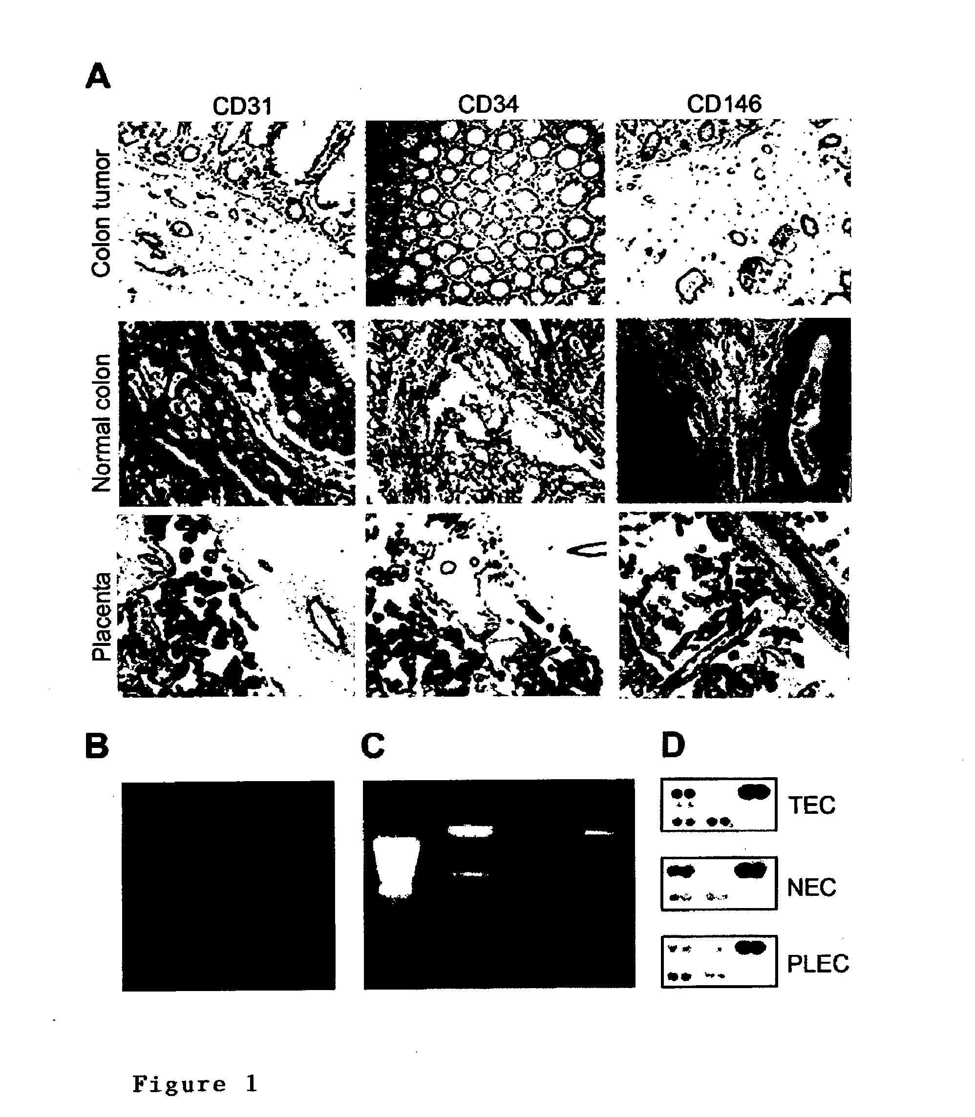
[0279]A suppression subtractive hybridization (SSH) was performed in combination with cDNA array screening to identify novel tumor specific endothelial markers in an unbiased manner. Tumor endothelial cells (TEC) were successfully isolated from colon tumors (n=5) and patient-matched normal endothelial cells (NEC) from normal colon tissue samples (n=5), as well as from placenta tissues (PLEC, n=5) (FIG. 1). RNA was isolated (FIG. 1) and used to create subtraction repertoires of genes overexpressed in TEC. In addition, HUVEC were stimulated in vitro with tumor cell conditioned medium and used to create additional subtraction repertoires. A total of 2746 inserts, 1781 derived from the TEC subtractions and 965 derived from the HUVEC subtractions were amplified and spotted onto duplicate arrays that were probed with 33P-dCTP labeled cDNA derived from TEC, NEC, PLEC and HUVEC. Phospho-imaging and pair-wise comparisons of spot intensities w...

example 3

Gene Expression of Tumor Endothelial Cells is Closely Related to Gene Expression During Physiological Angiogenesis

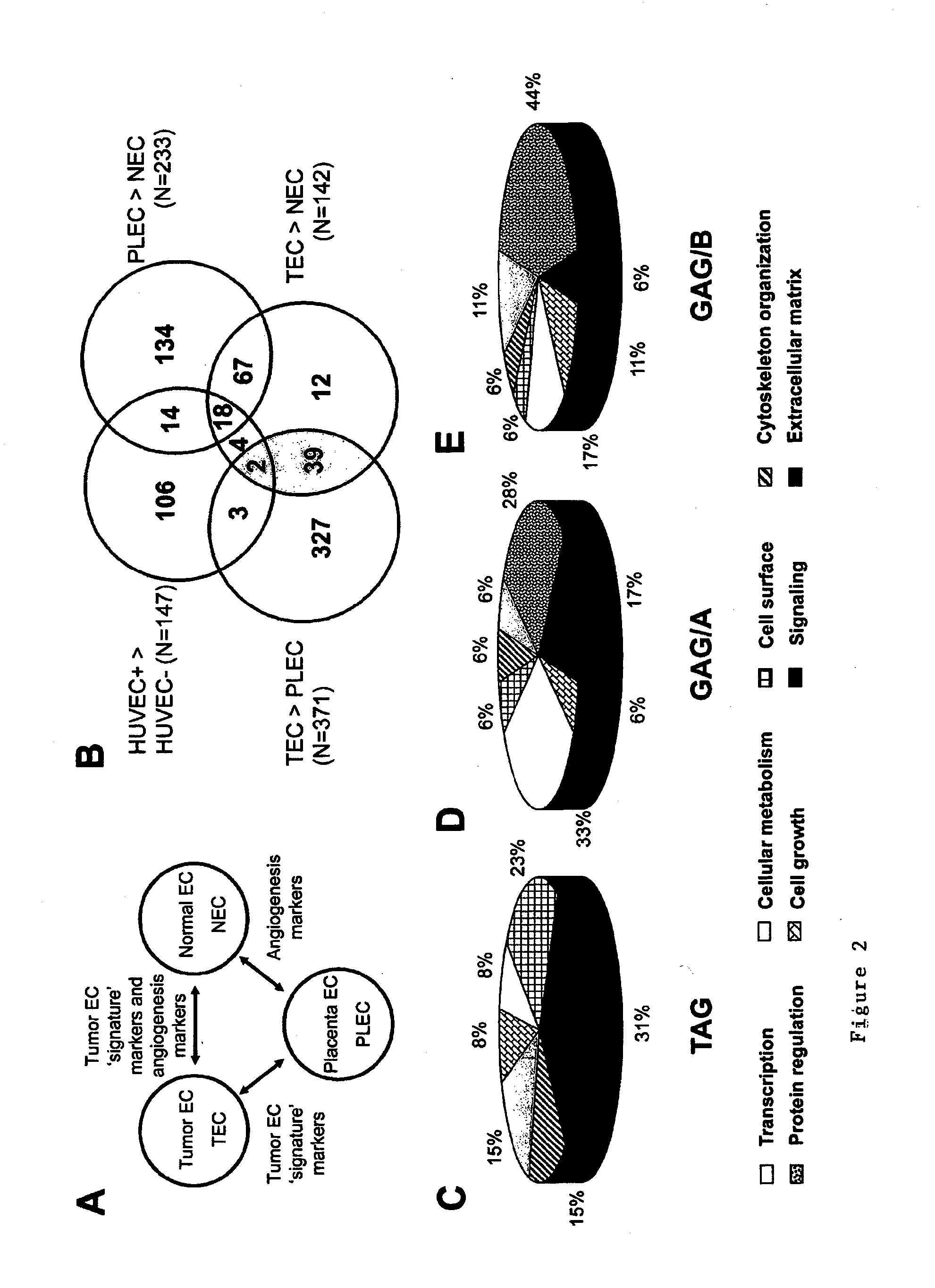
[0282]It emerged that the majority of TEC overexpressed transcripts (85 / 142=60%) are also associated with angiogenesis under physiological conditions in vivo, and are therefore not specific for tumor angiogenesis in vivo (FIG. 2B). These 85 GAG / A transcripts represent 46 different genes, including genes that have been associated with angiogenesis such as matrix metalloproteinases (MMPs) (Pepper, 2001), integrin β1 (Senger et al., 2002) and endothelial cell specific molecule-1 (Aitkenhead et al., 2002) (Table 3).


PUM

PropertyMeasurementUnit
critical volumeaaaaaaaaaa
concentrationaaaaaaaaaa
densityaaaaaaaaaa

Abstract

Crucial to designing anti-angiogenic and vascular targeting approaches is the identification of specific target molecules. We compared transcriptional profiles of tumor endothelial cells with that of normal resting endothelial cells, normal but angiogenically activated placental endothelial cells, and cultured endothelial cells. Although the majority of transcripts were classified as general angiogenesis markers, we identified 17 genes that show specific overexpression in tumor endothelium. Antibody targeting of four cell-surface expressed or secreted products (vimentin, CD59, HMGB1 and IGFBP7) inhibited angiogenesis in vitro and in vivo. Finally, targeting endothelial vimentin in a mouse tumor model significantly inhibited tumor growth and reduced microvessel density. Our results demonstrate the utility of the identification and subsequent targeting of specific tumor endothelial markers for anticancer therapy.

Description

BACKGROUND OF THE INVENTION[0001]Tumor progression and the development of distant metastases require the presence of an extensive vasculature. Active angiogenesis is a hallmark of most malignancies and inhibition of this process is considered to be a promising strategy for the treatment of tumors. In order to develop the most specific and effective anti-angiogenic therapies for treating cancer, it is of importance to have a fundamental understanding of the molecular differences between tumor endothelial cells and their normal counterparts. Since angiogenesis is not limited to pathological conditions, careful evaluation of the putative targets is necessitated to prevent side effects associated with impaired physiological angiogenesis.Angiogenesis[0002]Angiogenesis occurs in the healthy body for healing wounds and for restoring blood flow to tissues after injury. In females, angiogenesis also occurs during the monthly reproductive cycle, e.g. to rebuild the uterus lining and to mature...

Claims


Application Information

Patent Timeline
Patent Type & Authority Applications(United States)
IPC IPC(8): A61K39/395A61K38/16A61P35/04C12Q1/68G01N33/566C12Q1/02
CPCC07K14/82C12Q2600/136C12Q1/6886A61P35/00A61P35/04
Inventor GRIFFIOEN, ARJAN WILLEMVAN BEIJNUM, JUDITH ROSINA
Owner MAASTRICHT UNIVERSITY
Why Eureka
Try Eureka