Magnetic resonance imaging mediated radiofrequency ablation

radiofrequency ablation technology, applied in the field of radiofrequency ablation using a magnetic resonance imaging scanner, can solve the problems of poor image quality, limited utility of us to monitor treatment, and irreversible thermal damag

Inactive Publication Date: 2011-12-08
HUE YIK KIONG +3
View PDF8 Cites 51 Cited by
  • Summary
  • Abstract
  • Description
  • Claims
  • Application Information

AI Technical Summary

Benefits of technology

[0014]Unless otherwise defined, all technical and scientific terms used herein have the same meaning as commonly understood by one of ordinary skill in the art to which this invention belongs. Although methods and materials similar or equivalent to those described herein can be used in t

Problems solved by technology

The techniques involve heating the tissue above 60° C., leading to protein denaturation and membrane breakdown, and resulting in irreversible thermal damage.
US guidance is inexpensive and rapid because of its inherent real-time capability, but it has poor image quality.
Additionally, gas bubbles sometimes produced by tissue vaporization during heating limit the utility of US to monitor the treatment.
CT is capable of multi-planar imaging, but its poor soft tissue contrast requires the administration of an exogenous contrast agent to provide clear delineation of tumor tissue and may not permit visualization of induced coagulation.
Ionizing radiation exposure of both the patient and the physician fur

Method used


Image

  • Magnetic resonance imaging mediated radiofrequency ablation
  • Magnetic resonance imaging mediated radiofrequency ablation
  • Magnetic resonance imaging mediated radiofrequency ablation

Examples


examples

I. Simulations

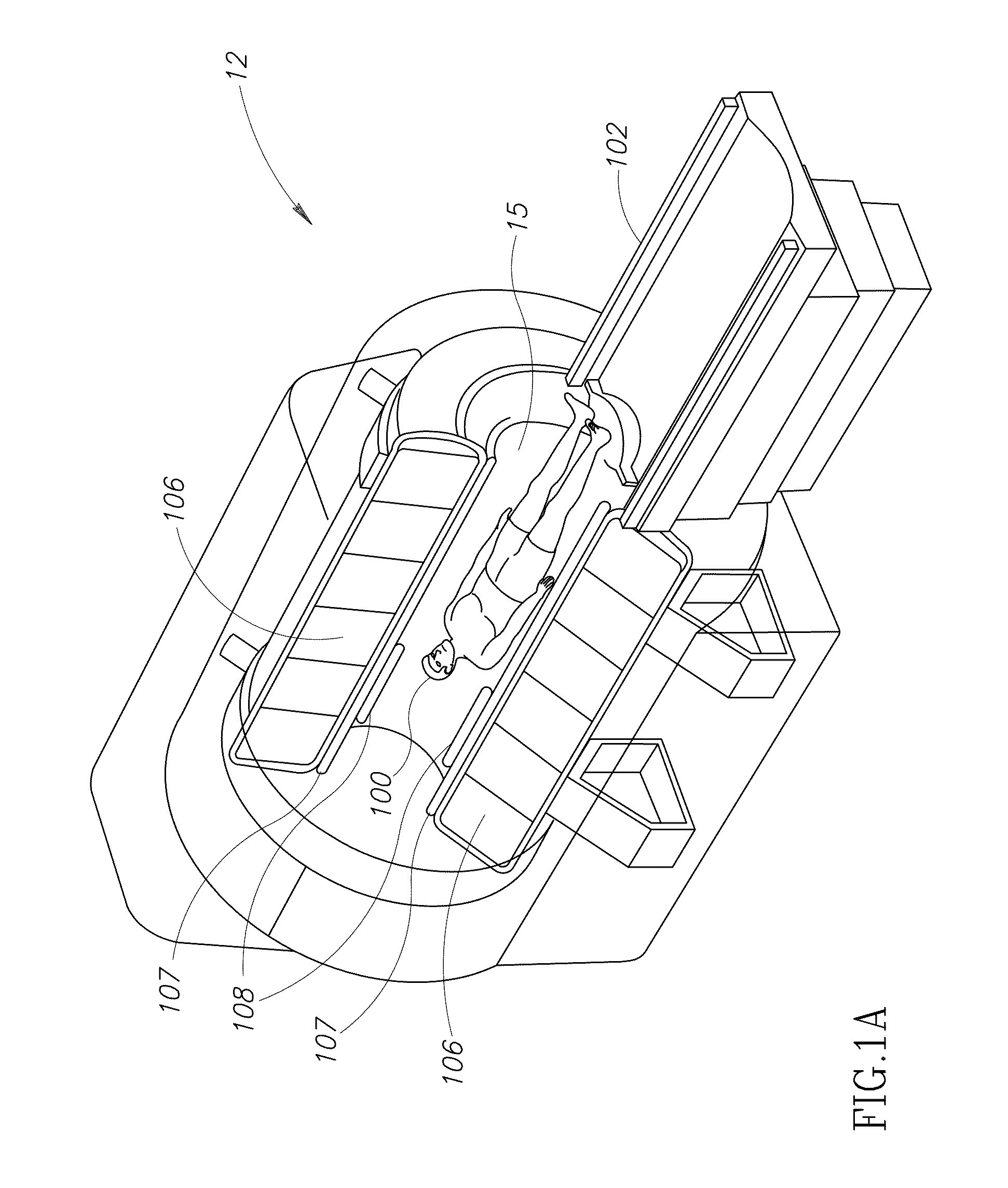
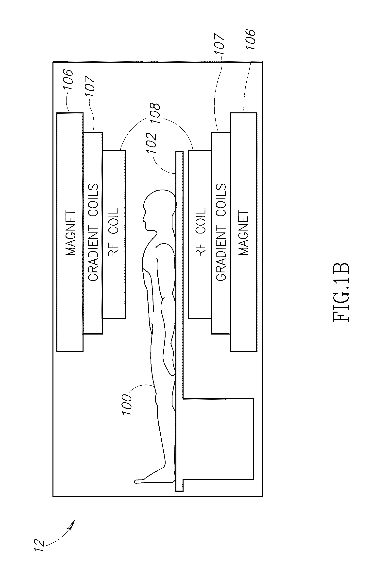
[0057]In order to better understand the safety and performance issues related to the proposed invention, we simulated the operation of an MR-driven RFA device in the body RF coil of a 1.5 T MRI scanner. The simulation was carried out using the Remcom, Inc. (State College, Pa., USA) XFDTD 7.0 (XF7) 3D electromagnetic simulation software package, which is based on the FDTD (finite difference time domain) method. The modeled body coil had dimensions of 60 cm long and 60 cm diameter, and was a 16 rung highpass birdcage coil. It was first tuned to 1.5 T so that the field within the center of the body coil was homogenous. Then, its performance with a rectangular solid (box phantom) 7 cm tall, 31 cm long, 23 cm wide, with the electrical properties of liver tissue (dielectric constant 70.62, conductivity 0.55 S / m) placed at the isocenter was recorded as a reference. Finally, the RFA device, modeled as a simple wire which captures RF energy from the body coil by electromagnetic...


PUM


Abstract

Radiofrequency ablation (RFA) may be used as a minimally invasive treatment of solid tumors, typically cancers of the liver, lung, breast, kidney and bone, most often via a percutaneous approach. In RFA tumor tissue is killed by heating. RFA requires guidance using an imaging method to correctly position the RF applicator. Magnetic resonance imaging (MRI) can be used for guidance, and offers the additional advantage of the ability to image tissue temperature. Because MRI employs high power RF fields, the MRI scanner could serve as the source of RF energy for ablation. Described herein are an MRI-driven RF ablation device and method. The device has minimal electrical circuitry, and uses the MR scanner radio frequency field as the energy source to generate heat in tissue using an antenna and a needle. Based on the Faraday induction law, different embodiments for coupling the body coil RF energy into tissue are disclosed.

Description

CROSS-REFERENCE TO RELATED APPLICATIONS[0001]This application claims priority benefit from U.S. Provisional Patent Application No. 61 / 343,591, filed Apr. 30, 2010, the contents of which is incorporated herein by reference in its entirety.FIELD OF THE INVENTION[0002]The present invention relates to radiofrequency ablation using a magnetic resonance imaging scanner, and, more particularly to a system and method for performing radiofrequency ablation of tissue using minimal electrical circuitry and using a magnetic resonance scanner radiofrequency field as an energy source.BACKGROUND OF THE INVENTION[0003]A variety of thermal tissue ablation techniques, including radiofrequency, microwave, laser and focused ultrasound, have been described which cause cell death by coagulation necrosis and / or apoptosis. The techniques involve heating the tissue above 60° C., leading to protein denaturation and membrane breakdown, and resulting in irreversible thermal damage. Among these techniques, perc...

Claims


Application Information

Patent Timeline
IPC IPC(8): A61B5/055A61B6/00
CPCA61B5/055A61B18/1206A61B2019/5236A61B2018/00577A61B18/14A61B2090/374
Inventor HUE, YIK-KIONGROTH, ABRAHAMACKERMAN, JEROME LEONARDNEVO, EREZ
Owner HUE YIK KIONG
Why Eureka
Try Eureka