Laparoscopic hifu probe

a hifu probe and laparoscopic technology, applied in the field of instruments, can solve the problems of damage to the kidney and possible organ death, and achieve the effects of facilitating the introduction of components, facilitating the passage of fluid, and facilitating the passage of components

Inactive Publication Date: 2012-02-09
FOCUS SURGERY INC
View PDF0 Cites 124 Cited by
  • Summary
  • Abstract
  • Description
  • Claims
  • Application Information

AI Technical Summary

Benefits of technology

[0008]According to an aspect of the invention, an apparatus and method employ first, second and third devices for introduction of equipment into, and removal of equipment from, a body region, an optical imaging system, a source of a relatively non-reactive fluid for expanding the body region to facilitate the introduction of components of the apparatus into the body region and manipulation of the introduced components of apparatus, and an ultrasound apparatus for at least one of visualization and treatment of the body region. A first of the devices facilitates passing of the component of the optical imaging system into and out of the body region. A second of the devices facilitates passing the fluid between the fluid source and the body region. A third of the devices facilitates passing the ultrasound visualization and/or treatment apparatus into and out of the body region.
[0009]The laparoscopic probe of the present invention is targeted for minimally invasive laparoscopic tissue treatments. However, the probe may also be used for non-laparoscopic procedures as discussed below. The probe is light weight, easy to use, and adaptable to the current Sonablate® 500 HIFU system. The laparoscopic probe, with the Sonablate® 500 system, illustratively provides laparoscopic ultrasound imaging, treatment planning, treatment and monitoring in a single probe. The probe fits through a trocar (illustratively an 18 millimeter diameter trocar). A coup

Problems solved by technology

If the surgeon takes too long, damage to th

Method used


Image

  • Laparoscopic hifu probe
  • Laparoscopic hifu probe
  • Laparoscopic hifu probe

Examples


Embodiment Construction

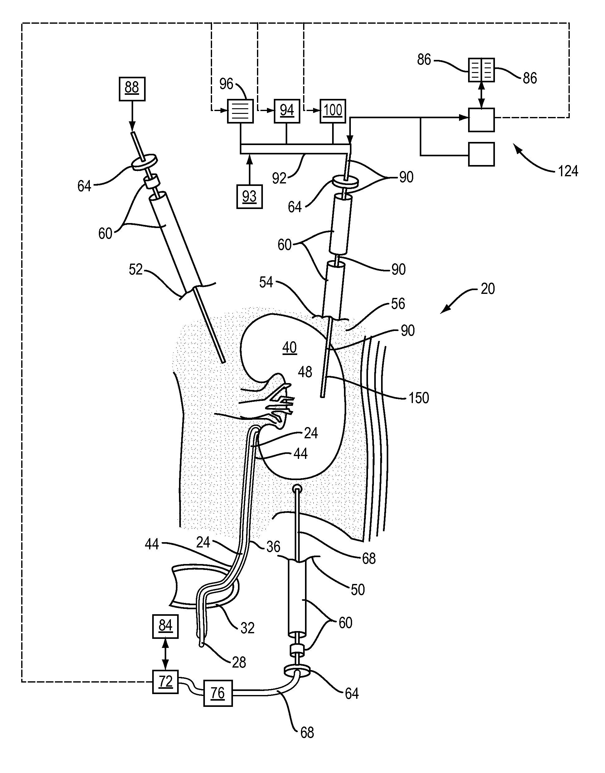
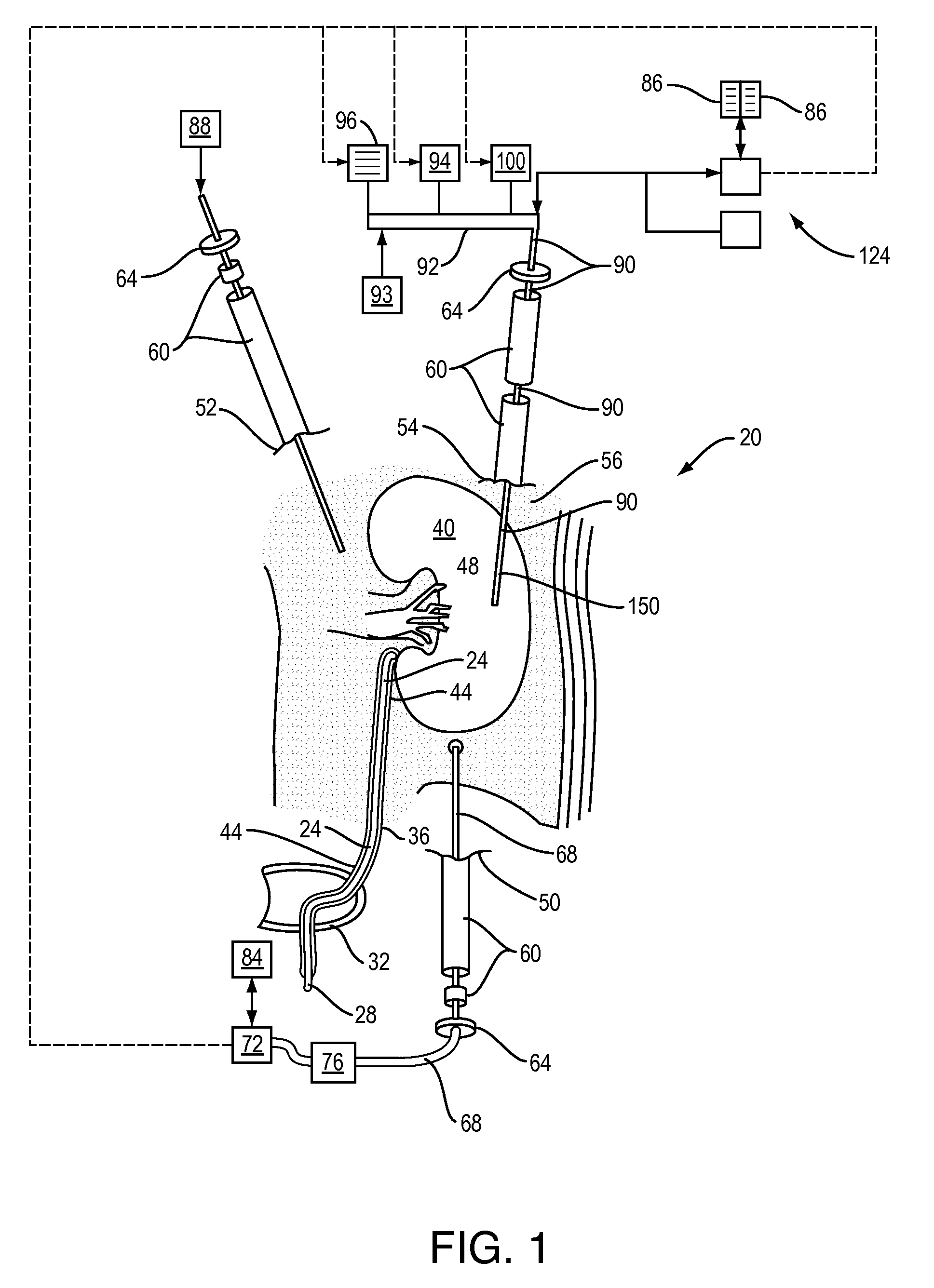
[0028]Although the illustrated embodiment is shown in connection with treatment of a kidney 40, the illustrated probe, water circulation system and treatment is not limited to kidneys. The present invention is presently believed to be applicable equally readily to the ablation of tissue of the liver, the pancreas, the urinary bladder 32, the gall bladder, the stomach, the heart, lungs, uterus or any other organ suitable for treatment by HIFU Therapy. In addition, the probe assembly and other features of the present invention described herein may be used in conventional non-laparoscopic HIFU Therapy of, for instance, the prostrate, esophagus, vagina, or the like.

[0029]In an illustrated minimally invasive, HIFU-based procedure, the patient 20 is first prepared by the insertion of a guide wire 24 through the urethra 28 and bladder 32 into the ureter 36 of a diseased kidney 40. The guide wire 24 is, of course, radiopaque, so that its progress to the surgical field can be straightforward...


PUM


Abstract

A high-intensity focused ultrasound ablation of tissue using minimally invasive medical procedures is provided.

Description

CROSS REFERENCE TO RELATED APPLICATIONS[0001]This application is a continuation-in-part of U.S. application Ser. No. 10 / 380,031, filed on Sep. 19, 2001, which is expressly incorporated by reference herein. This application also claims the benefit of U.S. Provisional Application Ser. No. 60 / 686,499, filed on Jun. 1, 2005, which is also expressly incorporated by reference herein.BACKGROUND AND SUMMARY OF THE INVENTION[0002]The present invention relates to instruments to conduct minimally invasive medical procedures with the aid of laparoscopic techniques, and to such procedures themselves. More particularly, the present invention relates to high-intensity focused ultrasound ablation of tissue using minimally invasive medical procedures. It is disclosed in the context of high-intensity focused ultrasound ablation of kidney tissue, but is believed to be useful in other applications as well.[0003]Several minimally invasive and non-invasive techniques for the treatment of living tissues a...

Claims


Application Information

Patent Timeline
IPC IPC(8): A61N7/00A61B8/14
CPCA61N7/022A61N2007/0052A61N2007/0065A61B8/481A61N7/00A61B8/12A61B8/4281A61B2090/378A61N2007/0091A61B2017/2253A61B17/2251
Inventor SANGHVI, NARENDRA T.SEIP, RALFFEDEWA, RUSSELL J.CARLSON, ROY F.CHEN, WO-HSINGKATNY, ARTUR P.
Owner FOCUS SURGERY INC
Why Eureka
Try Eureka