
Method for determining biologically active hgf

a biologically active and hgf technology, applied in the field of determining biologically active hgf, can solve the problems of cumbersome and laborious methods, inability to find therapeutic failures, and still no golden standards to be used

Active Publication Date: 2012-04-19
PEAS COMMLIZING ENTITY AB
View PDF2 Cites 1 Cited by
  • Summary
  • Abstract
  • Description
  • Claims
  • Application Information

AI Technical Summary

Problems solved by technology

Further, monitoring of HGF levels before and after treatment during infectious diseases has been shown to possibly reveal therapeutic failure at an early stage.
One major clinical problem is determining whether infection or other inflammatory disorders cause the disease.
However, there are still no golden standards to be used.
Problems in establishing correct diagnosis occurs daily while treating inflammatory disorders in bowel, ulcers, joint diseases, CNS disorders, peritoneal, pleural and pericardial effusions, among others.
However the methods are cumbersome and laborious.

Method used


Image

  • Method for determining biologically active hgf
  • Method for determining biologically active hgf

Examples


example 1

Production of Gels Comprising HGF Binding Components

[0057]Recipe 1, Dextran Sulphate Gel

[0058]Remark: Sterile process

[0059]5 mg Dextran sulphate sodium salt (Sigma Aldrich)

[0060]50 mg Agarose powder (Sigma aldrich)

[0061]5 ml (270 ml MQ+30 ml PBS pH 7.4)

[0062]Heat in microwave oven

[0063]Divide 12-15 μl in 1-2 ml tubes (eppendorf)

[0064]Keep in 4-8° C. until usage

[0065]Recipe 2, Dextran Sulphate Gel with Chitosan[0066]1—100 mg agarose gel is solved in 9 ml deionized sterile water+1 ml PBS[0067]2—100 mg chitosan is solved in 4 ml glycin 2.0[0068]3—1 ml of proteoaminoglycan (preferably dextran sulphate or heparan sulphate proteoglycan) solution containing 10 mg / ml dextran sulphate[0069]4—Add all and boil in microwave[0070]5—Separate the clump[0071]6—Divide the clear liquid in wells 25-100 μl and let it solidify during several minutes

[0072]The dextran sulphate gel is ready to use.

[0073]Recipe 3, HSPG Gel

[0074]100 μl heparan sulfat proteoglycan (HSPG) a: 400 μl(ml) Sigma Aldrich (H7640)

[00...

example 2

Analysis of Body Fluid

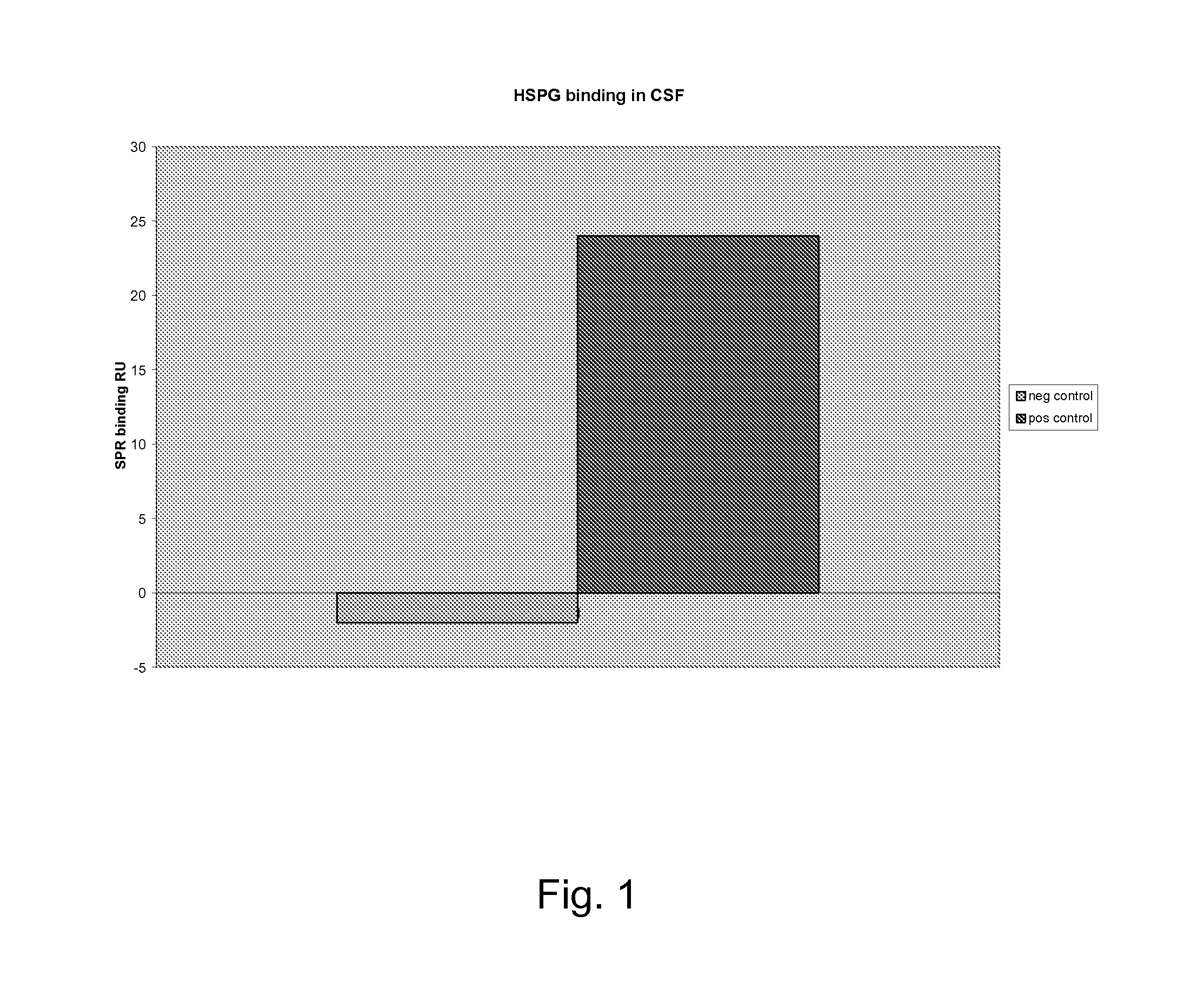
[0082]Sample: Lumbar puncture and 1 ml cerebrospinal fluid. Centrifuged 3000 g for 5 minutes.

[0083]Gel: The gel according to Recipe 1[0084]Add 100 μl CSF to the tube[0085]Wait for 2 minutes[0086]Remove excess fluid, e.g. by sterile cotton tip applicator[0087]Add 200 μl Toluidine blue[0088]Observe the colour change by eye or read by a table spectrophotometer.

[0089]A red colour indicates a negative result and a blue colour indicates a positive result.

[0090]Optionally, one or more reference solutions of known HGF content is used to evaluate the result. A negative reference may be e.g. water or PBS. A positive reference may be a body fluid sample or a commercial HGF containing product, with known HGF content.

example 3

Analysis of Commercial Product

[0091]Sample: Antithrombin III UF2 (Octapharma, Sweden)

[0092]Gel: The gel according to recipe 3

[0093]50 μl sample was added to the wells.

[0094]100 μl Toluidine blue indicator solution was added.

TABLE 1Spectropho-DilutiontometerUF2ColourResult(filter 620)1:1BluePos++0.1101:2BluePos++0.1071:4BluePos++0.0931:8Light bluePos+0.0851:16Redneg0.0801:32Redneg0.080MQRedneg0.079

[0095]Addition of HSPG or fragmin prior to addition of indicator solution gave negative results.

TABLE 2ColourResultDilution UF2 + 2μl HSPG1:1redneg1:2redneg1:4redneg1:8redneg1:16redneg1:32rednegDilution UF2 + 2μl low molecularheparin 5000 E / ml1:1redneg1:2redneg1:4redneg1:8redneg1:16redneg1:32redneg


PUM

PropertyMeasurementUnit
colouraaaaaaaaaa
distanceaaaaaaaaaa
concentrationsaaaaaaaaaa

Abstract

The present invention relates to a for determining the presence, absence or amount of biologically active or inactive HGF in a sample, comprising the steps: bringing the sample in contact with a gel comprising a HGF binding component of the extracellular matrix or cell membrane, adding toluidine blue to the gel, correlating the colour of the gel and / or a liquid in contact with the gel with the presence, absence or amount of biologically active HGF in the sample. It also relates to a kit of parts comprising toluidine blue and a gel comprising at least one HGF-binding component of the extracellular matrix or cell membrane, such as a proteoaminoglycan or a glucosaminoglycan, and optionally buffers, vials and sampling instruments and to a gel comprising at least one HGF-binding component of the extracellular matrix or cell membrane, such as a proteoaminoglycan or a glucosaminoglycan, toluidine blue and HGF.

Description

FIELD OF THE INVENTION[0001]The present invention is in the technical field of methods for detecting presence of growth factors such as hepatocyte growth factor (HGF) in biological samples. It also relates to products for performing such methods.BACKGROUND OF THE INVENTION[0002]Hepatocyte growth factor (HGF) is a unique growth factor, which is unrelated to other well-known polypeptide mitogens. It is a protein expressed in the mesenchymal cells such as lung macrophages and fibroblasts, Kupffer cells in the liver and leukocytes. HGF is secreted in response to cell damage and appears to be important for the regeneration of certain organs and healing of wounds. It is a heterodimer, having disulphide bonded heavy and light chains of approximately 60 and 30 kDa respectively, first synthesized as an inactive precursor. The precursor is cleaved to an active protein in the damaged organ by a specific activator. HGF acts paracrinally, i.e. it affects adjacent cells, as well as endocrinally, ...

Claims


Application Information

Patent Timeline
Patent Type & Authority Applications(United States)
IPC IPC(8): G01N33/566G01N33/567
CPCC08L5/02C08L5/08C08L5/10C08L5/12G01N33/74G01N2400/40G01N2333/4753C08L2666/26
Inventor NAYERI, FARIBA
Owner PEAS COMMLIZING ENTITY AB
Learn More