Novel EGFR-Binding Molecules and Immunoconjugates Thereof

a technology of immunoconjugates and egfr, which is applied in the field of antibodies, antigen-binding fragments thereof, polypeptides, and immunoconjugates, can solve the problems of skin toxicity, decreased life quality of patients, and imperfect anti-egfr therapies

Inactive Publication Date: 2012-06-21
IMMUNOGEN INC
View PDF4 Cites 27 Cited by
  • Summary
  • Abstract
  • Description
  • Claims
  • Application Information

AI Technical Summary

Benefits of technology

[0027]In one embodiment, the invention provides a method for inhibiting the growth of a cell expressing EGFR comprising contacting the cell with an immunoconjugate or the pharmaceutical composition described herein. In another embodiment, the cell is a tumor cell.
[0028]In one embodiment, the invention provides a method for treating a patient having a neoplasm comprising administering to said patient a therapeutically effective amount of an immunoconjugate or pharmaceutical composition described herein. In another embodiment, the neoplasm is selected from the group consisting of: abdominal, bone, breast, digestive system, liver, pancreas, peritoneum, adrenal, parathyroid, pituitary, testicles, ovary, thymus, thyroid, eye, head and neck, central nervous system, peripheral nervous system, lymphatic system, pelvic, skin, soft tissue, spleen, thoracic region, and urogenital system. In another embodiment, the method comprises administering a second anti-cancer agent to the subject. In a further embodiment, the second anti-cancer agent is a chemotherapeutic agent.
[0029]In one embodiment, the invention provides a method for treating a cell proliferative disorder in a patient comprising administering to said patient a therapeutically effective amount of an immunoconjugate or pharmaceutical composition described herein. In another embodiment, the cell proliferative disorder is selected from the group consisting of: adrenal cortex hyperplasia (Cushing's disease), congenital adrenal hyperplasia, endometrial hyperplasia, benign prostatic hyperplasia, breast hyperplasia, intimal hyperplasia, focal epithelial hyperplasia (Heck's disease), sebaceous hyperplasia, compensatory liver hyperplasia, and any other cell proliferation disease, besides neoplasia.

Problems solved by technology

Anti-EGFR therapies are not perfect.
EGFR therapies also result in skin toxicity.
Although rarely life-threatening, the skin toxicities cause significant physical and psycho-social discomfort that decrease the patient's life quality.
Additionally, in around 10% of patients, the skin toxicity is so severe that it requires treatment interruption or discontinuation that impairs the clinical outcomes of EGFR inhibitors.

Method used


Image

  • Novel EGFR-Binding Molecules and Immunoconjugates Thereof
  • Novel EGFR-Binding Molecules and Immunoconjugates Thereof
  • Novel EGFR-Binding Molecules and Immunoconjugates Thereof

Examples


example 1

[0259]Production of Murine EGFR Antibodies

[0260]To produce murine anti-EGFR antibodies, head and neck squamous carcinoma cell lines such as CA922 (Japanese Collection of Research Bioresources (JCRB), Shinjuku, Japan) and HSC4 (JCRB, Shinjuku, Japan) were injected subcutaneously into Balb / c female mice (Charles River Laboratory, Wilmington, Mass.) at the dose of 5×10⁶ cells per mouse every 2 weeks for 5 times. Three days before being sacrificed for hybridoma generation, the immunized mice received intraperitoneal injection of another dose of antigen. The spleen from the mouse was collected according to standard animal protocols and was ground between two sterile, frosted microscopic slides to obtain a single cell suspension in RPMI-1640 medium. After the red blood cells were lysed with ACK lysing buffer, the spleen cells were then mixed with murine myeloma P3X63Ag8.653 cells (P3 cells) (J. F. Kearney et al. 1979, J Immunol, 123:1548-1550) at ratio of 1 P3 cells:3 spleen cells. The mi...

example 2

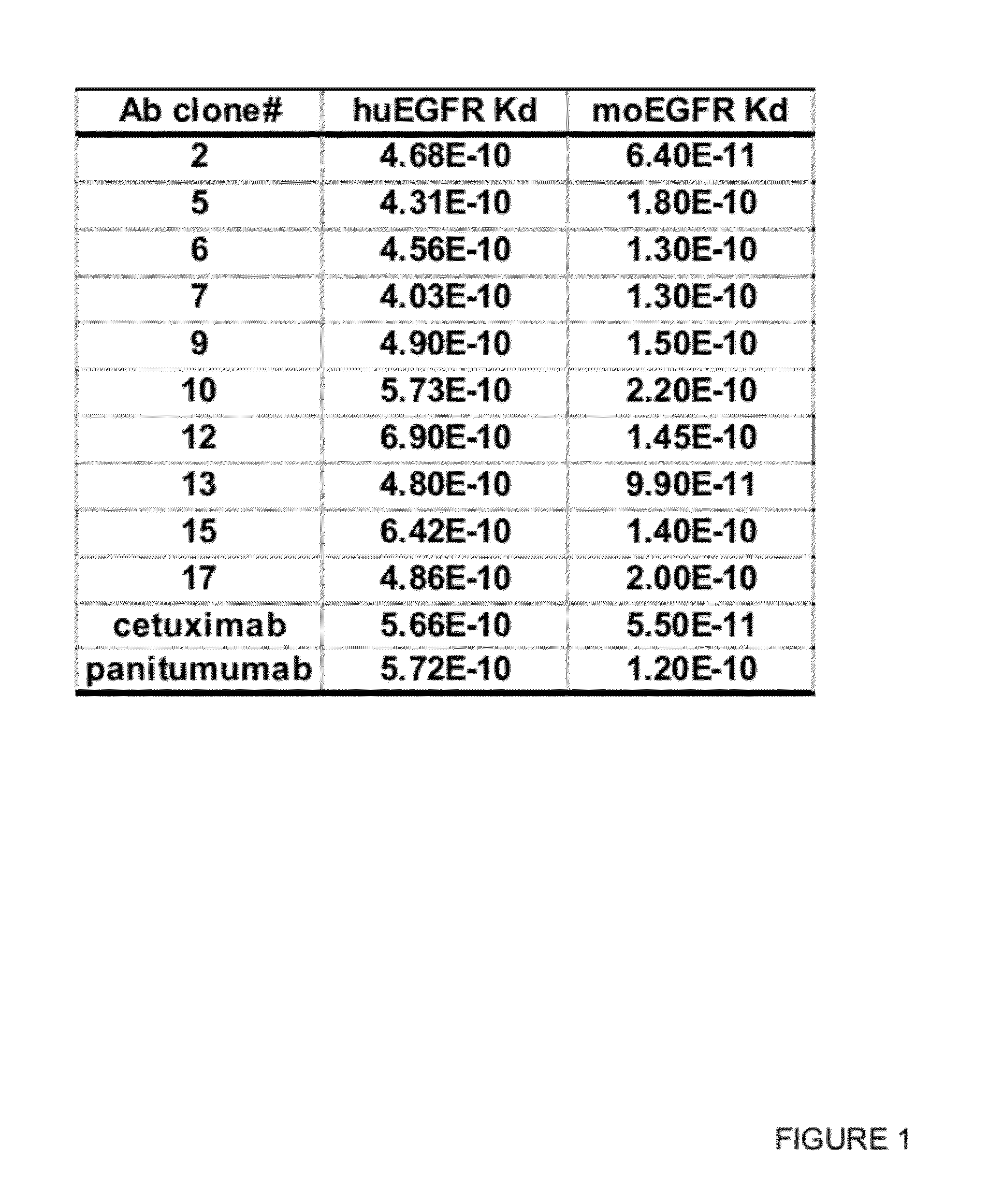
Binding Affinity to Human and Monkey EGFR Antigen

[0265]To determine the binding affinity of the EGFR antibodies to the human and monkey antigens, EGFR expressing human tumor cell lines such as MDA-MB468 (ATCC) and A431 (ATCC), and Vero monkey kidney cell line (ATCC) were used in a flow cytometric binding assay. In brief, the cells were incubated with various concentration of EGFR antibody for 1 h at 4° C. The cells were washed and incubated with PE-conjugated secondary antibody (Jackson Immunoresearch) for 1 h at 4° C. The cells were then washed, fixed in formalin and analyzed in FACSarray (BD Bioscience). To determine the binding affinity of these antibodies, geometric mean fluorescence intensity was plotted against the antibody concentration in a semi-log plot. A dose-response curve was generated by non-linear regression and the EC50 value of the curve, which corresponds to the apparent dissociation constant (Kd) of each antibody, was calculated using GraphPad Prism v4 (GraphPad s...

example 3

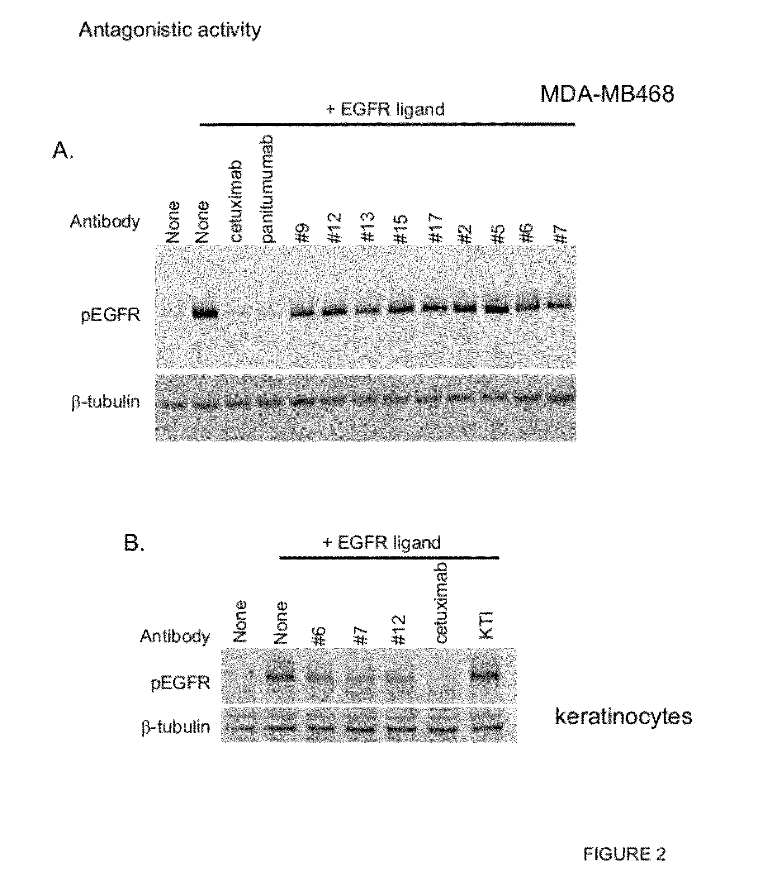
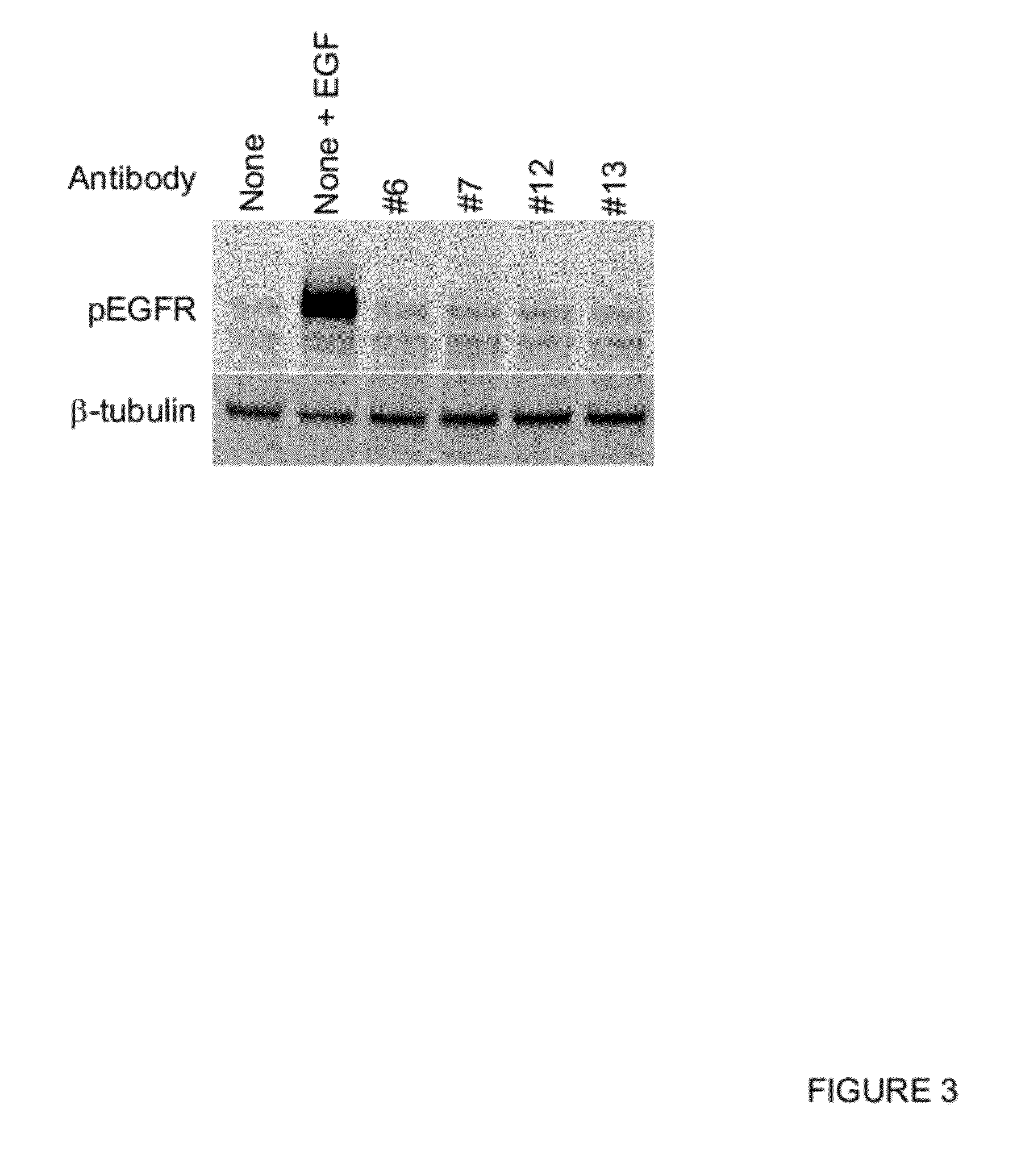
Inhibition of Ligand-Induced EGFR Activation

[0266]EGFR ligand binding induces EGFR phosphorylation followed by activation of downstream signaling pathways. To investigate the effect of anti-EGFR antibodies in ligand-induced EGFR activation, Western blot analysis was performed using MDA-MB468 tumor cell line and human primary adult keratinocytes. In brief, cells were seeded at 1e6 cells / well in a 6 well plate and cultured in normal media for overnight. Cells were washed and starved in serum free media containing 0.1% BSA for 2 hours at 37° C. 10 μg / ml antibody was added to the cells and the mixture was incubated for 3 hours at 37° C. 50 ng / ml EGF (R&D Systems) was added to the mixture and incubated for 15 minutes at 37° C. Cells were then washed with ice-cold PBS and lysed in RIPA buffer containing protease and phosphatase inhibitors. The protein lysates were separated in SDS-PAGE and transferred to a nitrocellulose membrane. The membrane was blocked with 5% BSA and incubated with an...


PUM

PropertyMeasurementUnit
dissociation constantaaaaaaaaaa

Abstract

Novel anti-cancer agents, including, but not limited to, antibodies and immunoconjugates, that bind to EGFR are provided. Methods of using the agents, antibodies, or immunoconjugates, such as methods of inhibiting tumor growth are further provided.

Description

CROSS-REFERENCE TO RELATED APPLICATIONS[0001]This application claims the priority benefit of U.S. Provisional Application No. 61 / 408,497, filed Oct. 29, 2010, and U.S. Provisional Application No. 61 / 477,086, filed Apr. 19, 2011, each of which is hereby incorporated by reference herein in its entiretyFIELD OF THE INVENTION[0002]The present invention generally relates to antibodies, antigen-binding fragments thereof, polypeptides, and immunoconjugates that bind to EGFR. The present invention also relates to methods of using such EGFR-binding molecules for diagnosing and treating diseases, such as malignancies.BACKGROUND OF THE INVENTION[0003]The epidermal growth factor receptor (EGFR or ErbB1 or HER1) is a transmembrane glycoprotein of 170 kDa that is encoded by the c-erbB1 proto-oncogene located in the 7q22 chromosome. EGFR is a member of the human epidermal growth factor receptor (HER) family of receptor tyrosine kinases (RTK) which includes HER2 (ErbB2), HER3 (ErbB3) and HER4 (ErbB...

Claims


Application Information

Patent Timeline
Patent Type & Authority Applications(United States)
IPC IPC(8): A61K39/395C07K19/00A61P35/00C12N5/10C12P21/00C12N5/071C12N15/63C12N1/21C07K16/22C12N15/13
CPCA61K47/48384A61K47/48561A61K2039/505C07K16/2863C07K2317/24C07K2317/33A61K45/06C07K2317/732C07K2317/76C07K2317/92C07K2317/34A61K39/39558C07K2317/73A61K47/6803A61K47/6849A61P1/16A61P13/08A61P15/14A61P17/00A61P35/00A61P43/00A61P5/38A61K39/395C07K16/28C07K2317/565
Inventor SETIADY, JULIANTOPARK, PETER U.RUI, LINGYUNCHITTENDEN, THOMAS
Owner IMMUNOGEN INC
Why Eureka
Try Eureka