


Quick Research
Generate reliable direction feasibility study reports for your R&D in just a few steps.

Technical Q&A
Discover and master advanced knowledge NOW. Basics, ideas, possibilities, all at once.

Find Solutions
As an expert in R&D theories, this can generate solutions to your technical problems instantly.

Evaluate Feasibility
Analyze your overall solution with one click, know your potential R&D risks in advance.

Monitor Landscape
Get weekly tech updates, stay abreast of the latest tech innovations and key insights.
Flap Endonuclease-1 As A Marker For Cancer
- Summary
- Abstract
- Description
- Claims
- Application Information
AI Technical Summary
Benefits of technology
Problems solved by technology
Method used
Image

Examples
example 1
[0171]Identification of FEN1 as a Potential Marker for Lung Cancer
[0172]Sources of Tissue:
[0173]Two different kinds of tissue, using proteomics methods, are used for identifying tumor specific proteins as diagnostic markers for lung cancer in accord with the disclosure provided herein.
[0174]In total, tissue specimens from 20 patients suffering from lung cancer (LC) are analyzed. From each patient two different tissue types are collected from therapeutic resections: tumor tissue (>80% tumor) (T) and adjacent healthy tissue (N). The latter one serves as matched healthy control sample. Tissues are immediately snap frozen after resection and stored at −80° C. before processing. Tumors are diagnosed by histopathological criteria.
[0175]Tissue Preparation:
[0176]0.8-1.2 g of frozen tissue are cut into small pieces, transferred to the chilled grinding jar of a mixer ball mill and completely frozen by liquid nitrogen. The tissue is pulverized in the ball mill, dissolved in the 10-fold volume ...
example 2
[0182]Generation of antibodies against the cancer marker protein FEN1
[0183]Polyclonal antibody to the lung cancer marker protein FEN1 is generated for further use of the antibody in the measurement of serum and plasma levels or concentrations in other body fluids of FEN1 by immunodetection assays, e. g. Western Blotting and ELISA.
[0184]Recombinant protein expression in E. coli:
[0185]In order to generate antibodies against FEN1, the recombinant antigen is produced in E. coli: Therefore, the FEN1-encoding region is PCR amplified from a full-length cDNA clone obtained from the German Resource Center for Genome Research (RZPD, Berlin, Germany) using the following primers:
[0186]Forward primer (SEQ ID NO 3:) 5′ -cacacacaattgattaaagaggagaaattaactATGAGAGGATCGCATCACCAT CACCATCACATTGAAGGCCGTGGAATTCAAGGCCTGGCC-3′ (MunI-site is underlined, coding nucleotides in capital letters).
[0187]Reverse primer (SEQ ID NO 4):
[0188]5′ -acgtacgtaagcttTCATTATTTTCCCCTTTTAAACTTC-3′ (HindIII-site is underlined, ...
example 3
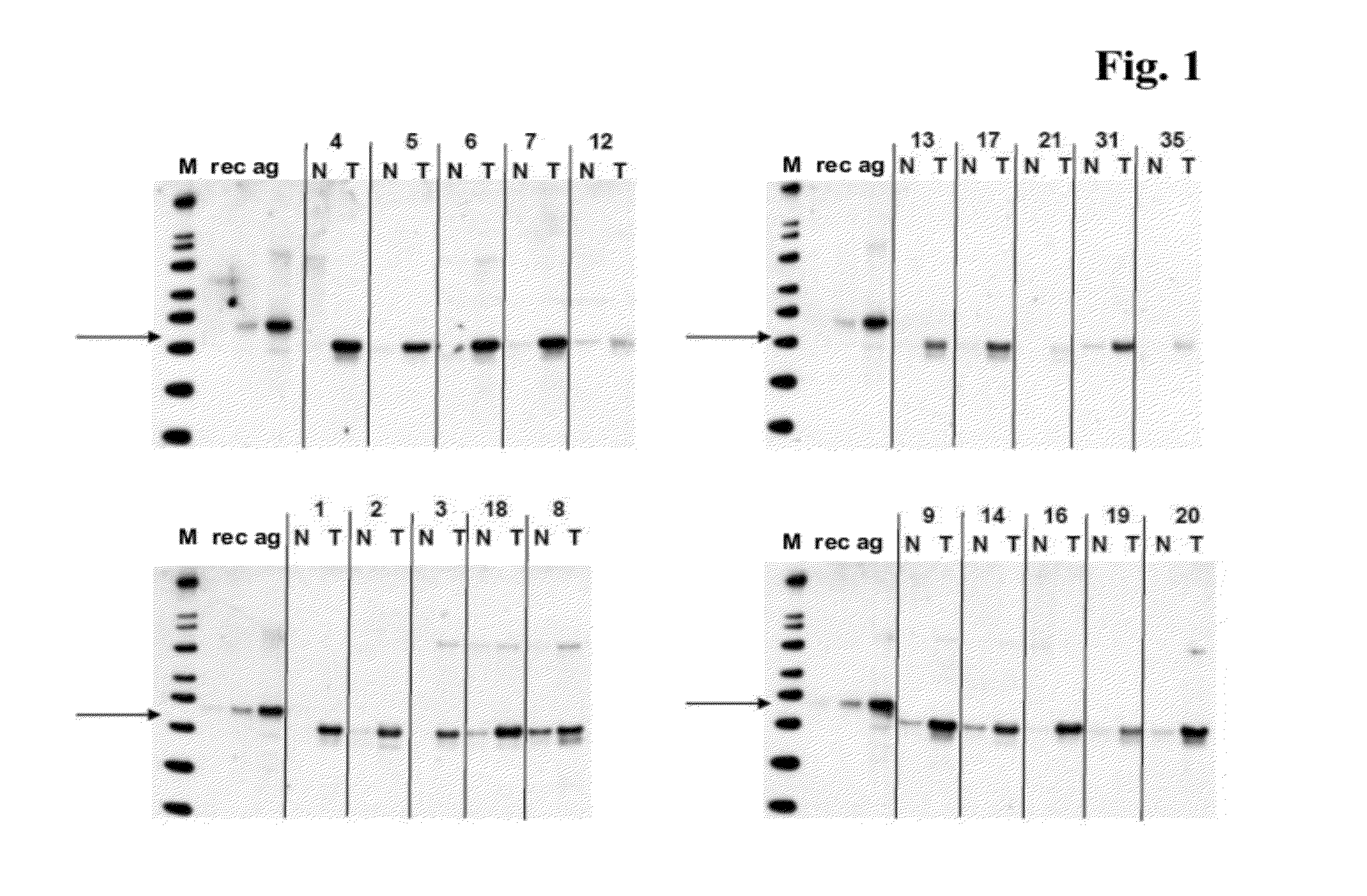
[0203]Western Blotting for the detection of FEN1 in human lung cancer (LC) tissue using polyclonal antibody as generated in Example 2
[0204]Tissue lysates from tumor samples and healthy control samples are prepared as described in Example 1, “Tissue preparation”.
[0205]SDS-PAGE and Western-Blotting are carried out using reagents and equipment of Invitrogen, Karlsruhe, Germany. For each tissue sample tested, 15 μg of tissue lysate are diluted in reducing NuPAGE® (Invitrogen) SDS sample buffer and heated for 10 min at 95° C. Samples are run on 4-12% NuPAGE® gels (Tris-Glycine) in the MES running buffer system. The gel-separated protein mixture is blotted onto nitrocellulose membranes using the Invitrogen XCell II™ Blot Module (Invitrogen) and the NuPAGE® transfer buffer system. The membranes are washed 3 times in PBS / 0.05% Tween-20 and blocked with Roti®-Block blocking buffer (A151.1; Can Roth GmbH, Karlsruhe, Germany) for 2 h. The primary antibody, polyclonal rabbit anti-FEN1 serum (ge...
PUM
| Property | Measurement | Unit |
|---|---|---|
| Concentration | aaaaa | aaaaa |
Abstract
Description
Claims
Application Information
 - R&D Engineer
- R&D Manager
- IP Professional
- Industry Leading Data Capabilities
- Powerful AI technology
- Patent DNA Extraction
Browse by: Latest US Patents, China's latest patents, Technical Efficacy Thesaurus, Application Domain, Technology Topic, Popular Technical Reports.
© 2024 PatSnap. All rights reserved.Legal|Privacy policy|Modern Slavery Act Transparency Statement|Sitemap|About US| Contact US: help@patsnap.com