Method and apparatus for quantitative microimaging

a micro-imaging and quantitative technology, applied in the field of labonachip type imaging and assays, can solve the problems of limiting the application of optical analysis methods to truly portable total analysis systems and methods, affecting and limiting the application of optical analysis methods. , to achieve the effect of facilitating micro-operation and simplifying the design of cytometers

Inactive Publication Date: 2012-09-06
BOARD OF RGT THE UNIV OF TEXAS SYST +1
View PDF9 Cites 89 Cited by
  • Summary
  • Abstract
  • Description
  • Claims
  • Application Information

AI Technical Summary

Benefits of technology

[0011]In one embodiment of the invention, truly portable, low-cost, and easy to operate microscale analysis systems are provided by adapting digital image sensors as quantitative optical detectors in a microfluidic assay system. In one aspect, static contact images of biomarker-labeled cell populations are analyzed using digital image processing to identify and count individual target cells. By eliminating the need for sheath flows and dynamic particle focusing, the cytometer design is greatly simplified. Quantitative microfluidic bioassays are also provided using these sensors. In one aspect, a chip-scale complementary metal-oxide-semiconductor (CMOS) image sensor is utilized via contact imaging to quantify formed elements such as microbial and mammalian cells in sub-nanoliter reagent droplets.
[0012]In certain aspects a cytometer is provided through the use of contact imaging whereby the cell sample to be analyzed is contained in a disposable volume-calibrated reservoir that is placed in direct proximity to the digital imaging array. The cell sample reservoir can be fabricated as part of a microfluidic sample preparation cartridge that will facilitate fluid handling and minimize the volumes of sample and reagent needed for each assay, but will offer much of the simplicity and economy of a traditional hemocytometer, enabling a relatively unskilled worker to quickly perform biomarker labeling on cell samples.

Problems solved by technology

Although inexpensive, portable point-of-need assay systems would have immediate applications in clinical diagnostics, global health, environmental monitoring, and forensics, few commercial examples presently exist.
However, while microfluidic implementations of optical methods have been demonstrated, detection is typically achieved off-chip using conventional microscope optics and digital camera systems or custom and relatively expensive chip-scale prototype optoelectronics.
Thus, translation of optical analysis methods into truly portable total analysis systems and methods has been hindered by a lack of reasonably priced, sensitive and compact detectors that can easily be integrated with microscale sample handling and processing.

Method used


Image

  • Method and apparatus for quantitative microimaging
  • Method and apparatus for quantitative microimaging
  • Method and apparatus for quantitative microimaging

Examples


example 1

Initial Proof of Principal Studies

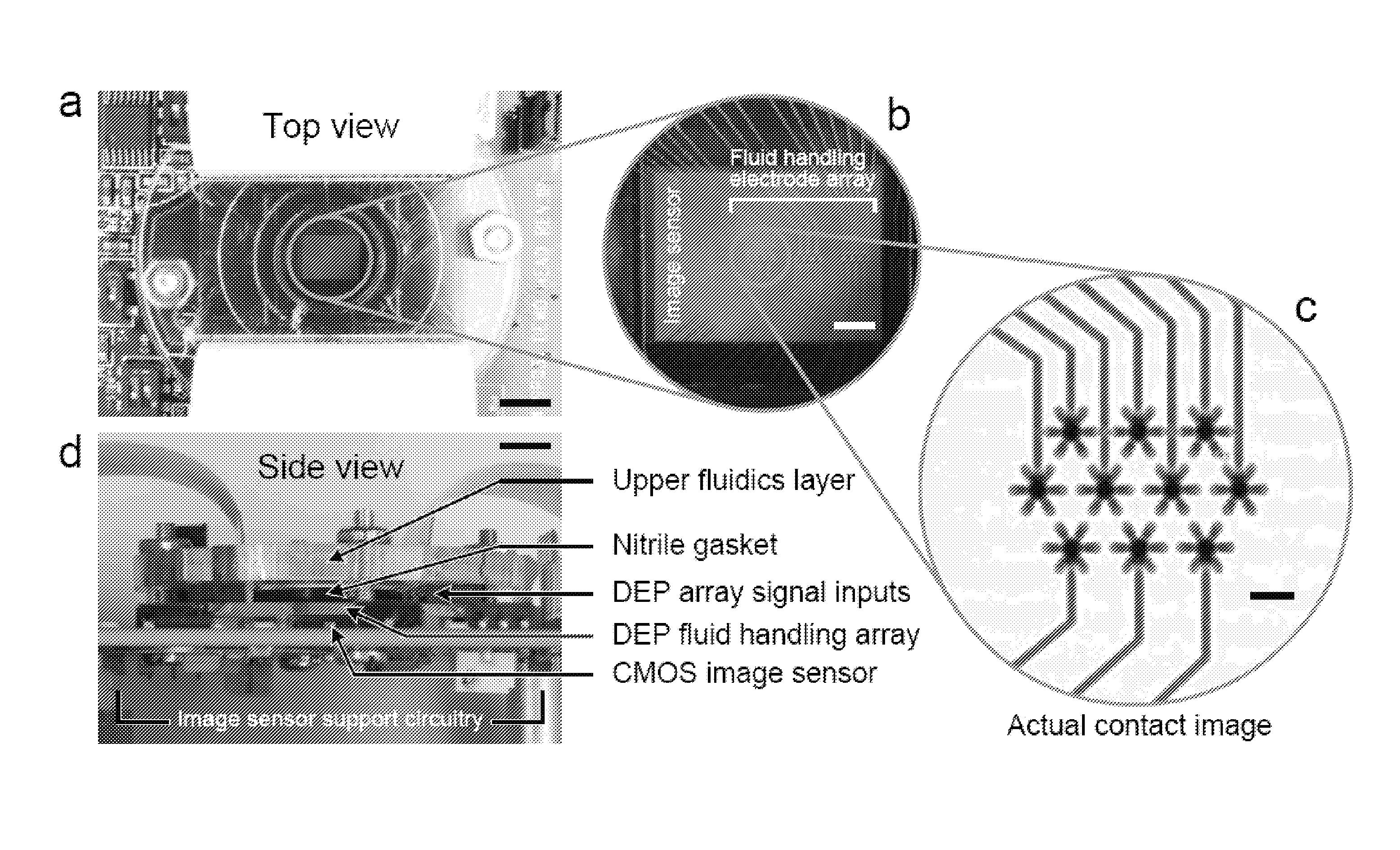
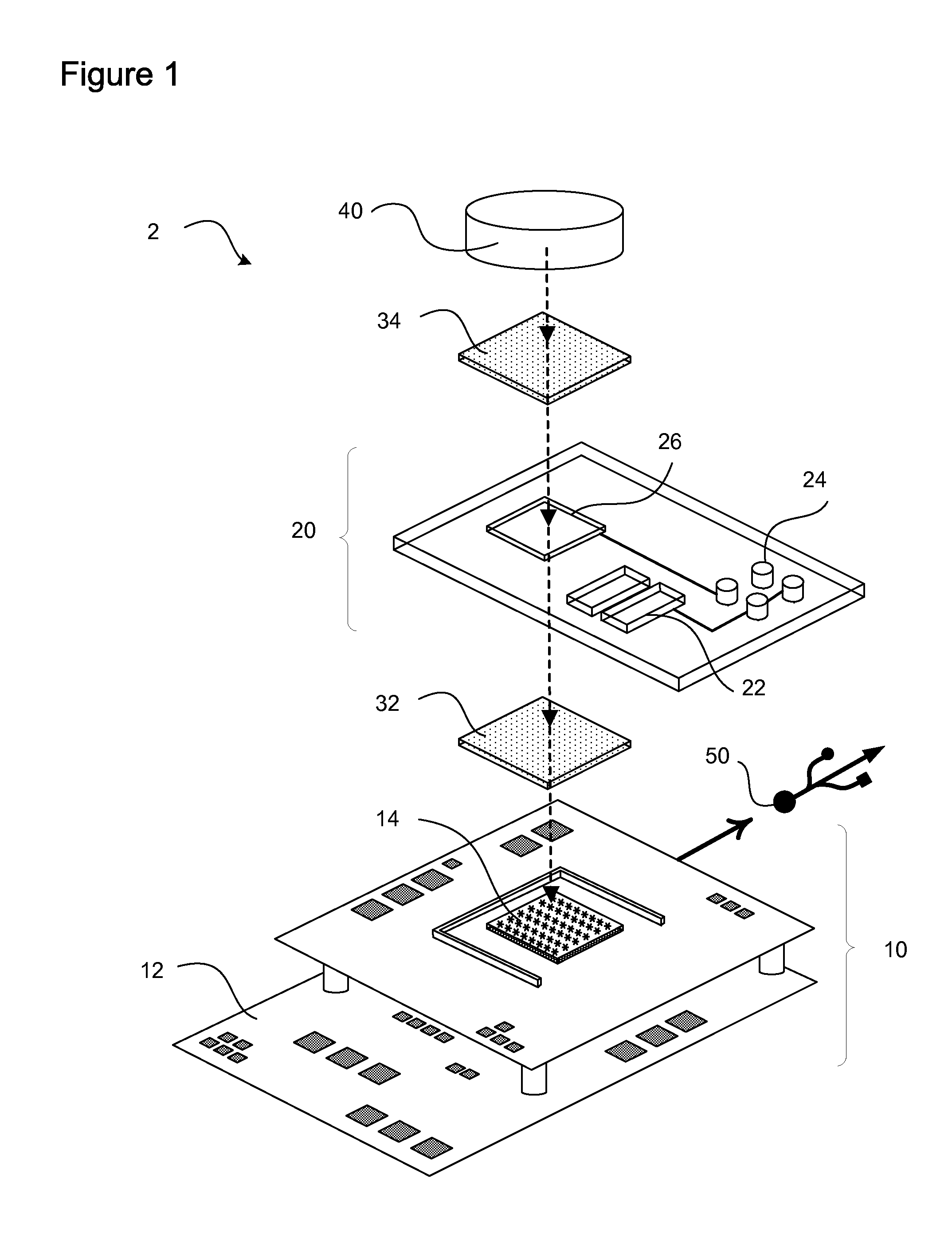
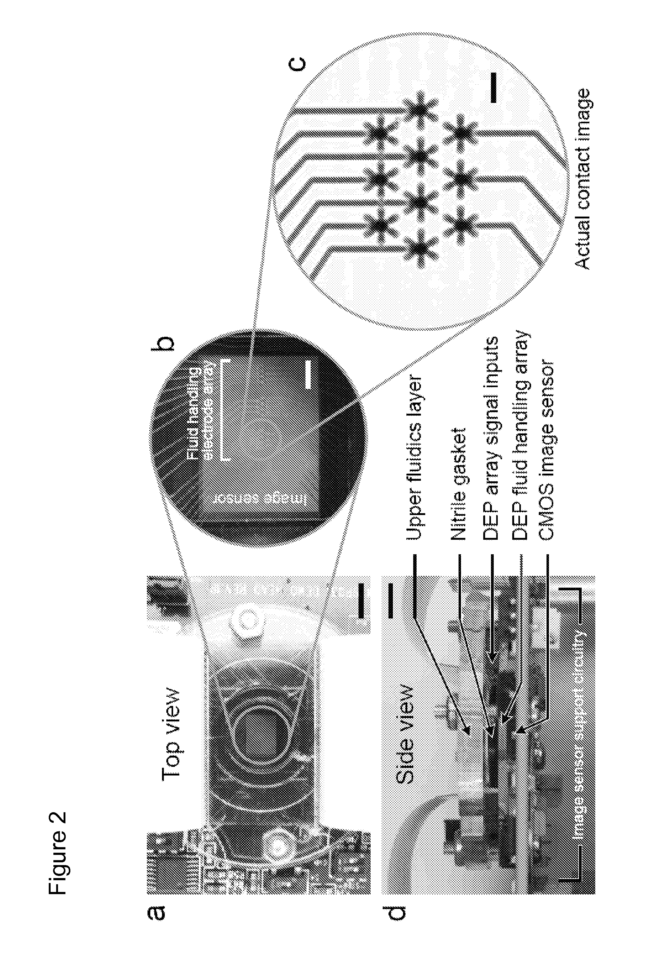
[0060]Reagents were imaged in the digital microfluidic device depicted in FIGS. 2a-d including a CMOS image sensor integrated with digital microfluidics. FIG. 2a is a top view, while FIG. 2d is a side view. Scale bars of 5 mm are shown on FIGS. 2a and d. FIG. 2b provides a magnified view of the CMOS image sensor overlayed by a dielectrophoresis (DEP) fluid handling electrode array. The scale bar is 1 mm. The DEP microfluidic device utilizes electrically generated forces to manipulate discrete reagent droplets. The reagents are not confined to channels but are instead manipulated using an addressable electrode array.

[0061]The fluid handling microelectrode array was fabricated using standard microlithographic wet etch processing from thin film Au / Ti (2500 Å / 500 Å) on 1 mm-thick Pyrex substrates. The upper fluidics layer was laser-machined in-house (VersaLASER, Universal Laser Systems, Inc., Scottsdale, Ariz.) from a cast acrylic sheet (Acrylite G P, E...

example 2

Cell Quantitation

[0072]In one embodiment of the invention, apparatus and methods are provided for quantitation of relative cell populations in a mixed cell population. In ordinary manual cell counting assays using Neubauer haemocytometers, cells are loaded into a fixed volume 3 mm×3 mm×0.1 mm chamber that contains 900 nL of cell suspension. Contact imaging such reservoirs is feasible as these dimensions are within the active area of typical digital image sensor formats, including a 1 / 3.6″ format which comprises a 4.00 mm×3.00 mm imaging area and is the smallest of the standard image sensor formats. In one embodiment, the sample preparation cartridge for the point-of-need image cytometer will include an integrated volume-calibrated reservoir similar to that found in a haemocytometer, enabling cell counts to be expressed in terms of concentration, an important consideration for diagnostic assays. In particular embodiments, all fluid handling is integral to the sample preparation cartr...

example 3

Personalized Medicine

[0086]There are numerous examples in medicine where selection of a particular drug from the large class of drugs available to treat a particular condition is empirical. For any given drug, efficacy and side effects are essentially averaged over populations of treated patients while the activity of the drug in a given individual in unknown until it is administered. Often different drugs from a class must be serially administered to an individual until a particular drug from the class is identified that is both safe and efficacious in that individual. Individual drug actions cannot be translated from one individual to another. With certain drugs and in certain disease the empirical approach is dangerous and prolongs the period of uncontrolled disease. For example, selection of a safe and effective drug for an individual patient from powerful and potentially dangerous classes of drugs has heretofore been largely empirical. Such drug classes include anti-platelet dr...


PUM


Abstract

Optical detection platforms are described as well as methods of using such platforms to perform quantitative assays.

Description

CROSS REFERENCE TO RELATED APPLICATIONS[0001]This application claims priority to U.S. Provisional Application Ser. No. 61 / 187,669 filed on Jun. 17, 2009, and U.S. Provisional Application Ser. No. 61 / 220,002, filed on Jun. 24, 2009. The disclosures of the prior application are considered part of (and are incorporated by reference in) the disclosure of this application.STATEMENT REGARDING GOVERNMENT INTERESTS[0002]This work was supported in part by the following United States Government grants: National Institutes of Health / National Institute of Biomedical Imaging and Bioengineering grant 5R01EB006198. The Government may have certain rights in this invention.FIELD OF THE INVENTION[0003]This invention relates to lab-on-a-chip type imaging and assays and methods for both qualitative and quantitative optical detection and microimaging.BACKGROUND OF THE INVENTION[0004]Without limiting the scope of the invention, its background is described in connection with existing microimaging apparatu...

Claims


Application Information

Patent Timeline
Patent Type & Authority Applications(United States)
IPC IPC(8): H04N7/18
CPCB01L3/502715B01L2300/0654B01L2300/0816G01N2201/062G01N15/1463G01N21/6454G01N2201/0221B01L2400/0406
Inventor VYKOUKAL, JODYVYKOUKAL, DAYNENE M.STONE, GREGORY P.ALT, ECKHARD U.
Owner BOARD OF RGT THE UNIV OF TEXAS SYST
Why Eureka
Try Eureka