Neural Regeneration

a neuronal injury and neuronal regeneration technology, applied in the field of neuronal injury treatment, can solve the problems of inability of adult cns to regenerate, limited ability for collateral sprouting of spared fibers, and inability to improve neuronal function, so as to enhance the ability of axons, reduce inhibition, and high affinity

Inactive Publication Date: 2012-09-13
CASE WESTERN RESERVE UNIV +1
View PDF5 Cites 9 Cited by
  • Summary
  • Abstract
  • Description
  • Claims
  • Application Information

AI Technical Summary

Benefits of technology

[0007]The present invention provides for a transmembrane protein tyrosine phosphatase, protein tyrosine phosphatase sigma (PTPσ), that binds with high affinity to neural CSPGs. Thus, PTP is the first-identified receptor for the scar tissue-associated regeneration inhibitors. CSPG binding involves the CS chains and a specific site on the first immunoglobulin-like domain of PTPσ. In culture, PTPσ− / − neurons show reduced inhibition by CSPG. A PTPσ fusion protein probe can detect cognate ligands upregulated specifically at neural lesion sites. After spinal cord injury, PTPσ gene disruption enhancesd the ability of axons to penetrate regions containing CSPG. These results indicate that PTPσ can act as a receptor for CSPGs, and may provide new therapeutic approaches to neural regeneration.

Problems solved by technology

A fundamental obstacle facing efforts to improve neuronal function after injury is the inability of the adult CNS to regenerate.
The inhibitory nature of CSPG is not only reflected in the formation of dystrophic axonal retraction bulbs that fail to regenerate through the lesion, but also in the limited ability for collateral sprouting of spared fibers.
Although it has been known for nearly two decades that sulfated proteoglycans are major contributors to the repulsive nature of the glial scar, the precise inhibitory mechanism was poorly understood.

Method used


Image

  • Neural Regeneration
  • Neural Regeneration
  • Neural Regeneration

Examples


example 1

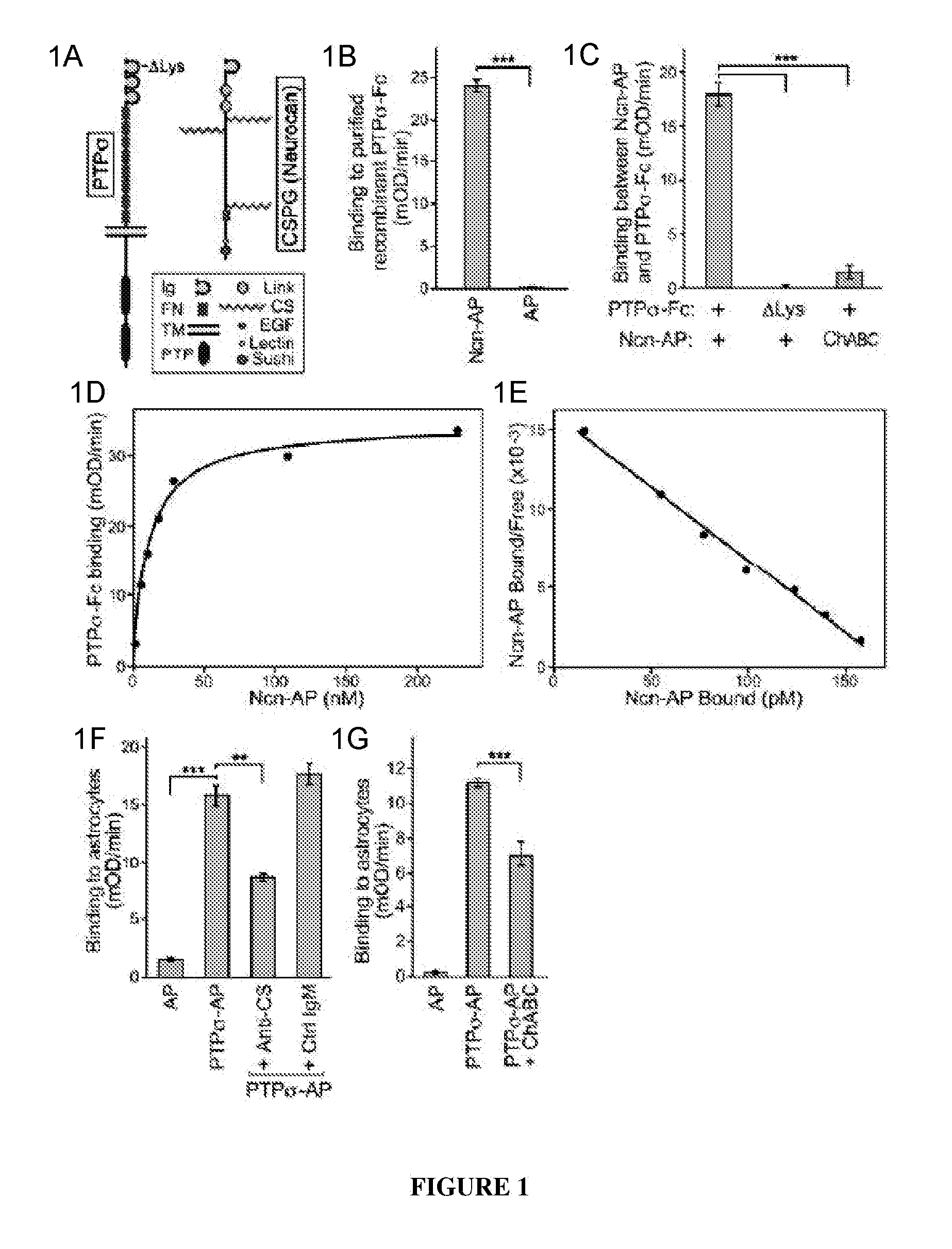
PTPσ Interacts with the CS Chains of CSPGs

[0135]The Ncn-AP fusion protein construct was generated by PCR amplification of nucleotide 1-2889 of a full-length mouse neurocan clone (Accession: BC065118, IMAGE:6853253) and subcloning the neurocan fragment into the NheI and HindIII sites of the APtag5 vector. See Flanagan et al., 327 Meth. Enzymol. 19 (2000). The resulting fusion protein includes neurocan amino acids 1-963 (N-terminal Ig domain, tandem link domains and central chondroitin sulfate attachment domain) fused to placental alkaline phosphatase. PTPσ-Fc and PTPσ-AP fusion protein constructs were generated by PCR amplification of nucleotide 1-2538 of a full-length mouse PTPσ clone (Accession BC052462, IMAGE 6834684), encoding the short isoform of PTPσ, which differs from the long isoform by alternative splicing to give 4 or 8 fibronectin domains. Chagnon et al., 82 Biochem. Cell Bio. 664 (2004); Sajnani-Perez et al., 22 Mol. Cell. Neurosci. 37 (2003). The PTP fragment was subclo...

example 2

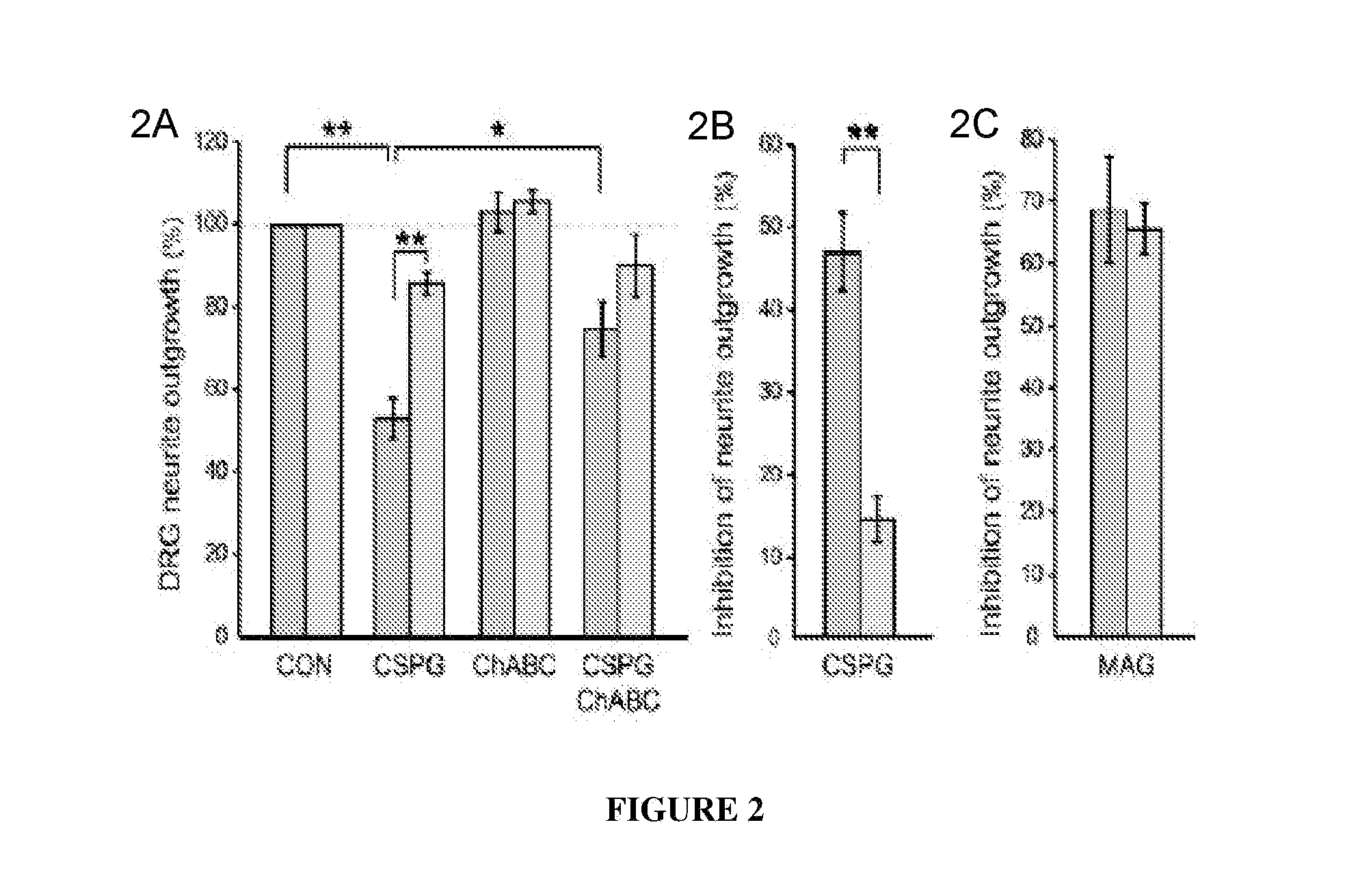
PTPσ Mediates the Inhibitory Effects of CSPG on Neuronal Regeneration

[0157]Having identified a binding interaction between PTPσ and CSPGs, studies were undertaken to test whether PTPσ is functionally involved in the inhibitory effects of CSPG on neurons. DRG neurons express high levels of PTPσ throughout life. Haworth et al., 12 Mol. Cell. Neurosci. 93 (1998). Postnatal day 8 (P8) dorsal root ganglion (DRG) neurons from mice with a targeted gene disruption of PTPσ (Elchebly et al., 21 Nat. Genet. 330 (1999)), or wild type controls, were cultured in the presence of a neural CSPG mixture (FIG. 2—DRG neurons from P8 mice were grown for 18 hours, then treated for 24 hours with or without CSPG, and visualized by GAP-43 immunolabeling.) or purified neurocan (FIG. 5 and data not shown). The CSPG mixture reduced control DRG neurite outgrowth by approximately 50% (FIGS. 2A and 2B) but had far less effect on neurons from PTPσ− / − mice (p<0.01; 2A and 2B), showing a functional involvement of PT...

example 3

PTPσ as a Marker for Neuronal Injury

[0161]Tests were carried out to determine whether PTPσ has appropriate binding specificity to detect endogenous CSPG at sites of neural injury. In particular, experiments focused on whether PTPσ could preferentially recognize injured versus uninjured adult CNS tissue. The answer could not be deduced simply from its ability to bind CSPGs (FIG. 1), because PTPσ also binds HSPGs and potentially other ligands. Receptor ectodomain fusion proteins can be used to detect the distribution of their cognate ligands in tissues. Flanagan et al., 327 Meth. Enzymol. 19 (2000).

[0162]Immunofluorescence was performed on sections from the spinal cord lesion site and from unlesioned spinal cord. The sections were double fluorescence labeled with anti-neurocan antibody (Ncn), together with either PTPσ-Fc probe, or Fc control. In the unlesioned spinal cord, anti-neurocan labeled a thin line at the pia. PTPσ-Fc showed no labeling noticeably above Fc control. In the spin...


PUM

PropertyMeasurementUnit
timeaaaaaaaaaa
timeaaaaaaaaaa
timeaaaaaaaaaa

Abstract

A method of promoting neural cell regeneration is carried out by contacting a neural cell with a compound that inhibits the binding of a chondroitin sulfate proteoglycan (CSPG) to a cellular (e.g., trans-membrane) PTPσ protein. The neural cell is associated with an injury or neurodegenerative condition.

Description

RELATED APPLICATIONS[0001]This application claims the benefit of priority of U.S. Patent Application Ser. No. 61 / 274,520, filed 18 Aug. 2009, and Ser. No. 61 / 251,521, filed 14 Oct. 2009, the contents of which are incorporated herein by reference in their entirety.GOVERNMENTAL SUPPORT[0002]This invention was made with Government support under grants HD29417, EY11559 and NS40043, awarded by the National Institutes of Health. The U.S. Government has certain rights in the invention.FIELD OF THE INVENTION[0003]The invention relates to the treatment of neuronal injury.BACKGROUND OF THE INVENTION[0004]Spinal cord injury and other central nervous system (CNS) injuries can cause permanent disability or loss of movement (paralysis) and sensation below the site of the injury. Recovery after CNS injury is minimal, leading to substantial current interest in potential strategies to overcome this challenge. A fundamental obstacle facing efforts to improve neuronal function after injury is the inab...

Claims


Application Information

Patent Timeline
Patent Type & Authority Applications(United States)
IPC IPC(8): A61K39/395A61P25/00A61K38/46A61K38/17C12N5/0793C12N5/071
CPCC07K2319/30A61K38/1709A61P25/00A61P25/28
Inventor FLANAGAN, JOHN G.TENNEY, ALAN PETERSHEN, YINGJIESILVER, JERRY
Owner CASE WESTERN RESERVE UNIV
Why Eureka
Try Eureka