Apparatus and method for non-invasive diabetic retinopathy detection and monitoring

a diabetic retinopathy and non-invasive technology, applied in the field of computer-aided technology for diseases diagnosis, can solve problems such as suppressing interaction among

Active Publication Date: 2012-10-04
THE HONG KONG POLYTECHNIC UNIV
View PDF4 Cites 31 Cited by
  • Summary
  • Abstract
  • Description
  • Claims
  • Application Information

AI Technical Summary

Benefits of technology

[0009]Accordingly, one object of the present invention is to provide solutions to the aforementioned issues. This object is realized by providing a unique and special design of a new lighting and position alignment structure for fundus camera (hardware component) and a hierarchical retinal image analysis with new algorithms for multispectral image representation of the retina, dynamic feature extraction, reliable image registration, dynamic image tracking, hierarchical image matching and robust identification of changes (software component).

Problems solved by technology

First, it can suppress the interaction among features during the process of feature extraction.

Method used


Image

  • Apparatus and method for non-invasive diabetic retinopathy detection and monitoring
  • Apparatus and method for non-invasive diabetic retinopathy detection and monitoring
  • Apparatus and method for non-invasive diabetic retinopathy detection and monitoring

Examples


Embodiment Construction

Hardware Aspect

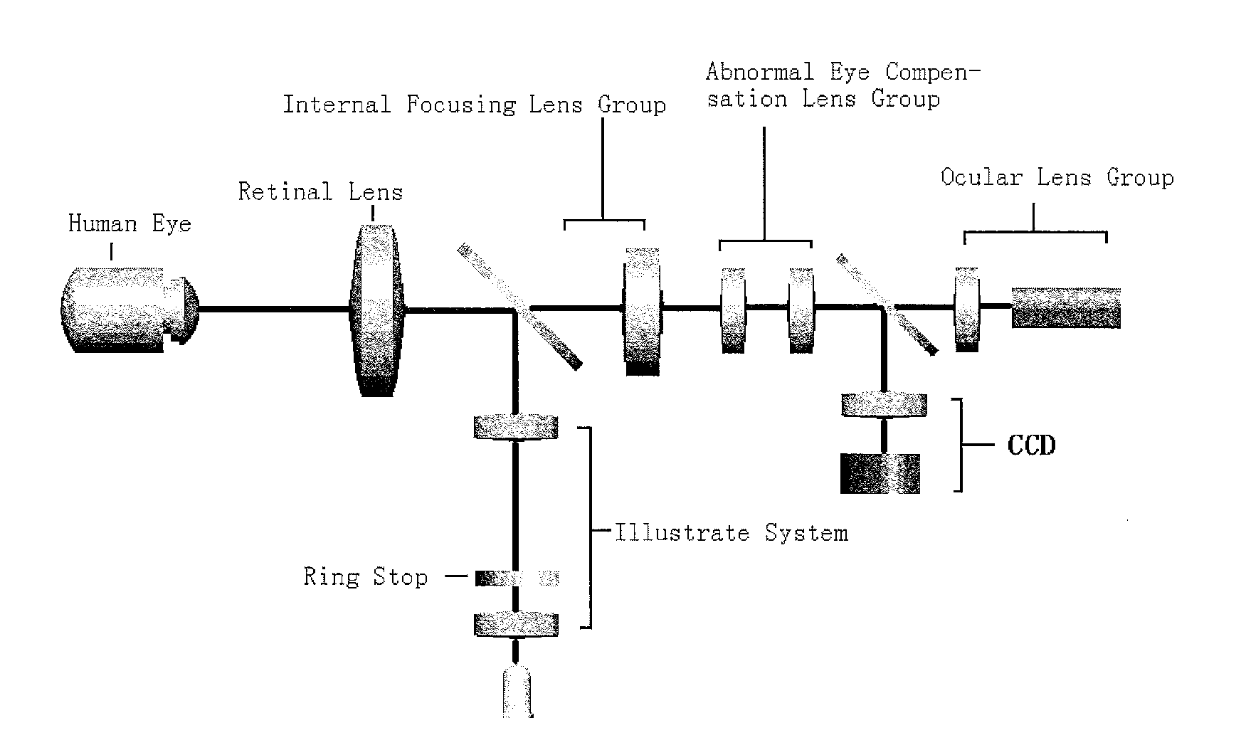
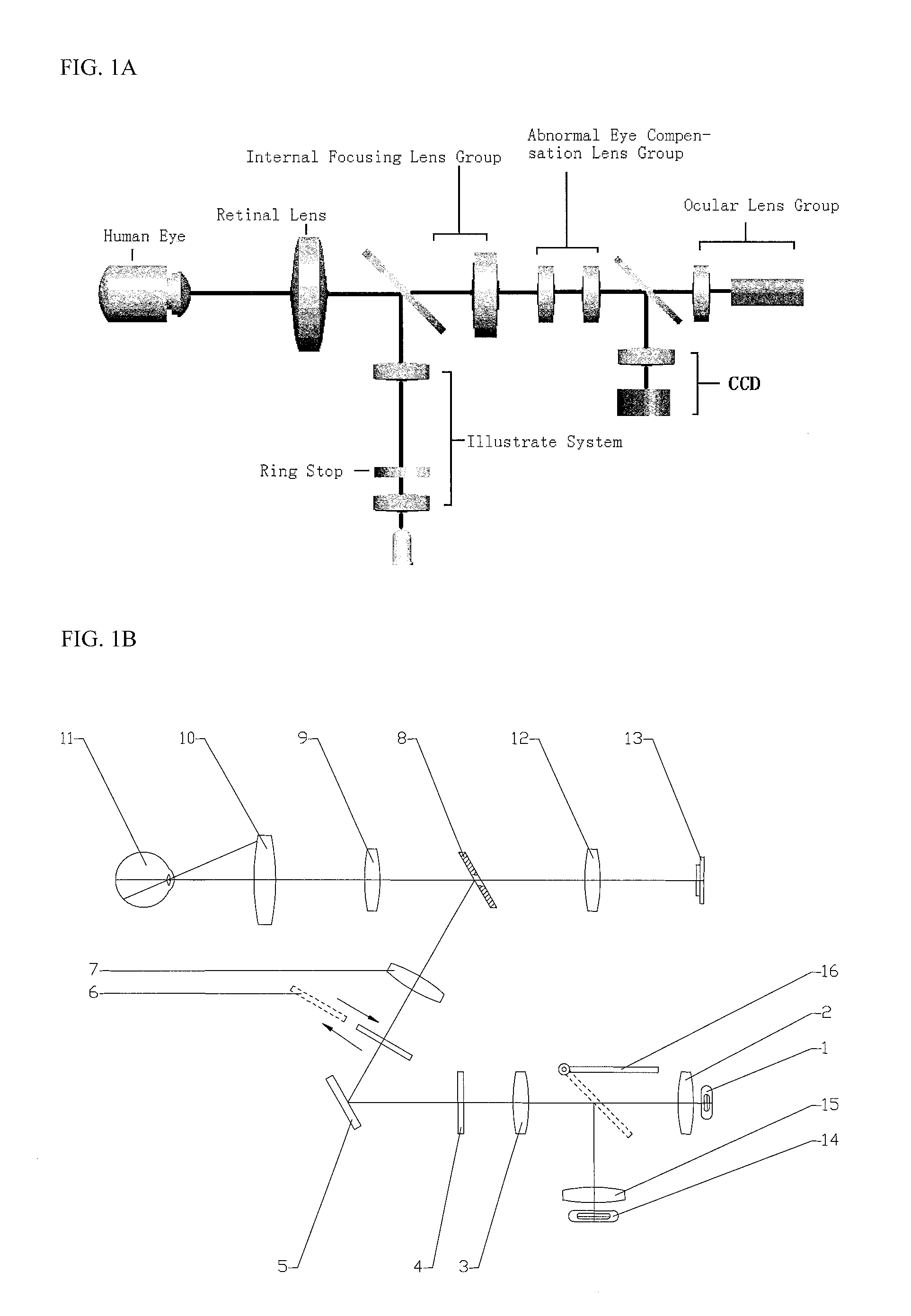
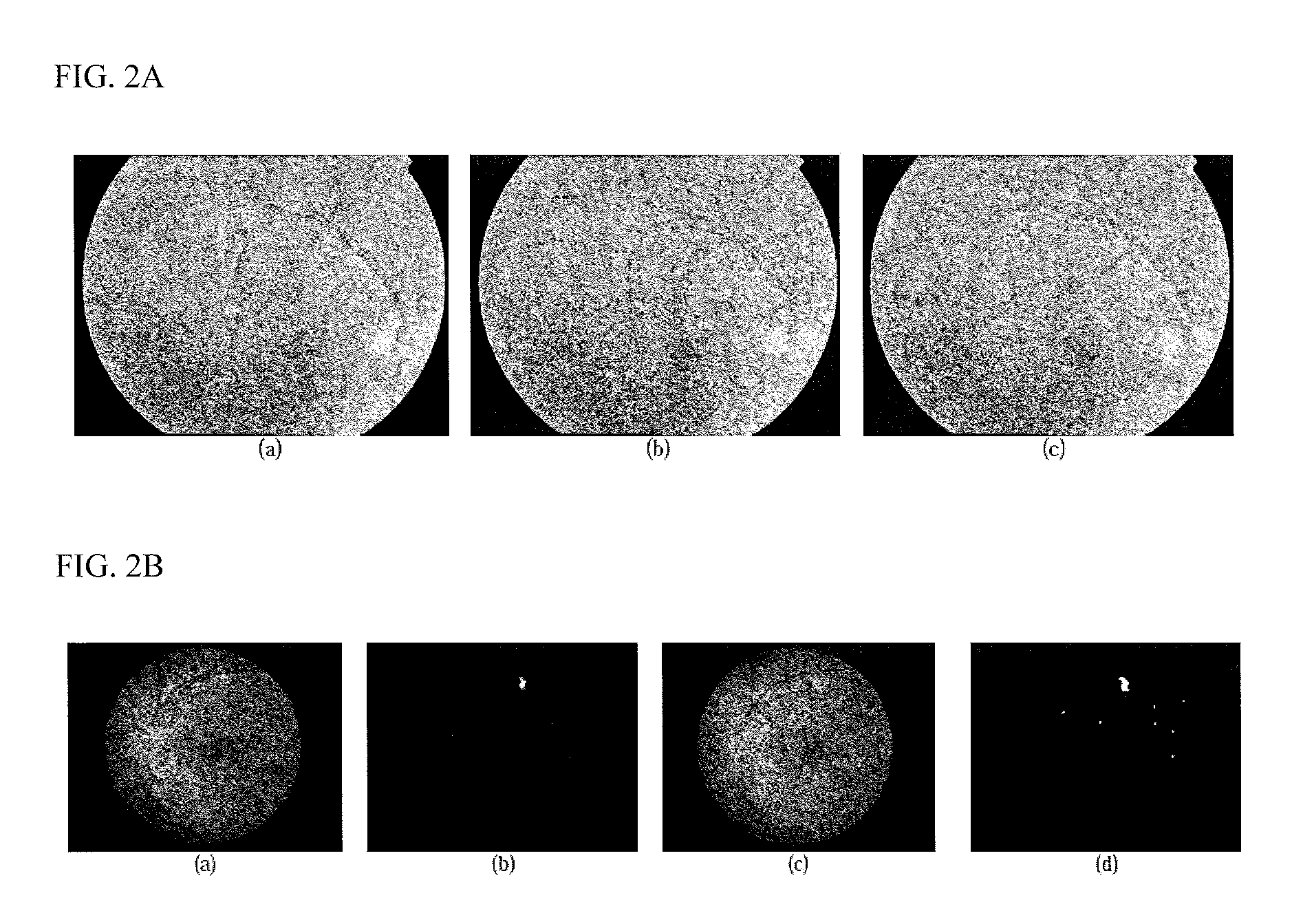
[0062]Although recent advances in optical instrumentation such as adaptive optics (AO), scanning laser ophthalmoscopy (SLO), optical coherence tomography (OCT) and non-mydriatic retinal cameras provide high resolution photos of retinal structure, the conventional ophthalmology-based method is time consuming because it requires extraction of lots of ophthalmological parameters, such as visual acuity, shape and size of optic disc, status of the anterior segment and fundus, thickness of vessels, and the distribution of retinal nerves by the ophthalmologist manually for decision making in diagnosis. Fundus camera has been adopted by ophthalmologists as one of the commonly used medical device to capture retinal images for clinical examination and analysis. It plays an important role for the quality of images. The first fundus camera in the world was developed by German company CAR ZEISS in 1925. The optical design and system structure have been improved significantly as th...


PUM


Abstract

A fundus camera using infrared light sources, which included an imaging optical path and an optical path for focusing and positioning and two optical paths share a common set of retina objective lens, and a computer-assisted method for retinal vessel segmentation in general and diabetic retinopathy (DR) image segmentation in particular. The method is primarily based on Multiscale Production of the Matched Filter (MPMF) scheme, which together the fundus camera, is useful for non-invasive diabetic retinopathy detection and monitoring.

Description

CROSS REFERENCE OF RELATED APPLICATION[0001]This application claims benefit from U.S. Provisional Application No. 61 / 469,824, filed Mar. 31, 2011, the content of which is incorporated herewith by reference in its entirety.FIELD OF THE INVENTION[0002]This invention relates to a computer-assisted technology for diseases diagnosis. Particularly, it relates to a computer-assisted apparatus and method for non-invasive diabetic retinopathy detection and monitoring.BACKGROUND OF THE INVENTION[0003]The human retina offers a window into the health of a person. Retinal microvasculature is the only part of the human circulation that can be visualized non-invasively in vivo, readily photographed and quantitatively analyzed. The content of a retinal image with high resolution structure of photoreceptors and vascular flow offers detailed information that would enable doctors to make early and accurate diagnosis of diseases. Clinical signs discernible in retinal fundus images include microaneurysm...

Claims


Application Information

Patent Timeline
Patent Type & Authority Applications(United States)
IPC IPC(8): A61B3/12
CPCA61B3/12G06K9/4609G06T7/0079G06K9/00597G06T7/0081G06K9/4652G06K9/36G06K9/46G06K9/0061G06V40/18G06V40/193
Inventor YOU, JIALI, QINGUO, ZHENHUASUN, QIANGLI, CHUN
Owner THE HONG KONG POLYTECHNIC UNIV
Why Eureka
Try Eureka