Unlock instant, AI-driven research and patent intelligence for your innovation.
Immunochemical detection of single target entities
Active Publication Date: 2012-10-25
AGILENT TECH INC
View PDF2 Cites 8 Cited by
Summary
Abstract
Description
Claims
Application Information
AI Technical Summary
This helps you quickly interpret patents by identifying the three key elements:
Problems solved by technology
Method used
Benefits of technology
Benefits of technology
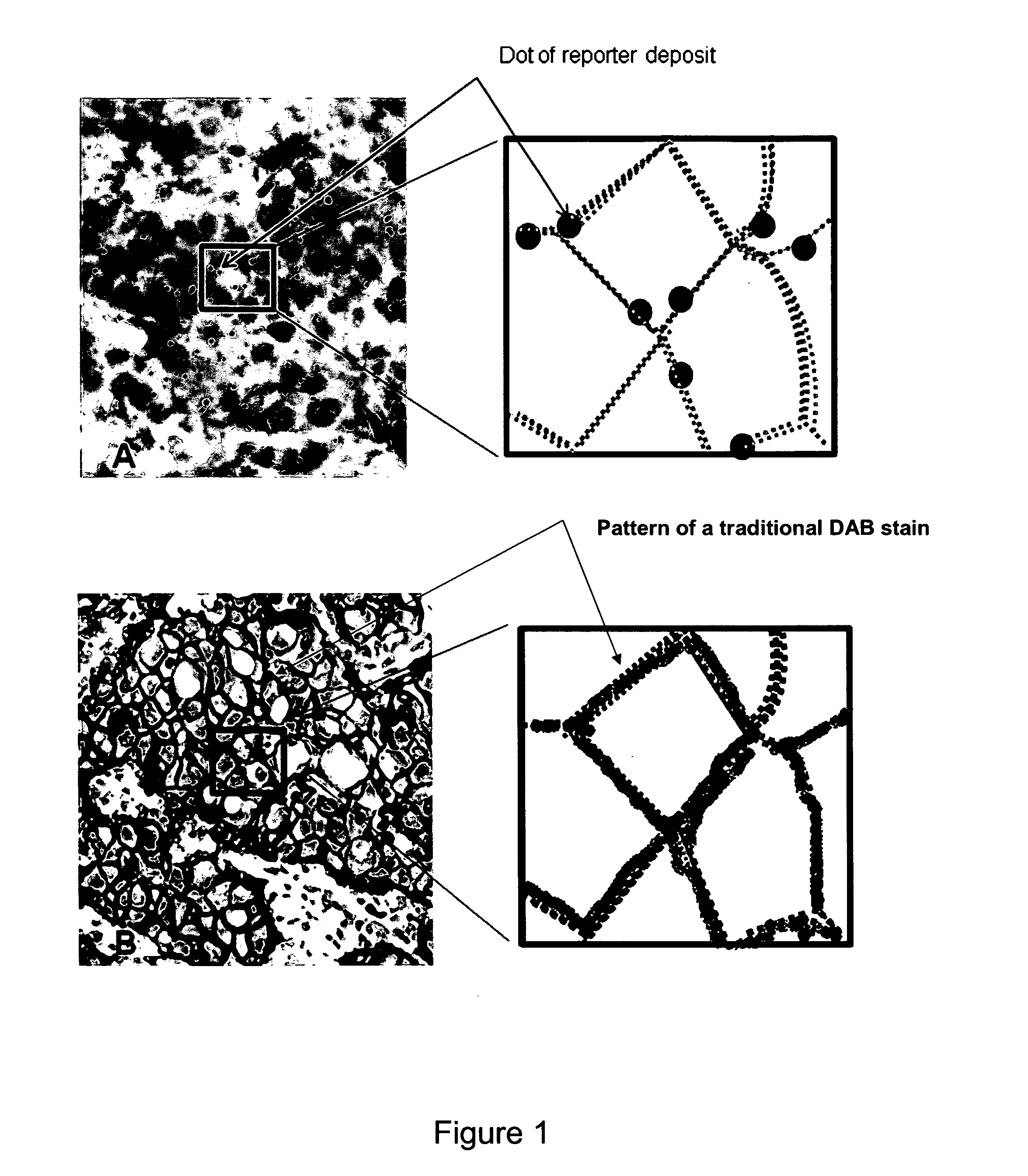
[0015]This invention provides rapid, simple and robust methods for visualization, detection and quantification of single entities a variety of targets in different samples, wherein the targets are immobilized. The methods are particular advantageous for evaluation of complex biological samples, such as histological samples.
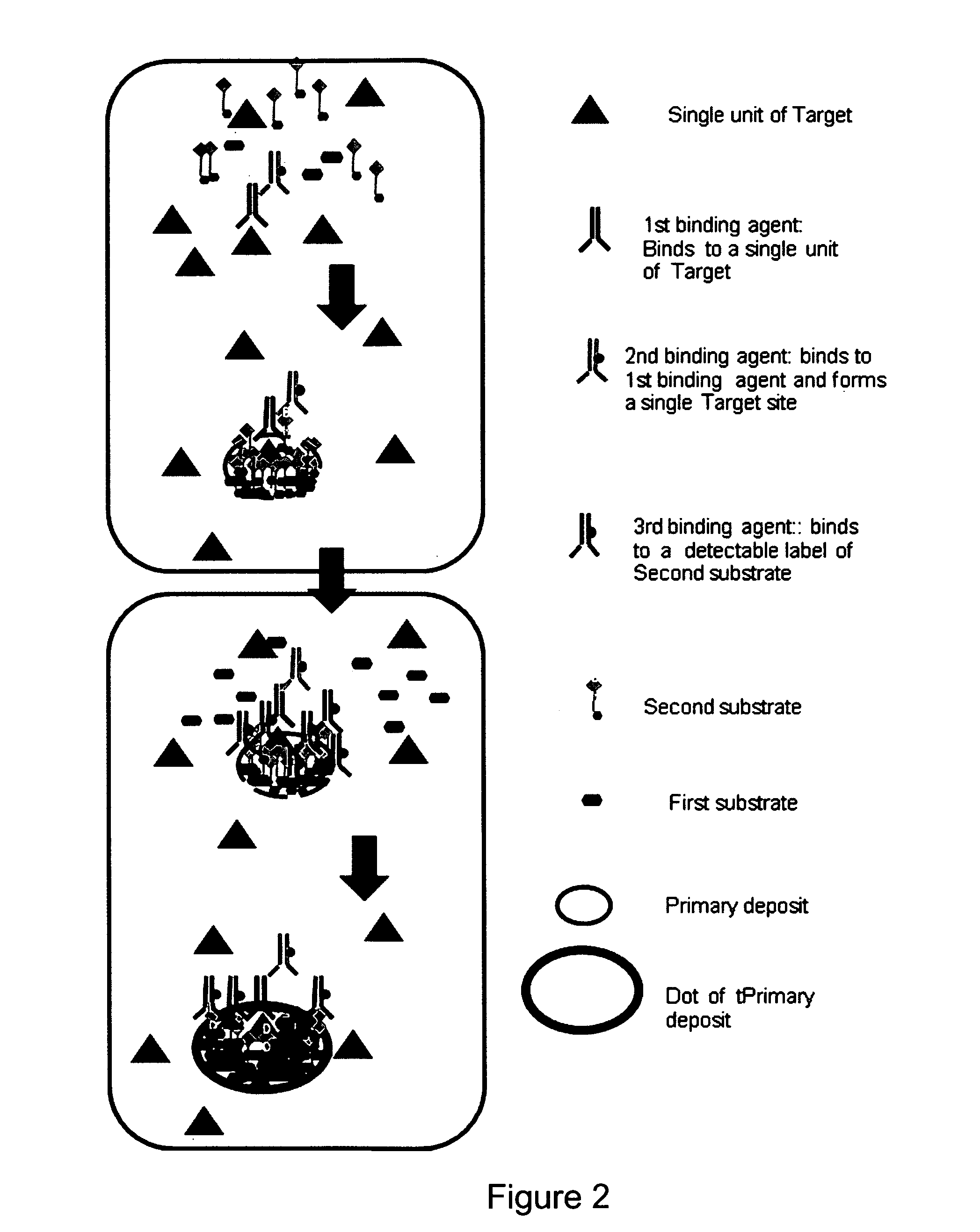
[0016]Methods of the invention comprises a novel powerful signal amplification system that makes possible visualizing individual single entities of targets, such as single molecules, single molecular structures, single molecular complexes, single particles etc., in a very wide dynamic concentration range in a host variety of samples. The term “single entity of target” is interchangeably used herein with the term “single / individual unit of target”.
[0069]The amplification system of the invention being very powerful and robust is at the same time flexible and easily controllable. It vastly expands the limits of current detection methods, in particular detection methods using a regular bright field or fluorescent microscopy for evaluation of samples. In particular, using detection methods comprising the amplification system of the invention(i) single entities of an immobilized target can be visualized and quantified in complex samples such as histological samples;(ii) single entities of an immobilized target can be detected and quantified using a variety assay formats;(iii) single entities of an immobilized target can be detected and quantified very rapidly, such as within 10-20 min, however, if necessary, the visualization and detection procedures may be prolonged or interrupted for longer periods of time without compromising quality of the results;(iv) blocking, typically used to reduce background labeling, is unnecessary;(v) temperature control is unnecessary;(vi) single entities of an immobilized target can be detected and quantified in a very broad dynamic range,(vii) single entities of multiple immobilized targets can be detected and quantified in the sample in one procedure.
[0070]Thus, great advantages of the SMD visualization system of the invention are that it is simple, rapid, robust, reliable and flexible. It allows visualization and quantification of single entities of a variety of targets in a variety of samples using a variety of assays. Additional advantages are that the methods utilize compounds that are well-defined chemical compounds which are either commercially available or easy to produce. A further advantage is that all procedures of the methods can be carried out both manually and automatically.
Problems solved by technology
The application of antibodies to cells and tissues presents specific difficulties beyond those encountered when these reagents are applied to purified proteins immobilized onto solid supports in or solution.
An additional difficulty is the ability to detect targets present at low levels.
The method is not especially sensitive and therefore suitable for detection of relatively abundant target proteins.
Other drawbacks to mention are that the method demands rather high amounts of target specific antibodies to saturate all target sites and it is relatively slow.
Furthermore, the method provides a uniform staining pattern that appears to the microscopist as a homogeneous color with intracellular resolution of cellular structures, e.g. membrane, cytoplasm, and nucleus, which makes it impossible to quantify the staining accurately.
However, due to a strong background staining and difficult interpretation of the results of staining, in particular of Fluorescent in-situ hybridization (FISH) and immunohistochemistry (IHC) samples, CSA has never been widely accepted as a routine approach for evaluation of histological samples in clinical histopathology.
However, the problem of the previous methods, namely assessment of quantity of the target in IHC samples that is based on the assessment of the quantity of detected stain, has not been solved.
This stain pattern does not allow direct approximating the quantity of the target to the quantity of the stain in a sample, because the correlation between these two quantities is not linear.
Thus, whilst quality assurance schemes for the methodology have been improved and raised the standards of IHC staining, the schemes that relate to interpretation of the staining results have not been changed.
Such currently used assessment is inevitably associated with errors which may be of crucial importance in medical diagnostic.
Unfortunately, the number of available techniques allowing visualizing single molecules of target proteins in histological samples is presently very limited and they are rather laborious and long procedures.
Some of the substantial drawbacks of these SMD approaches to mention are that(i) synthesis of the antibody-DNA hybrids can be problematic as controlling the location and number of DNA conjugates per protein is not always straightforward, often leading to heterogeneous ratios of DNA tags per antibody; amplification reaction is difficult to control; amplification step is temperature sensitive; labeling is not stable-the label will defuse from the target over time; etc.
Despite of recent developments in site-specific conjugation of oligonucleotide tags to proteins using inteinchemistry (or chemical ligation) have been very successful, conjugate preparation still remains laborious;(ii) steps of the methods require the temperature control;(iii) detection procedures comprise too many steps; and(iv) the whole process of detection takes a relatively long time.
Method used
Image
Click on the blue label to locate the original text in one second.
Reading with bidirectional positioning of images and text.
Examples
example 1
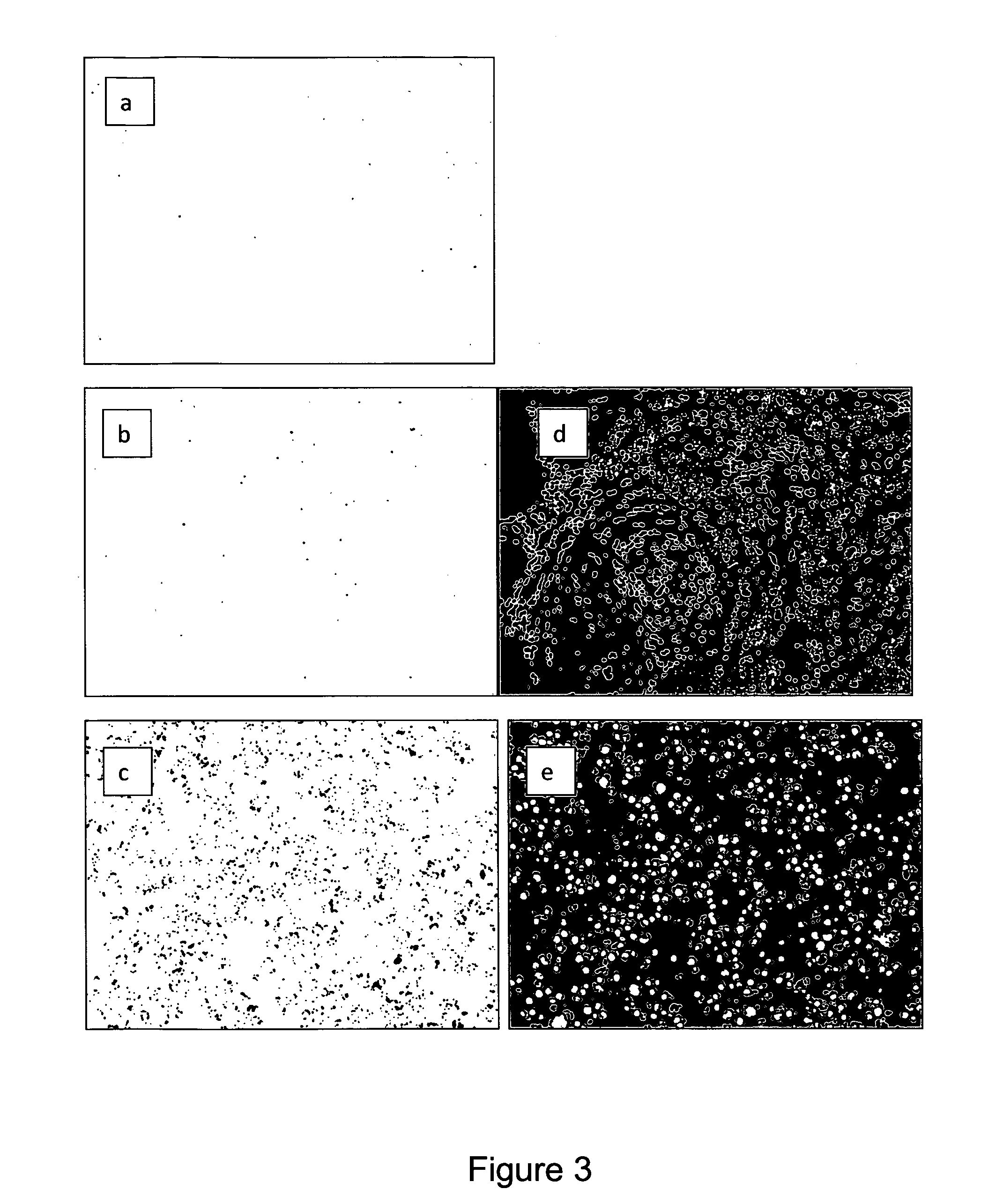
Dot Size as Function of the Amount of Reporter, DAB and H2O2 in Incubation Media or Incubation Time
General Procedure:
[0602]Staining experiments were run on formalin fixedparaffin embedded tissue. As pretreatment slides were de-paraffinized in 2 baths of xylene (5 min each), two baths of 96% ethanol, 2 baths of 70% ethanol (2 min each). The slides were then boiled in a microwave oven for 10 min in target retrieval solution (Dako S 1699). The slides were allowed to cool, endogenous peroxidase activity was quenched with 10% hydrogenperoxide for 2 min
experiment 1
[0603]All slides were: incubated with an experimental monoclonal mouse antibody directed at the C-terminal of the HER2 protein was used, “clone 6C2”, The following protocol was used: Incubation with clone 6C2, 55 picoM for 3 min, washed with wash buffer (Dako S3306) then incubation with GaM / HRP (D20052 frac. 8) in concentration 370 picoM for 3 min, then washed. Then the slides were subjected to a deposition step, a reporter binding agent step (all reporters, D19112, D20068, D20086, D20118, D20120 were used in concentration 10 μM) and a staining step with LPR chromogen as detailed in table 1. Washing steps were used between each step.
TABLE 1Detection (step c)DepositionReporter binding agentStain media (step b)(step c′)(step c″)Slide 1D19112: DAB 0.28 mM,D20036 50 nM,LPR 10 minH2O2 1.5 mM, 10 min10 minSlide 2D19112: DAB 0.28 mM,D20036 50 nM,LPR 5 minH2O2 1.5 mM, 10 min10 minSlide 3D19112: DAB 0.28 mM,D20036 50 nM,LPR 3 minH2O2 1.5 mM, 10 min10 minSlide 4D19112: DAB 0.28 mM,D20036 50 n...
experiment 2
[0604]All slides were treated as in example 1, then subjected to deposition, reporter binding agent and chromogen stain as detailed in table 2.
PUM
Abstract
The present invention relates to immunochemical visualization and quantification of single target entities, such as single molecules, single molecular structures, single particles, etc. in samples wherein said single entities are immobilized. In particular, the invention relates to methods for visualization and quantification of single units of biological or chemical targets, in particular to immunochemical visualization of single molecules of biological targets in histological samples. The methods of the invention comprise a step of forming discrete deposits of detectable molecules at single target sites of sample mediated by an enzyme with oxydoreductase activity, wherein a single target site comprises a single unit of a target. The invention also relates to assays comprising the present visualization and quantification methods and diagnostic applications of said methods.
Description
FIELD OF THE INVENTION[0001]The present invention lies in the field of immunochemical visualization and quantification of single target entities, such as single molecules, single molecular structures, single particles, etc. in samples wherein said single entities are immobilized. In particular, the invention relates to methods for visualization and quantification of single units of biological or chemical targets, in particular to immunochemical visualization of single molecules of biological targets in histological samples. The methods of the invention comprise a step of forming discrete deposits of detectable molecules at single target sites of sample mediated by an enzyme with oxydoreductase activity, wherein a single target site comprises a single unit of a target.BACKGROUND OF THE INVENTION[0002]Immunochemistry is a common tool in medical diagnostics and it is also usual for the assessment of therapeutic biomarkers. The latter, in particular, often require a quantitative evaluat...
Claims
Application Information
Patent Timeline
Application Date:The date an application was filed.
Publication Date:The date a patent or application was officially published.
First Publication Date:The earliest publication date of a patent with the same application number.
Issue Date:Publication date of the patent grant document.
PCT Entry Date:The Entry date of PCT National Phase.
Estimated Expiry Date:The statutory expiry date of a patent right according to the Patent Law, and it is the longest term of protection that the patent right can achieve without the termination of the patent right due to other reasons(Term extension factor has been taken into account ).
Invalid Date:Actual expiry date is based on effective date or publication date of legal transaction data of invalid patent.
