Fluorescent silica-based nanoparticles

a technology of fluorescence silica and nanoparticles, applied in the direction of powder delivery, granular delivery, dna/rna fragmentation, etc., can solve the problems of disproportionate number of invasive biopsies, difficult to visualize well-defined tumor margins with current imaging techniques, and single fluorophores that are usually not stable and limited for imaging

Active Publication Date: 2013-02-14
CORNELL UNIVERSITY +1
View PDF3 Cites 157 Cited by
  • Summary
  • Abstract
  • Description
  • Claims
  • Application Information

AI Technical Summary

Benefits of technology

[0018]The present invention further provides a method for targeting a tumor cell comprising administering to a cancer patient an effective amount of a fluorescent silica-based nanoparticle comprising a silica-based core comprising a fluorescent compound positioned within the silica-based core; a silica shell surrounding at least a portion of the core; an organic p

Problems solved by technology

However, well-defined tumor margins are often difficult to visualize with current imaging techniques.
This has led to a disproportionate number of invasive biopsies.
Single fluorophores are usually not stable and have limited brightness for imaging

Method used


Image

  • Fluorescent silica-based nanoparticles
  • Fluorescent silica-based nanoparticles
  • Fluorescent silica-based nanoparticles

Examples


example 1

Preparation and Characterization of PEG-Coated Nanoparticles

[0163]Nanoparticles containing an NIR-emitting dye (Cy-5) were synthesized and functionalized by PEGylation according to well-established protocols as disclosed in PCT / US2008 / 074894 and Stober et al. Controlled growth of monodispersed silica spheres in the micron size range. Colloid Interface Sci. 1968; 26:62-69. Ohnishi et al. J. Mol. Imaging 2005, 4:172-181. Cy5 malemide was reacted with a co-reactive organo silane compound, (3-Mercaptopropyl)tromethoxysilane to form a fluorescent silica precursor. This fluorescent silica precursor was co-condensed with tetraethylorthosilicate to form a fluorescent silica based core. A PEG-silane compound, with methoxy-terminated poly(ethylene glycol) chains (PEG, ˜0.5 kDa) Methoxy(Polyethyleneoxy) Propyl]-Trimethoxysilane, was added to the fluorescent silica based core to form a PEG coating on the core. PEG-coated nanoparticles were dialyzed to physiological saline (0.15M NaCl in H2O) th...

example 2

Renal Clearance of PEG Coated Nanoparticles

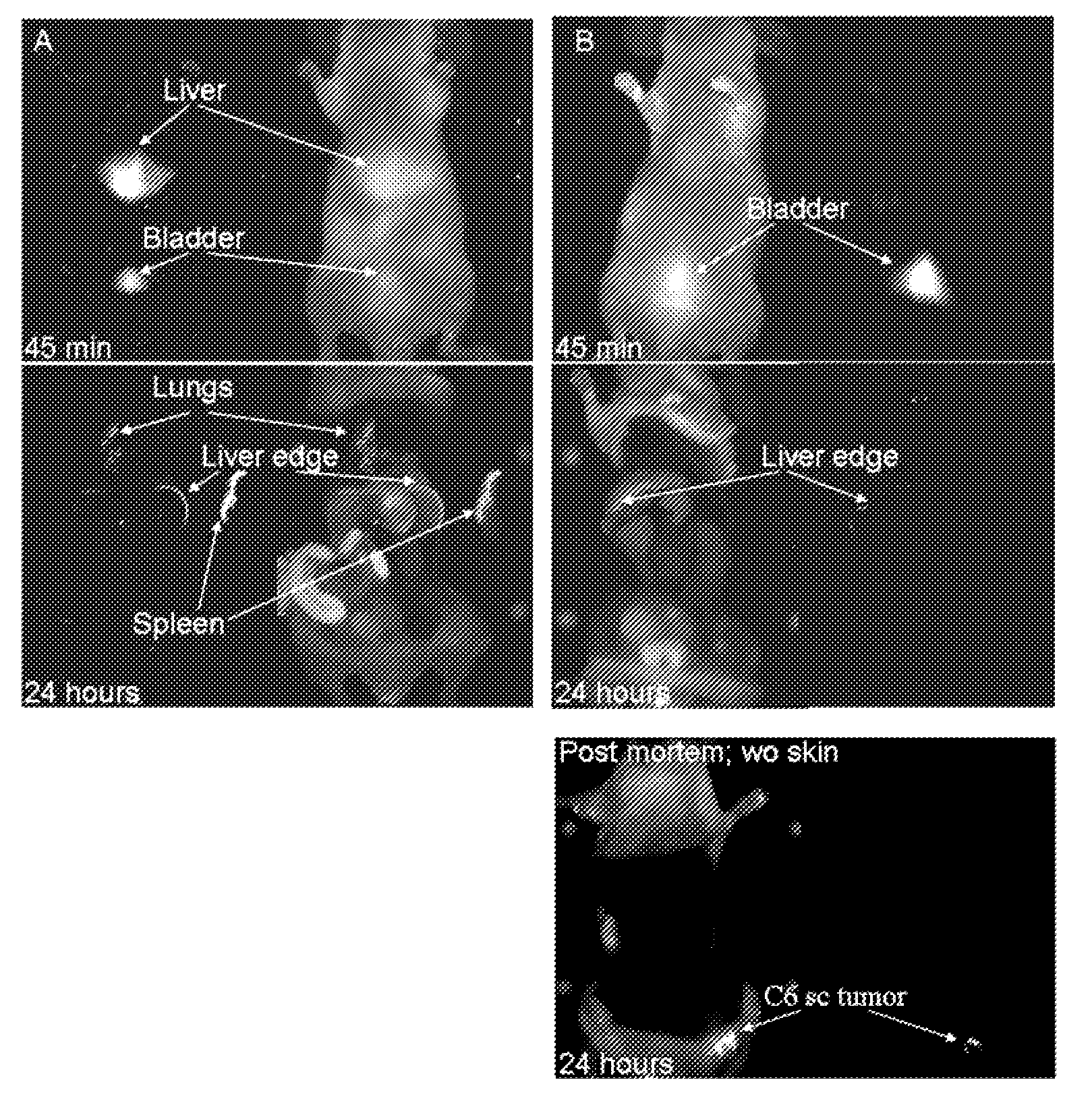
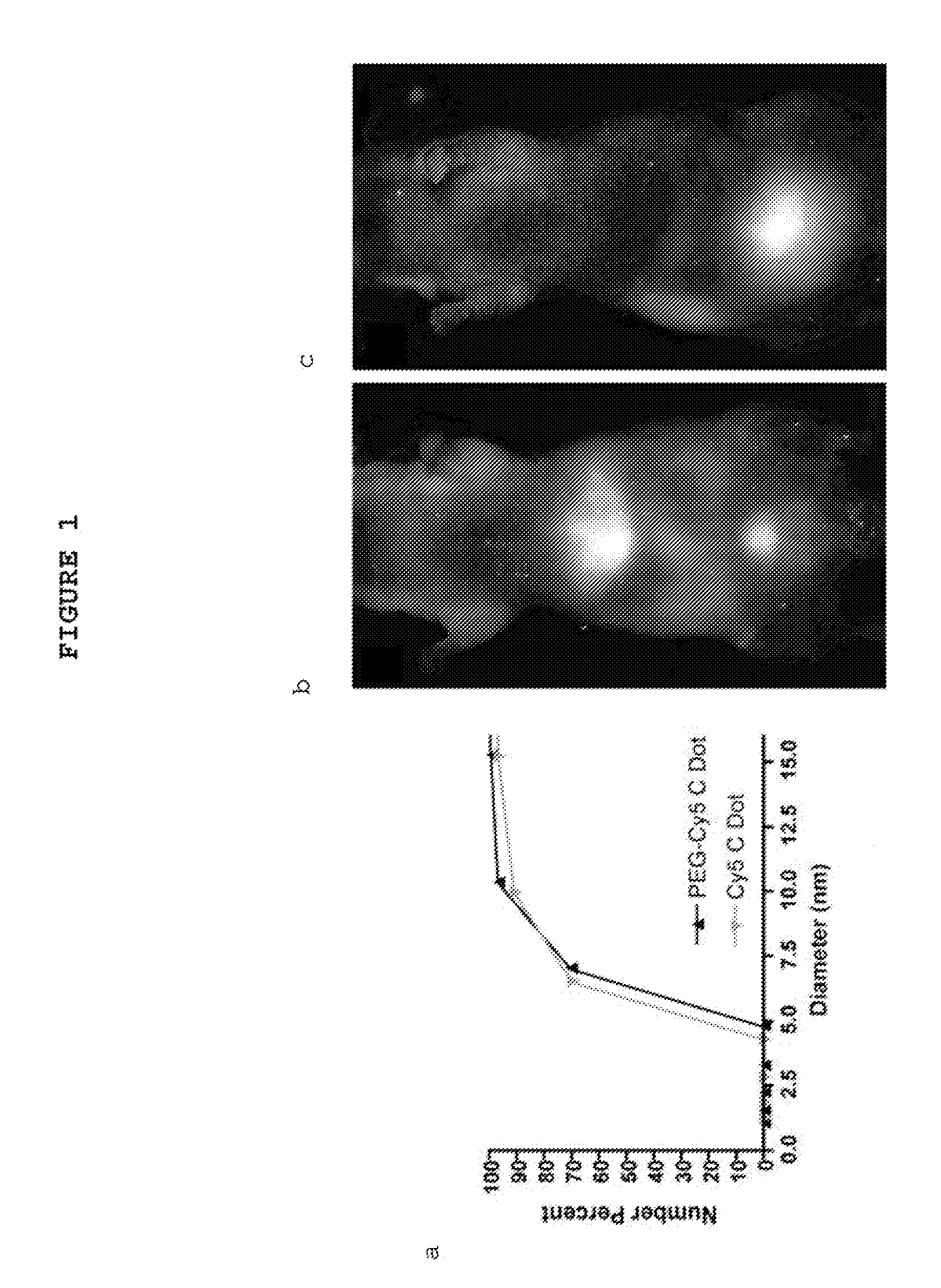
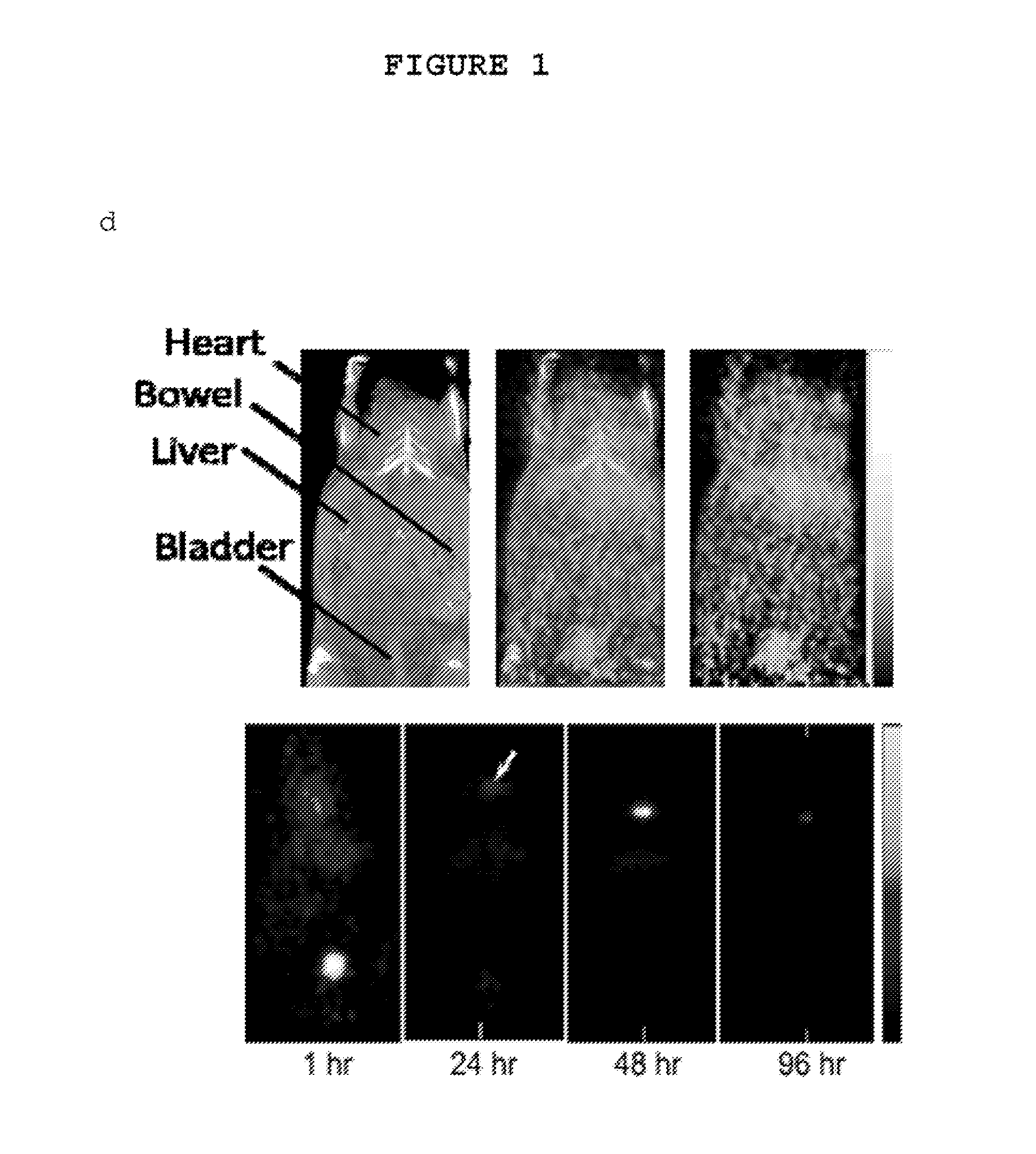
[0164]Fluorescent core-shell silica nanoparticles, having a hydrodynamic radius of about 3 nm, were synthesized. These nanoparticles were found to be in the 6-10 nm diameter range, as shown by dynamic light scattering (DLS) results (FIG. 1a). In vivo whole-body NIR fluorescence imaging of bare (no PEG coat) silica nanoparticles, on the order of 6-nm and 3.3-nm, in nude mice showed considerable renal clearance 45 min post-injection with a significant accumulation remaining in the liver (FIG. 1b). Eventual excretion into the enterohepatic circulation occurred during the ensuing 24 h. On the basis of these results, particles were covalently coated with methoxy-terminated poly(ethylene glycol) chains (PEG, ˜0.5 kDa), per protocols in PCT / US2008 / 074894, to prevent opsonization and further enhance particle clearance while maintaining a small hydrodynamic size. This treatment decreased liver retention and resulted in increased renal filtration int...

example 3

Fluorescent Silica Nanoparticles Conjugated with αvβ3 Integrin-Targeting Peptide (Melanoma Model)

[0167]To synthesize a multimodal nanoparticle with high affinity for tumor marker αvβ3 integrin, linear RGD hexapeptide (CGGRGD) was conjugated to the nanoparticle via a Cys-maleimide linkage. Male athymic nude mice were injected subcutaneously into their flanks with C6 rat glioma cells. At ˜0.5 cm in diameter, mice were IV-injected with either bare silica nanoparticles or PEG-ylated RGD nanoparticles (˜500 nm / kg). FIG. 4 shows the in vivo biodistribution in non-tumor-bearing and tumor-bearing mice.

[0168]In vitro binding characteristics of targeted (RGD-bound) and non-targeted (PEG-coated) nanoparticles to αvβ3-integrin-positive (M21 cells) and integrin-negative (M21L cells) human melanoma cell lines were investigated using flow cytometry (FIGS. 5a, 5b).


PUM

PropertyMeasurementUnit
Fractionaaaaaaaaaa
Fractionaaaaaaaaaa
Fractionaaaaaaaaaa

Abstract

The present invention provides a fluorescent silica-based nanoparticle that allows for precise detection, characterization, monitoring and treatment of a disease such as cancer The nanoparticle has a fluorescent compound positioned within the nanoparticle, and has greater brightness and fluorescent quantum yield than the free fluorescent compound To facilitate efficient urinary excretion of the nanoparticle, it may be coated with an organic polymer, such as polyethylene glycol) (PEG) The small size of the nanoparticle, the silica base and the organic polymer coating minimizes the toxicity of the nanoparticle when administered in vivo The nanoparticle may further be conjugated to a ligand capable of binding to a cellular component associated with the specific cell type, such as a tumor marker A therapeutic agent may be attached to the nanoparticle Radionuclides/radiometals or paramagnetic ions may be conjugated to the nanoparticle to permit the nanoparticle to be detectable by various imaging techniques.

Description

CROSS REFERENCE TO RELATED APPLICATIONS[0001]This application claims priority to U.S. Provisional Application Nos. 61 / 222,851 (filed Jul. 2, 2009) and 61 / 312,827 (filed Mar. 11, 2010).STATEMENT REGARDING FEDERALLY SPONSORED RESEARCH OR DEVELOPMENT[0002]This invention was made with government support under NIH-NRRA, Clinical and Translational Science Center Grant; NSF STC Program Agreement No. ECS-9876771; ICMIC P50 CA86438; NIH SAIRP Grant No R24CA83084; and NIH Center Grant No P30 CA08748.FIELD OF THE INVENTION[0003]The present invention relates to fluorescent silica-based nanoparticles, and methods of using the nanoparticles to detect, diagnose, or treat diseases such as cancer.BACKGROUND OF THE INVENTION[0004]Early tumor detection and treatment selection is paramount to achieving therapeutic success and long-term survival rates. At its early stage, many cancers are localized and can be treated surgically. However, well-defined tumor margins are often difficult to visualize with c...

Claims


Application Information

Patent Timeline
IPC IPC(8): A61K49/00A61K49/06A61K49/04A61K9/14A61K39/395A61K31/7088A61K48/00A61K31/56A61K31/713A61K31/7105G01N21/64A61P35/00A61P31/04A61P31/00A61P39/06A61P37/06A61P7/02A61P9/08A61P29/00A61K51/12B82Y5/00B82Y15/00
CPCA61K49/0019A61K49/0054A61K49/0056A61K49/0093A61K51/08A61K51/082B82Y5/00B82Y15/00G01N33/54346G01N33/582A61K51/1255Y10S977/773Y10S977/927A61P29/00A61P31/00A61P31/04A61P35/00A61P37/06A61P39/06A61P7/02A61P9/08A61K9/5115A61K9/5146A61K9/5169A61K49/0032A61K51/086A61K51/1244G01N33/552G01N33/574G01N33/587G01N33/60
Inventor BRADBURY, MICHELLEWIESNER, ULRICHPENATE MEDINA, OULAOW, HOOSWENGBURNS, ANDREWLEWIS, JASONLARSON, STEVEN
Owner CORNELL UNIVERSITY
Why Eureka
Try Eureka