Method for differentiation induction in cultured stem cells

a stem cell and differentiation induction technology, applied in the field of stem cell differentiation induction, can solve the problems of insufficient in vitro differentiation, cell and tissue damage, and extremely difficult to obtain human retinal tissue for such research, and achieve selectively inducing the differentiation of retinal cells, the effect of promoting cell differentiation and promoting cell proliferation

Active Publication Date: 2013-02-14
RIKEN +1
View PDF2 Cites 11 Cited by
  • Summary
  • Abstract
  • Description
  • Claims
  • Application Information

AI Technical Summary

Benefits of technology

[0053]According to the present invention, it is possible to induce the differentiation of a stem cell into a nerve progenitor cell, particularly into a retinal progenitor cell, efficiently. The method of the present invention also enables efficient differentiation induction into nervous system cells, particularly into retinal cells, a task that has been difficult to achieve by the conventional method of differentiation induction. Therefore, the method of the present invention is particularlyuseful in applying cytotherapy for diseases associated with abnormalities in a nerve tissue, particularly in retinal tissue.
[0054]According to the method of the present invention, it is also possible to selectively inducing the differentiation of retinal layer-specific neurons. The retinal tissue obtained by the method of the present invention has a laminar structure that is extremely similar to the living retina...

Problems solved by technology

However, because an extremely large number of kinds of nerve cells are present in the brain and surrounding tissues, there are still many types of nerve cells and tissues for which efficient in vitro differentiation has been unsuccessful.
For the treatment of retinopathies, etiologic research, drug discovery research, cell transplantation therapy research and the like have been conducted so far, but it is extremely difficult to obtain human retinal tissue for the sake of such research.
458(3):126-31], it has been ...

Method used


Image

  • Method for differentiation induction in cultured stem cells
  • Method for differentiation induction in cultured stem cells
  • Method for differentiation induction in cultured stem cells

Examples


example

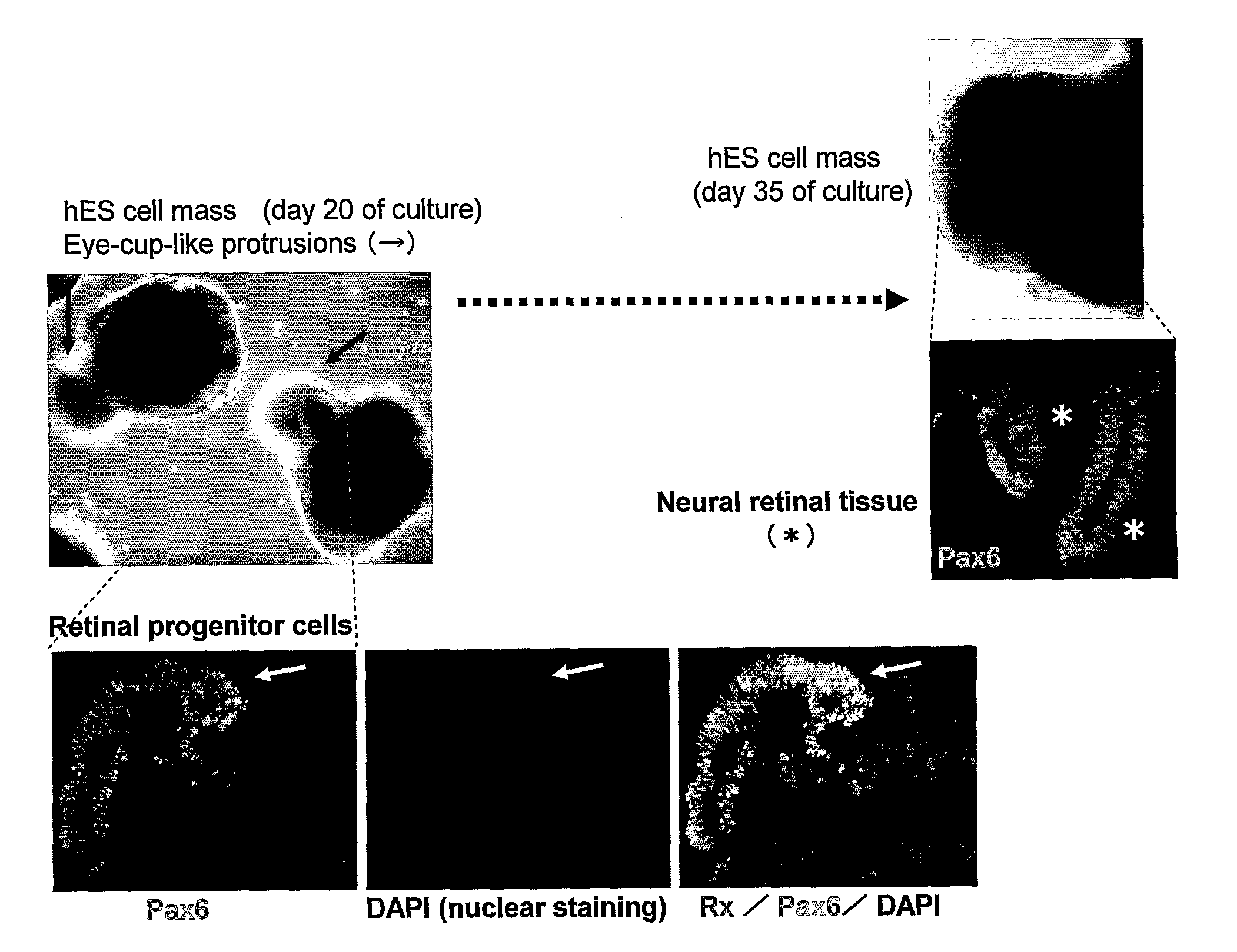
Highly Efficient Differentiation Induction to Retinal Progenitor Cells by the Modified SFEBq Method with High Concentrations of Matrix Components Added and Optimized KSR Concentration

(Method)

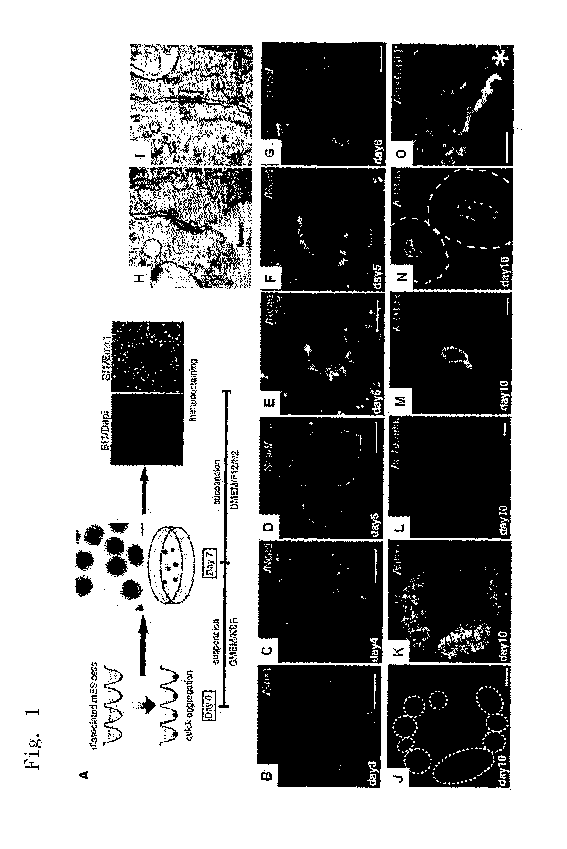
[0179]Rx-EGFP mES cells (mouse ES cells having EGFP knocked in at the early retinal progenitor cell marker gene Rx locus; Wataya et al, PNAS, 2008) were treated by the SFEBq method (a 96-well culture plate of low cell-binding ability) to quickly generate a homogenous aggregate at 3000 cells per well, which was cultured for differentiation. The differentiation induction medium used here was G-MEM medium (Invitrogen), 2% KSR, 2 mM glutamine, 0.1 mM non-essential amino acids, 1 mM pyruvic acid and 0.1 mM 2-mercaptoethanol. Starting one day later, Matrigel was added to the medium in a ratio by volume of 1 / 100 to 1 / 25, and the cells were subjected to suspension aggregate culture for 7 days 5% CO2, at 37° C. Subsequently, the cultivation was further continued under the nerve differentiation promoting ...

example 2

Self Formation of Optic Cup-Like Tissue from Retinal Progenitor Cells Using the Modified SFEBq Method

(Method)

[0182]The aggregate of Rx-positive cells obtained in Example 1 was cultured using a culture broth prepared by adding Matrigel in a 1 / 50 volume to 2% KSR medium for 7 days, after which it was transferred to a culture broth prepared by adding an N2 additive to the DMEM / F12 medium, and further suspension-cultured under 5% CO2 / 40% O2 conditions for 3 days.

(Results)

[0183]The strongly Rx-EGFP-positive portion formed a tissue as a protrusion from the cell mass (FIG. 3). Its histological profile was similar to that of the fetal optic cup (an early retinal tissue formed as a protrusion from thediencephalon), representing a structure wherein Rx-positive, Chx10-positive juvenile neural retinal tissue was wrapped by a layer of retinal pigment cells.

example 3

Self Assembly of Retinal Tissue from Retinal Progenitor Cells Using the Modified SFEBq Method

(Method)

[0184]Optic cup-like tissue as obtained in Example 2 (10 days of culture) was separated from the cell mass using microtweezers, and subjected to suspension culture with DMEM / F12 / N2+0.5 μM retinoic acid (known to promote the survival of photoreceptor cells).

(Results)

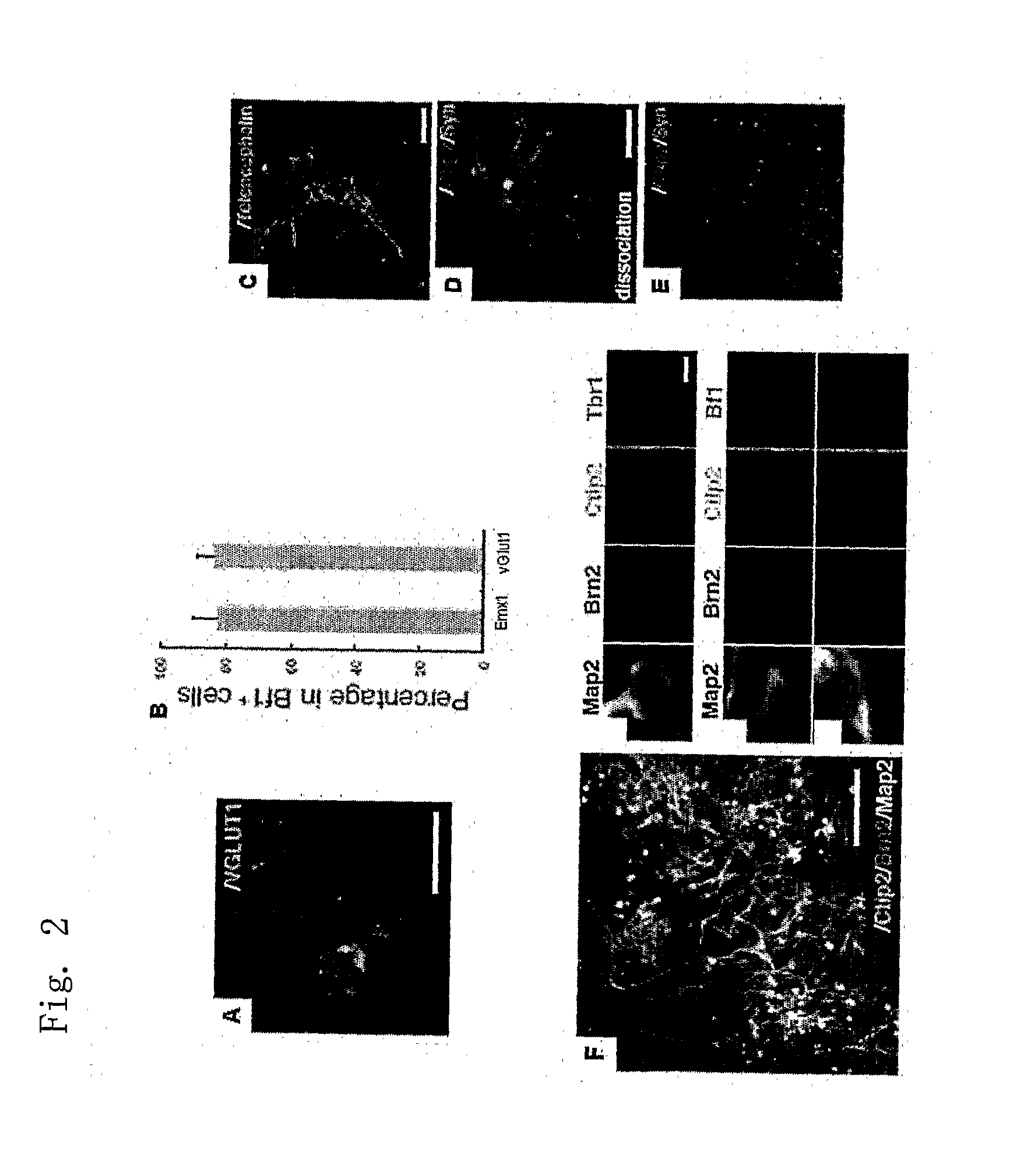
[0185]The suspension-cultured optic cup-like tissue had been induced to a laminar structure that is structurally extremely similar to the retina after birth (FIGS. 2 and 3). Regularly formed in the outermost layer was a planar structure of photoreceptor cells (rhodopsin-positive and having an outer segment structure), possessing the right cell polarity. Under this layer were a layer of Chx10-positive bipolar cells and a layer of Calbindin-positive horizontal cells, under which a layer of Calretinin-positive / Pax6-positive / TuJ1-negative amacrine cells was present, and the lowermost layer was a layer of Brn3-positive / TuJ1-pos...


PUM


Abstract

Provided is a method of inducing the differentiation of a stem cell into nerve progenitor cells, comprising the step (1) of forming a homogenous aggregate of stem cells in a serum-free medium (1) and the step (2) of suspension-culturing the homogenous aggregate of stem cells in the presence of a basement membrane reference standard.

Description

TECHNICAL FIELD[0001]The present invention relates to a method of differentiation induction of stem cells, more specifically to a method of differentiation induction of stem cells comprising combining quick re-aggregation and three-dimensional suspension culture in performing stem cell aggregate culture.BACKGROUND ART [0002]To date, some culturing methods for differentiation induction of nerves from pluripotent stem cells such as ES cells have been known, including those reported by the present inventors [Watanabe, K., Ueno, M., Kamiya, D., Nishiyama, A., Matsumura, M., Wataya, T., Takahashi, J.B., Nishikawa, S., Nishikawa, Muguruma, K. and Sasai, Y. (2007), A ROCK inhibitor permits survival of dissociated human embryonic stem cells. Nature Biotechnology 25, 681-686, Su, H.-L.; Muguruma, K., Kengaku, M., Matsuo-Takasaki, M., Watanabe, K., and Sasai, Y. (2006), Generation of Cerebellar Neuron Precursors from Embryonic Stem Cells. Developmental Biology 290, 287-296; Ikeda, H., Watanab...

Claims


Application Information

Patent Timeline
IPC IPC(8): C12N5/0789C12Q1/02
CPCC12N5/0621C12N5/0623C12N2533/52C12N2506/02C12N2501/16C12N2500/99A61P27/02C12N2500/90
Inventor SASAI, YOSHIKIEIRAKU, MOTOTSUGU
Owner RIKEN
Why Eureka
Try Eureka