
Method and Kit for Identifying Compounds Capable of Inhibiting Human Papilloma Virus Replication

a human papilloma virus and anti-hpv technology, applied in the field of virology, cell culturing, drug development, can solve the problems of malignant carcinoma progression and formation, cell modelling of these replication stages in cells is problematic, and most tissue culture cells do not support any mode of hpv genomic replication

Inactive Publication Date: 2013-06-13
ICOSAGEN CELL FACTORY
View PDF2 Cites 6 Cited by
  • Summary
  • Abstract
  • Description
  • Claims
  • Application Information

AI Technical Summary

Benefits of technology

The patent describes a method for identifying cell clones that carry different HPV copies per cell, and making cell banks from these clones. The method also involves seeding cells in a low density and cultivating them for a short period of time until they establish about 40% confulency, which maintains HPV DNA replication in the latent phase. The technical effects of this patent include improved methods for identifying and manipulating HPV-positive cells, which could help in the development of new treatments for HPV-related diseases.

Problems solved by technology

In some cases this infection may lead to progression and formation of malignant carcinomas.
Modelling of these replication stages in cells has been problematic in the case of human papillomaviruses.
Most of the tissue culture cells do not support any mode of HPV genomic replication.
Attempts to get viral genomic DNA replication going from transfected plasmids of β-papillomavirus types has completely failed in any keratinocyte cell lines or primary keratinocytes.
Also, it has been difficult to generate reproducible human cell lines that carry stable HPV replicating genomes, especially that of the “low risk”-HPV types.
However, it is not sufficient, because the vaccines target at best only for four subtypes of hundreds of papillomaviruses, including “high risk”-type of mucosal or cutaneous skin papillomaviruses.
However, this objective has been difficult to achieve due to the lack of an effective cellular system for screening for drug candidates.
However, these methods do not allow high-throughput screening for drug candidates, and a simpler and more convenient method is necessarily required.
However, maintenance of episomal HPV by itself is not sufficient for a high-throughput screening assay to identify possible HPV replication inhibitors.

Method used


Image

  • Method and Kit for Identifying Compounds Capable of Inhibiting Human Papilloma Virus Replication
  • Method and Kit for Identifying Compounds Capable of Inhibiting Human Papilloma Virus Replication
  • Method and Kit for Identifying Compounds Capable of Inhibiting Human Papilloma Virus Replication

Examples


example 1

Transient HPV DNA Replication in U2OS Cells

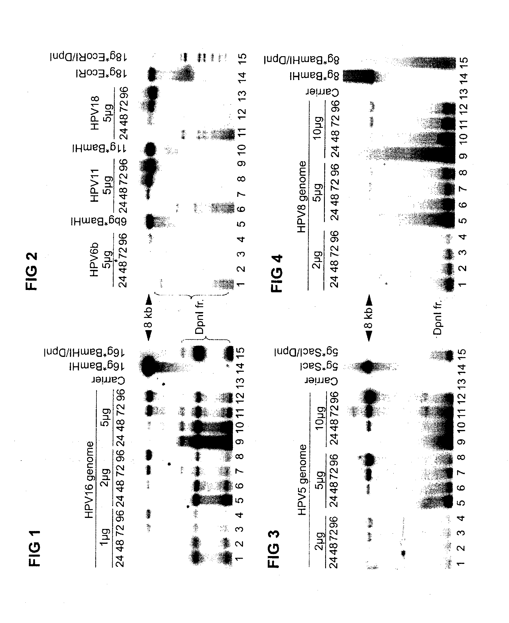
[0099]Human papillomaviruses show strong tropism for epithelial cells. It was discovered that human osteosarcoma cell line U2OS, exhibiting epithelial adherent morphology, although derived from a moderately differentiated osteosarcoma, supported very effectively the HPV E1 and E2 protein dependent viral DNA replication, when the expression-vectors for viral replication proteins were used together with reporter plasmids containing viral origin. U2OS cells encode wild-type pRb and p53.

[0100]Hereafter it was investigated, whether the viral trans factors (E1 and E2) could act in their native configurations supporting the replication of the viral genomes in U2OS monolayer cultures. A set of four different cutaneous type of papillomaviruses were included, two of them belonging to high-risk type (HR / HPV-18 and HR / HPV-16) and two to low-risk type (LR / HPV-11 and LR / HPV-6b) according to their prognosis for cancer development. Additionally, two subtyp...

example 2

HPV Stable Replication in U2OS Monolayer Cultures

Establishment of Persistent HPV Stable Maintenance in U2OS Cell Line

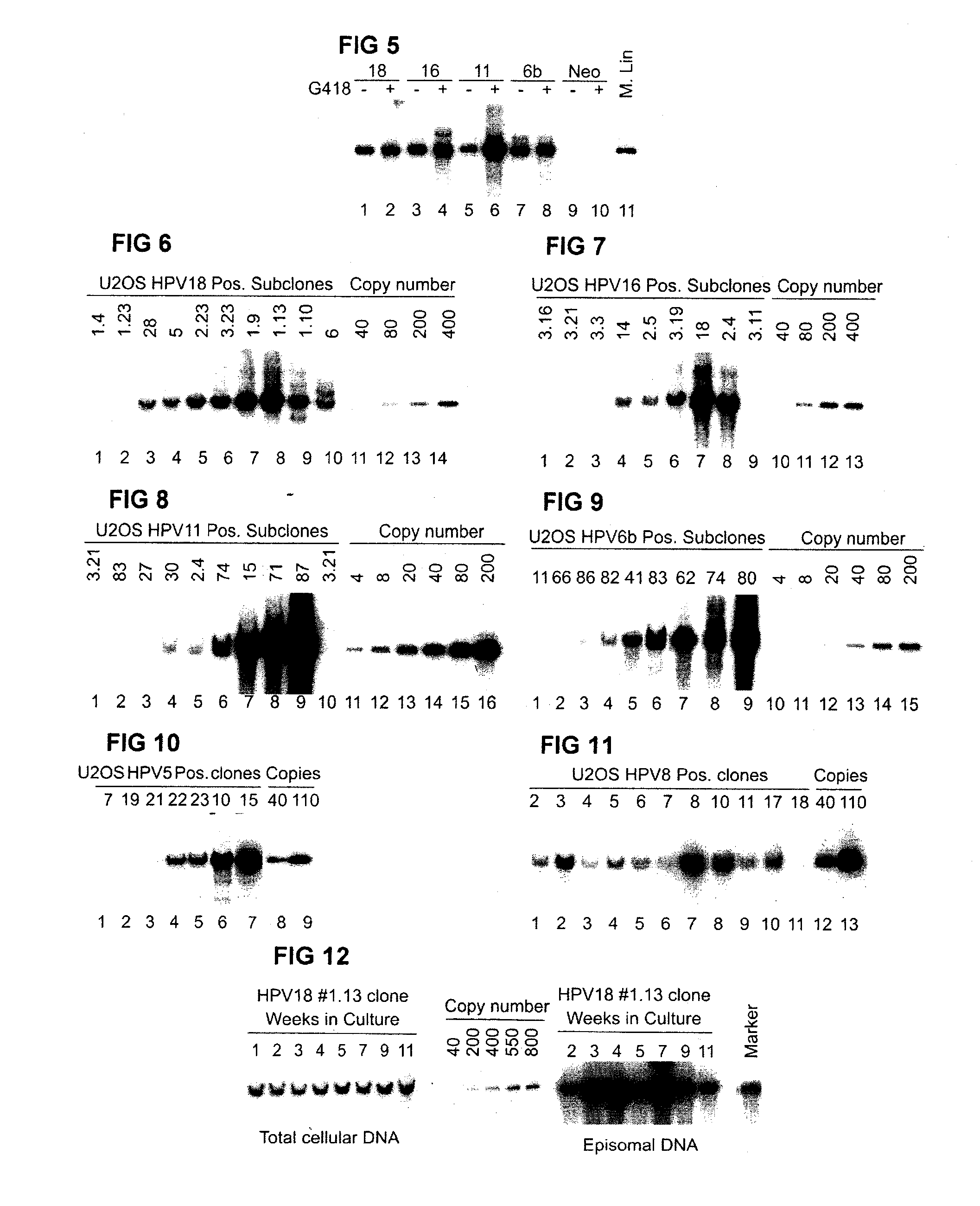
[0103]Quite strong HPV genomic DNA replication signal in U2OS cells in transient assays suggested further evaluation of the capacity of HR- and LR-HPV plasmids for stable episomal replication. For this purpose we co-transfected into U2OS cells 5 μg of HPV-6b, or HPV-11, HPV-16, HPV-18, HPV-5, HPV-8 circular plasmid together with 5 μg AraD carrier DNA and with 2 μg of Eco01091-linearized of pNeo-EGFP or EcoRI-linearized pBabeNeo plasmid, encoding antibiotic resistance marker, which would allow the selection for the transfected cells. 48 hrs after the transfection G418 selection was performed. After two to three weeks of cultivation with G418 selection, the low-molecular weight (LMW) Hirt extracts from whole cell population (“pool” DNA) were analyzed by Southern blotting with radioactively labelled probes against the appropriate HPV types. The analysis shows that all te...

example 3

Late Amplification of the HPV Genomes

Genome Amplification in a Manner Similar to Differentiation-Dependent Viral Amplification

[0108]In the productive stage of PV life cycle, amplification of the viral genome occurs in differentiated cells within the upper layer of epidermis. To study the productive stage of viral life cycle in tissue culture, the three-dimensional architecture of the epithelium has been usually tried to be reproduced with organotypic or raft cultures, suspension in methylcellulose, feeder cells, by using regulated culture and growth conditions.

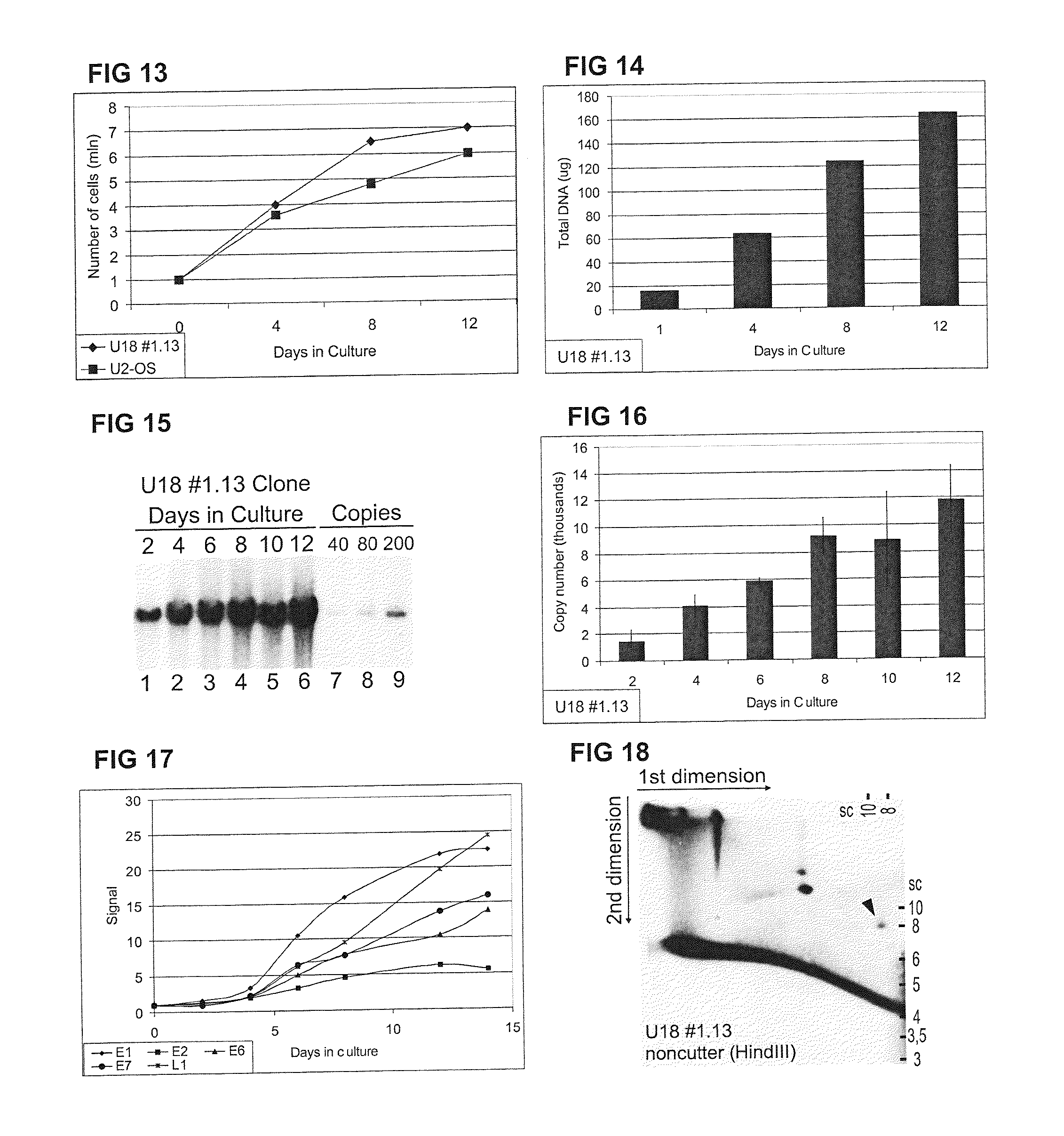
[0109]We used an alternative method, only dense cell cultures to imitate differentiation-dependent viral amplification. For this purpose equal number of cells (for example 1×10⁶ cells per 10 cm culture dish) of appropriate HPV-positive cell clone were split on several dishes (for example 6) and maintained as regular confluent monolayers grown up to high densities. The total DNA or low molecular weight (Hirt) DNA samples were c...


PUM

PropertyMeasurementUnit
concentrationsaaaaaaaaaa
volumeaaaaaaaaaa
volumeaaaaaaaaaa

Abstract

This invention provides a method, kit and an in vitro system for identifying compounds capable of inhibiting Human Papilloma Virus replication at all the stages of viral replication cycle. The method, kit and in vitro system is applicable to all types of Human Papilloma Virus. The method enables high throughput screening of compounds inhibiting HPV replication in one or more phases of the cycle.

Description

PRIORITY[0001]This application is a continuation in part application of International Application Number PCT / EE2010 / 000010 filed on May 19, 2010 which is incorporated herein by reference in its entirety.SEQUENCE LISTING[0002]This application contains sequence listing.TECHNICAL FIELD OF THE INVENTION[0003]The present invention relates to the fields of virology, cell biology, cell culturing, and drug development. More particularly the invention provides a method for screening for anti-HPV substances and a kit for screening for anti-HPV substances. The invention also provides plasmids for transfecting cell lines and cell lines capable of supporting all replication phases of Human Papilloma Virus.BACKGROUND OF THE INVENTION[0004]The continuous interest to study the human papillomaviruses (HPV) has been generated from their association with specific human cancers. HPV infects basal proliferating cells of the epithelium and induces the formation of benign tumors. In some cases this infect...

Claims


Application Information

Patent Timeline
Patent Type & Authority Applications(United States)
IPC IPC(8): C12Q1/70
CPCC12Q1/18G01N2333/025C12Q1/708
Inventor USTAV, MARTUSTAV, ENEGEIMANEN, JELIZAVETAPIPITS, REGINAISOK-PAAS, HELENREINSON, TORMIUSTAV, JR., MARTLAOS, TRIINORAV, MARITSALK, KRISTIINAMANNIK, ANDRESREMM, ANU
Owner ICOSAGEN CELL FACTORY
Learn More