System and method for using medical image fusion

Inactive Publication Date: 2013-08-15
CONVERGENT LIFE SCI
View PDF9 Cites 57 Cited by
  • Summary
  • Abstract
  • Description
  • Claims
  • Application Information

AI Technical Summary

Benefits of technology

[0024]It is noted that as an alternate, the MRI or CAT scan data may be used to provide a coordinate frame of reference for the procedure, and the TRUS image modified in real-time to reflect an inverse of the ultrasound distortion. That is, the MRI or CAT data typically has a precise and undistorted geometry. On the other hand the ultrasound image may be geometrically distorted by phase velocity variations in the propagation of the ultrasound waves through the tissues, and to a lesser extent, by reflections and resonances. Since the biopsy instrument itself is rigid, it will correspond more closely to the MRI or CAT model than the TRUS model, and therefore a urologi

Problems solved by technology

Such treatments are costly and can cause serious side effects, including incontinence and erectile dysfunction.
However, ultrasound imaging has relatively low tissue discrimination ability.
Accordingly, ultrasound imaging provides adequate imaging of the prostate organ, but it does not provide adequate imaging of tumors within the organ due to the similarity of cancer tissue and benign tissues, as well as the lack of tissue uniformity.
Due to the inability to visualize the cancerous portions within the organ with ultrasound, the entire prostate must be considered during the biopsy.
As with ultrasound imaging, MRI also has limitations.
For instance, it has a relatively long imaging time, requires specialized and costly facilities, and is not well-suited for performance by a urologist at a urology center.
Furthermore, performing direct prostate biopsy within MRI machines is not practical for a urologist at a urology center.
Until now, however, such systems have not been adequate for enabling MRI-ultrasound fusion to be performed by a urologist at a urology center.
Such MRI data, however, is not readily available to urologists and it would be commercially impractical for such MRI data to be generated at a urology center.
This is due to many reasons, including urologists' lack of training or expertise, as well as the lack of time, to do so.
Also, it is uncertain whether a urologist can profitably implement an image-guided biopsy system in his or her practice while contemporaneously attempting to learn to perform MRI scans.
Furthermore, even if a urologist invested the time and

Method used


Image

  • System and method for using medical image fusion
  • System and method for using medical image fusion
  • System and method for using medical image fusion

Examples


Example

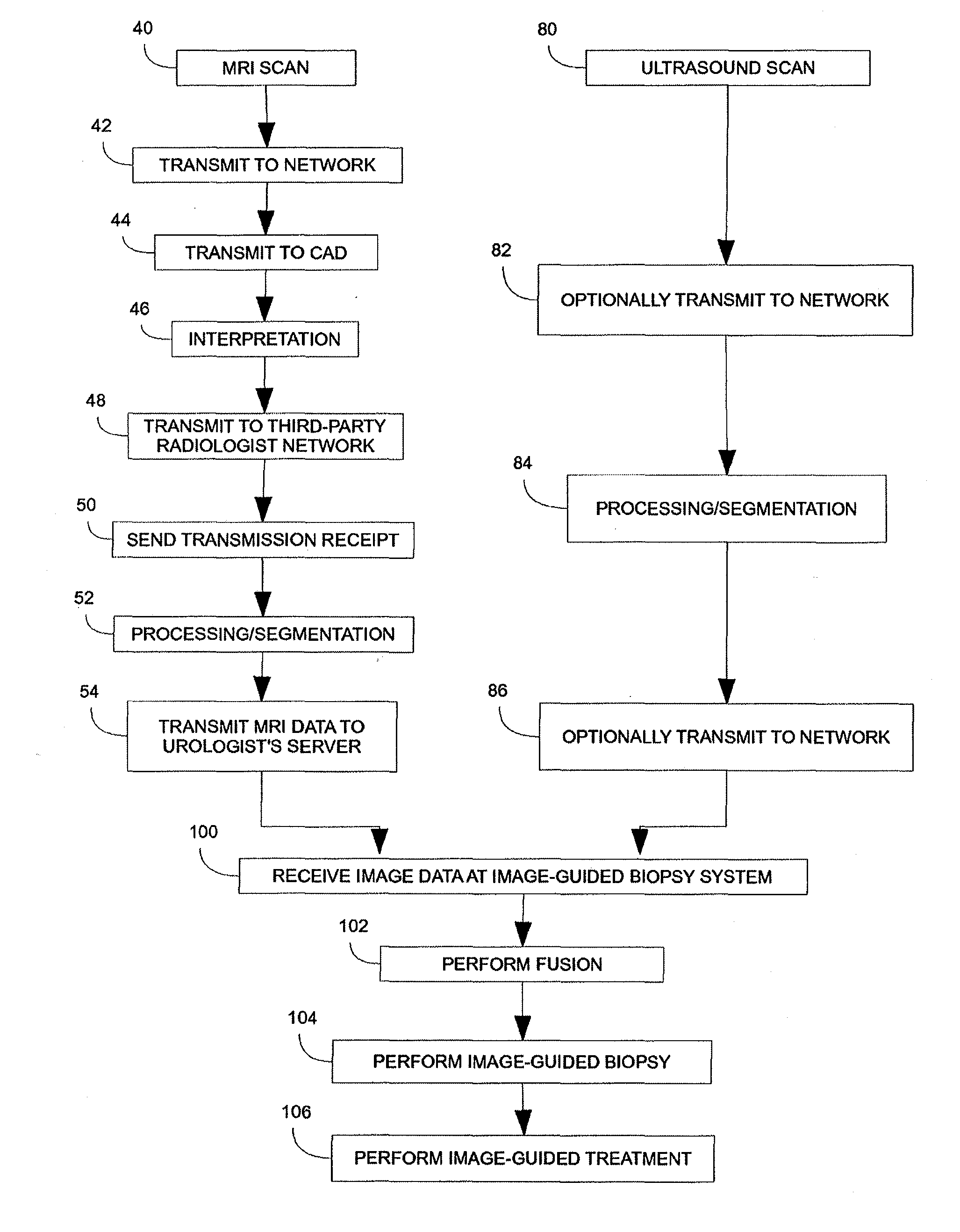
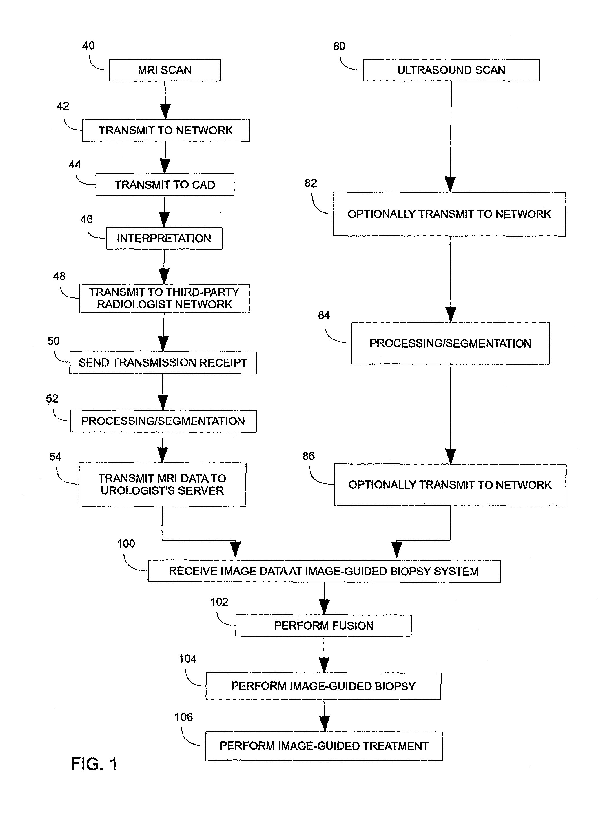
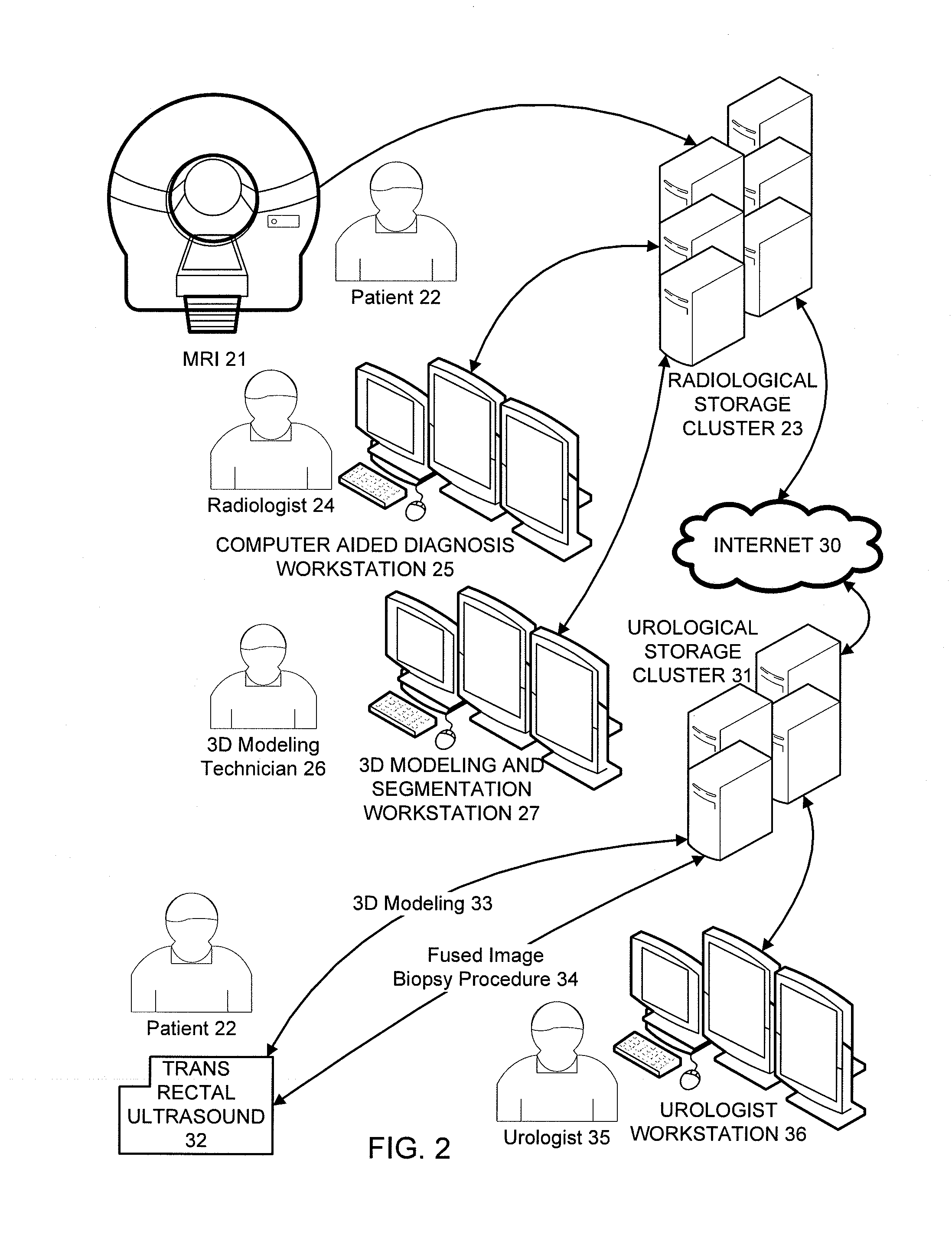
[0041]The present invention will be described with respect to a process, which may be carried out through interaction with a user or automatically, to generate a composite medical image made up of MRI and ultrasonic imaging data acquired separately at a radiology center and a urology center. One skilled in the art will appreciate, however, that imaging systems of other modalities such as PET, CT, SPECT, X-ray, and the like may be used in substitution for or in conjunction with MRI and / or ultrasound to generate the composite image in accordance with this process. Further, the present invention will be described with respect to the acquisition and imaging of data from the prostate region of a patient. One skilled in the art will appreciate, however, that the present invention is equivalently applicable with data acquisition and imaging of other anatomical regions of a patient.

[0042]The medical diagnostic and treatment system and a service networked system of the current invention incl...


PUM


Abstract

A method and system for diagnosis and treatment of medical conditions. The method includes communicating MRI, CT, PET and/or ultrasound image data, and fusing such data using an image-guided biopsy system. It further includes using such fused images in conjunction with the image-guided biopsy system for performing diagnosis and treatment procedures.

Description

CROSS REFERENCE TO RELATED APPLICATIONS[0001]The present application is a non-provisional of U.S. Provisional Patent Application 61 / 596,372, filed Feb. 9, 2012, the entirety of which is expressly incorporated herein by reference.BACKGROUND OF THE INVENTION[0002]1. Field of the Invention[0003]The present disclosure relates to medical imaging and surgical procedures.[0004]2. Description of the Art[0005]Prostate cancer is one of the most common types of cancer affecting men. It is a slow growing cancer, which is easily treatable if identified at an early stage. A prostate cancer diagnosis often leads to surgery or radiation therapy. Such treatments are costly and can cause serious side effects, including incontinence and erectile dysfunction. Unlike many other types of cancer, prostate cancer is not always lethal and often is unlikely to spread or cause harm. Many patients who are diagnosed with prostate cancer receive radical treatment even though it would not prolong the patient's li...

Claims


Application Information

Patent Timeline
IPC IPC(8): A61B8/08A61B8/13A61B6/03A61B5/055
CPCA61B8/12A61B8/468A61B8/565A61B5/4381A61B5/4836A61B8/085A61B8/5261A61B5/055A61B8/13A61B6/037A61B6/032A61B5/0035A61B5/0036
Inventor SPERLING, DANIEL S.
Owner CONVERGENT LIFE SCI
Why Eureka
Try Eureka