Therapeutic composition for treatment of glioblastoma
Active Publication Date: 2013-12-05
RGT UNIV OF MINNESOTA
0 Cites 6 Cited by
AI-Extracted Technical Summary
Problems solved by technology
Glioblastoma multiforme (GBM) is a common type of primary brain tumor in humans and is a very aggressive and devastating canc...
Benefits of technology
[0003]The present invention provides an anti-tumor composition comprising a purified glioblastoma GBM6-AD stem cell. In certain embodiments, the cell is an adherent cell line in serum free media that is useful to generate anti-tumor immune responses. In certain embodiments, the present invention provides a glioblastoma stem cell line (GBM6-AD), assigned ATCC® Patent Deposit Designation PTA-11498. In certain embodiments, the GBM6-AD cells are lysed. In certain embodiments, the GBM6-AD cells are irradiated. In certain embodiments, the anti-tumor composition furt...
Abstract
The present invention is directed to compositions and methods for treating an animal diagnosed with Glioblastoma multiforme (GBM).
Application Domain
BiocideOrganic active ingredients +12
Technology Topic
Individual animalGlioblastoma
Image
Examples
- Experimental program(4)
Example
EXAMPLE 1
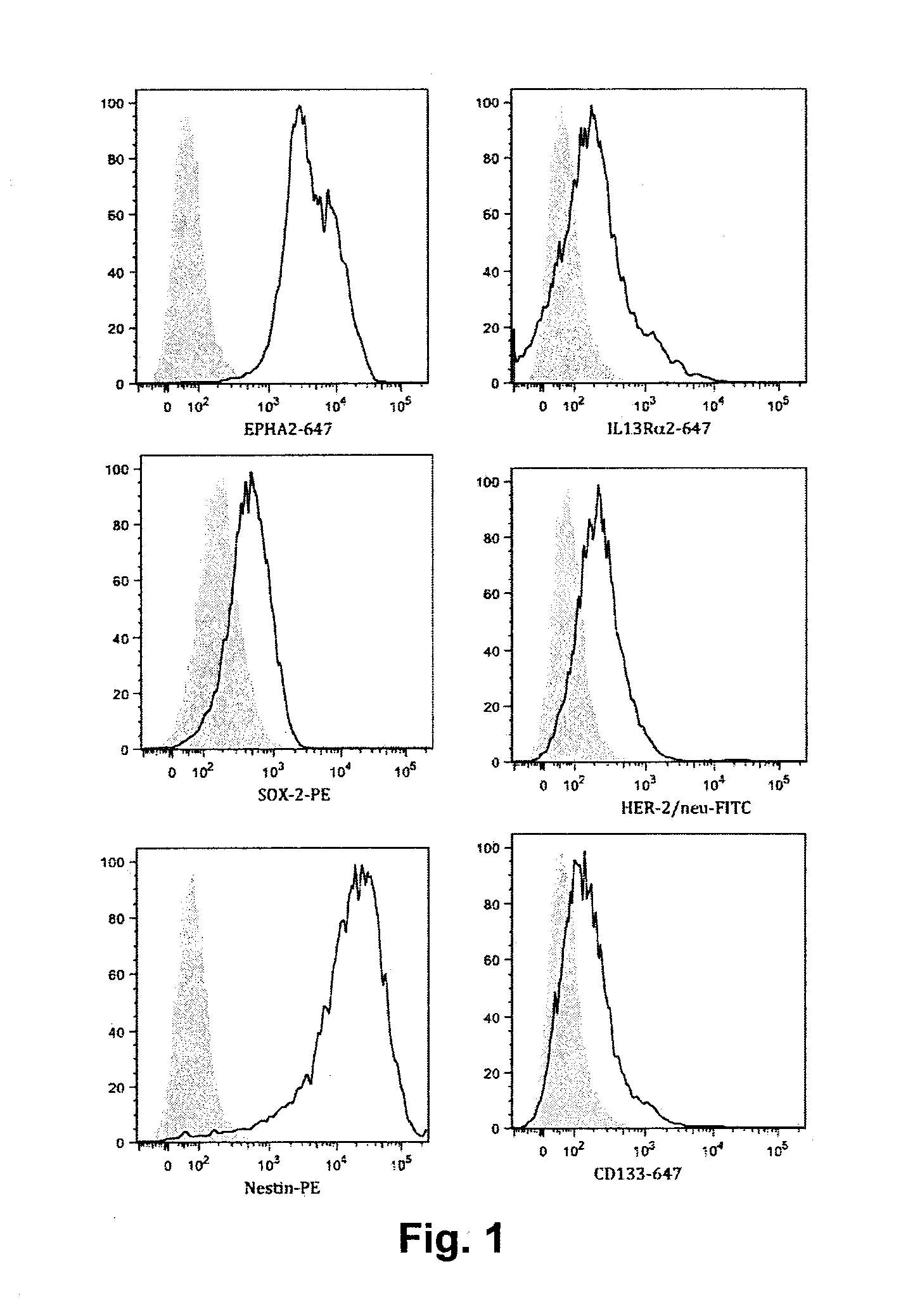
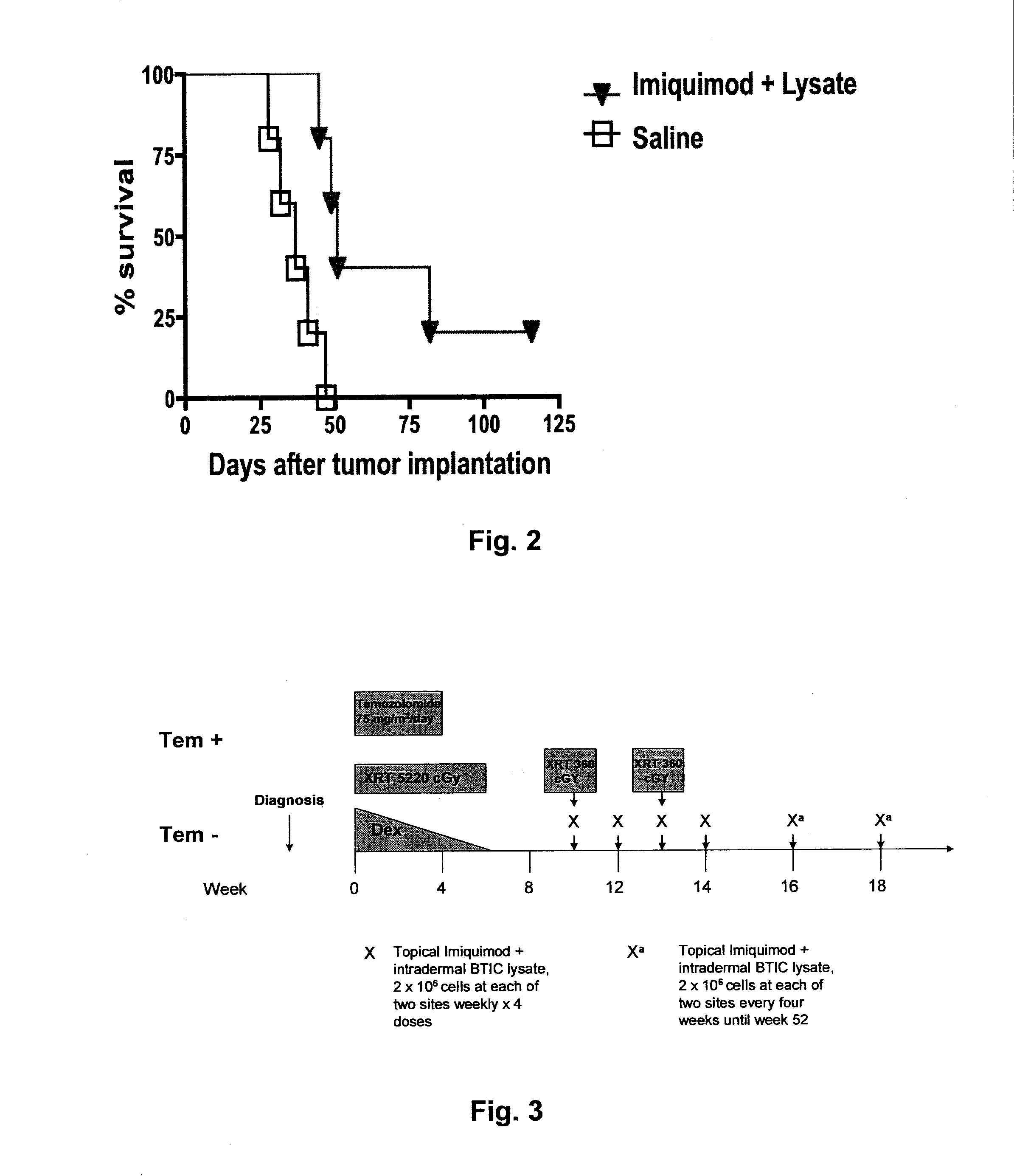
[0125]The present inventors have developed a novel therapy utilizing immunotherapy with Imiquimod and brain tumor initiating cell (BTIC) vaccine for the treatment of Diffuse Intrinsic Pontine Glioma (DIPG), a deadly brain tumor that occurs chiefly during childhood and the young adult years. The histology of the great majority of DIPGs is glioblastoma multiforme (GBM), WHO grade IV (Tsuchida T, Shimbo Y, Fukuda M, et al. Computed tomographic and histopathological studies of pontine glioma. Child's Nery Syst. 1985;1:223-229). Past experience has shown that DIPG responds to radiation therapy, but only transiently. The majority of children afflicted with this tumor die within two years of diagnosis. Attempts to significantly improve outcomes with adjuvant chemotherapy and/or concurrent radio-sensitizing agents have failed to date (Frazier J L, Lee J, Ulrich W T, et al. Treatment of diffuse brainstem gliomas: failed approaches and future strategies. J Neurosurg Pediatrics. 2009;3:259-269). Based on statistics from The Central Brain Tumor Registry of the United States (CBTRUS), the annual incidence of brain stem tumors in the 0 to19 year-old age group is expected to be over 450 in the coming years. Of these, over 360 will be children and young adults with malignant DIPGs (Central Brain tumor registry of the United States. Retrieved 2009 Jul. 16, from Reports and Tables web site: http://cbtrus.org/reports/2009-NPCR-04-05/CBTRUS-NPCR2004-2005-Report-.pdf.2009). Few of these young people are expected to survive. This therapy results in increased survival among the young people afflicted with this disease.
[0126]The discovery that dendritic cells (DCs) play a pivotal role in antigen presentation and evoking adaptive immune responses has opened the possibility of manipulating them to treat GBM. Liau et al. published the first use of tumor peptide pulsed DC vaccination for a glioma patient in 2000; this study documented tumor-reactive T cells despite disease progression (Liau L M, et al. Treatment of a patient by vaccination with autologous dendritic cells pulsed with allogeneic major histocompatibility complex class I-matched tumor peptides. Case Report. Neurosurg Focus. 2000;9:e8). Since then, vaccination of GBM patients with tumor lysate-pulsed autologous DC was able to elicit antigen-specific CD8+ T cells and increase median survival to 133 weeks compared to 30 weeks in historical control patients receiving standard therapy (Yu J S, et al. Vaccination with tumor lysate-pulsed dendritic cells elicits antigen-specific, cytotoxic T-cells in patients with malignant glioma. Cancer Research. 2004;64:4973-4979). Yamanaka et al. reported similar results in a phase I/II clinical trial following subdermal injection of tumor lysate-pulsed DCs or concurrent intratumoral and subdermal vaccination. Four patients showed a clinical response, ten showed tumor stabilization, and ten had tumor progression (Yamanaka R, et al. Clinical evaluation of dendritic cell vaccination for patients with recurrent glioma: results of a clinical phase I/II trial. Clin Cancer Res. 2005;11:4160-4167). In a recent phase II study, the ability of CTLs to elaborate IFN-γ in response to DC vaccinations was significantly correlated with clinical response and overall survival (Wheeler C J, et al. Vaccination elicits correlated immune and clinical responses in glioblastoma multiforme patients. Cancer Research. 2008;68:5955-5964). Despite these promising results, most patients ultimately succumb to their disease. Immunotherapy will be more successful if specifically designed to target radiation resistant, brain tumor initiating cells.
[0127]Brain Tumor Initiating Cells: Markers and Implications for Immunotherapy
[0128]Research over the last 15 years suggests that heterogeneous populations of cells comprise a tumor, and only a subset of them is capable of self-renewal, tumor initiation, and partial differentiation (reviewed in Beachy P A, Karhadkar S S, Berman D M. Tissue repair and stem cell renewal in carcinogenesis. Nature. 2004;432:324-331). The existence of tumor initiating cells was firmly established in the 1990s in acute mylogenous leukemia (Lapidot T, et al. A cell initiating human acute myeloid leukaemia after transplantation into SCID mice. Nature. 1994:367:645-648; Bonnet D, Dick J E. Human acute myeloid leukemia is organized as a hierarchy that originates from a primitive hematopoietic cell. Nature Medicine. 1997;3:730-737) and very recently in primary brain tumors such as glioblastoma and medullablastoma (Ignatova T N, et al. Human cortical glial tumors contain neural stem-like cells expressing astroglial and neuronal markers in vitro. Glia. 2002;39:193-206; Singh S K, et al. Identification of human brain tumour initiating cells. Nature. 2004;432:396-401; Singh S K, et al. Identification of a cancer stem cell in human brain tumors. Cancer Research. 2003;63:5821-5828; Galli R, et al. Isolation and characterization of tumorigenic, stem-like neural precursors from human glioblastoma. Cancer Research. 20044:7011-7021; Li M C, et al. Isolation and characterization of brain tumor stem cells in human medulloblastoma. Ai Zheng. 2006;25:241-246) is a 120-kDa transmembrane glycoprotein expressed in hematopoietic and neural stem cells that has been used as a marker for brain tumor initiating cells (BTIC). Singh et al. demonstrated that as few as 100 CD133+ glioma cells were capable of tumor formation in immunodeficient mice, whereas 100,000 CD133− tumor cells isolated from the same bulk tumor mass engrafted but did not form tumors. (Singh S K, et al. Identification of human brain tumour initiating cells. Nature. 2004;432:396-401). Moreover, the percentage of CD133+ cells in the tumor mass increased in recurrent patients, and CD133 expression has now been used in predicting disease progression in low-grade gliomas (Zeppernick F, et al. Stem Cell Marker CD133 Affects Clinical Outcome in Glioma Patients. Clin Cancer Res. 2008;14:123-129). However CD133 is not the only marker for BTICs since CD133− cells from a subset of gliomas are tumor initiating (Beier D, et al. CD133+ and CD133− Glioblastoma-Derived Cancer Stem Cells Show Differential Growth Characteristics and Molecular Profiles. Cancer Research. 2007;67:4010-4015). Lee et al. have identified nestin+Sox-2+ cells isolated from human GBM that meet important criteria for BTIC (tumor initiation at low cell number) (Lee J, et al. Tumor stem cells derived from glioblastomas cultured in bFGF and EGF more closely mirror the phenotype and genotype of primary tumors than do serum-cultured cell lines. Cancer Cell. 2006;9:391-403). These CD15+ BTICs formed very invasive tumors upon transplantation into mouse brain and their gene expression signature closely resembled that of the primary tumor (Lee J, et al. Tumor stem cells derived from glioblastomas cultured in bFGF and EGF more closely mirror the phenotype and genotype of primary tumors than do serum-cultured cell lines. Cancer Cell. 2006;9:391-403). Although CD133 and Sox-2 are useful markers, they cannot by themselves always predict tumor initiation. Nevertheless, cells expressing CD133 in particular seem to preferentially survive radiation and chemotherapy, making targeting them by immunotherapy very important. The key concept emerging from this research is that brain tumors are comprised of bulk tumor cells with limited tumorogenic potential and BTICs. BTICs may only account for a fraction of total tumor cells, but are the likely source of unrelenting tumor-renewal and disease progression.
[0129]Rationale for using radiation as a vaccine sensitizer. Bao et al. have shown that CD133+ cells exhibit enhanced resistance to ionizing radiation relative to their CD133− daughter cells by preferential activation checkpoint kinases Chk1, Chk2 (Bao S, et al. Glioma stem cells promote radioresistance by preferential activation of the DNA damage response. Nature. 2006;444:756-760). Although CD133+ and CD133− cells exhibited similar DNA damage, CD133+ cells repaired the damage more rapidly and were enriched after each dose of radiation relative to CD133− cells (Bao S, et al. Glioma stem cells promote radioresistance by preferential activation of the DNA damage response. Nature. 2006;444:756-760). In an elegant study, Gasser et al. demonstrated that the DNA damage pathway regulates innate immune system ligands of the NKG2D receptor (Gasser S, Orsulic, S., Brown, E. J. & Raulet, D. H. The DNA damage pathway regulates innate immune system ligands of the NKG2D receptor. Nature. 2005;436:1186-1190). They determined that the molecular basis for radiation-induced NKG2D ligand expression was induction of Chk1/2 activation. Moreover, pharmacological inhibition of Chk1 inhibited NKG2D ligand upregulation, clearly revealing that radiation increases cellular immunogenicity through the checkpoint kinase pathway. However, these studies were conducted using normal cells and sarcoma cells (Gasser S, Orsulic, S., Brown, E. J. & Raulet, D. H. The DNA damage pathway regulates innate immune system ligands of the NKG2D receptor. Nature. 2005;436:1186-1190). Nonetheless, if these results apply to glioma cells, then the same pathway used by CD133+ cells to survive radiation therapy (signaling through Chk1) is expected to preferentially sensitize them to Natural killer or T cell cell-mediated lysis by upregulation of NKG2D ligands. If true, this would represent an “Achilles heel” for CD133+ cells, which is defined as “a fatal weakness in spite of overall strength, actually or potentially leading to downfall.” In certain embodiments, the present invention uses the combination of radiation with BTIC-targeted immunotherapy for treating DIPG.
[0130]Targeting BTIC antigens: culture conditions of glioma cells have crucial implications for immunotherapy. For decades, research done to understand glioma biology and investigate potential therapies has relied on using glioma cell lines cultured in serum. Accordingly, in recent clinical trials where GBM patients were vaccinated with tumor cell lysate or peptide-pulsed DCs, the tumor cells were cultured and expanded in serum (Yu J S, et al. Vaccination with tumor lysate-pulsed dendritic cells elicits antigen-specific, cytotoxic T-cells in patients with malignant glioma. Cancer Research. 2004;64:4973-4979; Yamanaka R, et al. Clinical evaluation of dendritic cell vaccination for patients with recurrent glioma: results of a clinical phase I/II trial. Clin Cancer Res. 2005;11:4160-4167; Yamanaka R, et al.
[0131]Vaccination of recurrent glioma patients with tumour lysate-pulsed dendritic cells elicits immune responses: results of a clinical phase I/II trial. Br J Cancer. 2003;89:1172-1179). Two well controlled studies have recently shown that culture of BTICs in serum causes differentiation, leading to enhanced genomic instability, accelerating genetic mutation and leading to global gene expression patterns and phenotypes that are drastically different from the primary tumor (Lee J, et al. Tumor stem cells derived from glioblastomas cultured in bFGF and EGF more closely mirror the phenotype and genotype of primary tumors than do serum-cultured cell lines. Cancer Cell. 2006;9:391-403; Gunther HS, et al. Glioblastoma-derived stem cell-enriched cultures form distinct subgroups according to molecular and phenotypic criteria. Oncogene. 2007). In contrast, culture of the primary tumor cells in neural stem cell media supplemented with EGF and FGF prevents differentiation. These neurosphere cultures recapitulate the genotype, gene expression pattern, and phenotype of the primary tumor when xenografted into mouse brain (Lee J, et al. Tumor stem cells derived from glioblastomas cultured in bFGF and EGF more closely mirror the phenotype and genotype of primary tumors than do serum-cultured cell lines. Cancer Cell. 2006;9:391-403; Gunther H S, et al. Glioblastoma-derived stem cell-enriched cultures form distinct subgroups according to molecular and phenotypic criteria. Oncogene. 2007). One should consider these recent findings as attempts are made to develop more effective immunotherapy. When DCs are pulsed with a tumor lysate and administered as a vaccine, the protein antigens in the lysate are processed within the DC and presented on MHC I and MHC II to prime naïve T cells that respond to such antigens. Therefore, it is likely that until now, lysate pulsed DC vaccines have been biased to targeting only a subset of antigens expressed on the primary tumor, and probably few (if any) of these were BTIC-specific antigens since the antigen source was cultured in serum. One study has now provided evidence that supports this hypothesis. Pellegata et al. have shown that mice vaccinated with DCs pulsed with neurosphere-cultured lysate had superior therapeutic response compared to mice vaccinated with DCs pulsed with serum cultured glioma cell lysate (Pellegatta S, et al. Neurospheres enriched in cancer stem-like cells are highly effective in eliciting a dendritic cell-mediated immune response against malignant gliomas. Cancer Research. 2006;66:10247-10252). In other words, the way DC vaccine trials have been conducted may create a problem of signal to noise. In the current project glioma cell cultures were derived from primary tumor and cultured in neural stem cell media to enrich for clinically relevant phenotype and expression of important BTIC antigens.
[0132]Imiquimod Plus Tumor Cell Lysate is a Potent Anti-Glioma Vaccine
[0133]Imiquimod is an FDA-approved immune response modifier administered as a cream on the skin for the treatment of cutaneous tumors. Imiquimod exerts its immunostimulatory effects through toll-like-receptor 7 (TLR7) expressed on DCs and B cells in humans. Imiquimod treatment causes release of proinflammatory cytokines including interferonα, interferonγ, IL-12, all important for priming a robust Th1 immune response associated with anti-tumor activity in animals. Topical Imiquimod has been used as a vaccine adjuvant with success in numerous studies targeting established tumors and viral infection (Adams S, et al. Immunization of malignant melanoma patients with full-length NY-ESO-1 protein using TLR7 agonist Imiquimod as vaccine adjuvant. J Immunol. 2008;181:776-784; Johnston D, Bystryn J C. Topical Imiquimod is a potent adjuvant to a weakly-immunogenic protein prototype vaccine. Vaccine. 2006;24:1958-1965; Craft N, et al. The TLR7 agonist Imiquimod enhances the anti-melanoma effects of a recombinant Listeria monocytogenes vaccine. J Immunol. 2005;175:1983-1990; Harrison C J, Miller R L, Bernstein D I. Reduction of recurrent HSV disease using Imiquimod alone or combined with a glycoprotein vaccine. Vaccine. 2001;19:1820-1826; Bernstein D I, Miller R L, Harrison C J. Adjuvant effects of Imiquimod on a herpes simplex virus type 2 glycoprotein vaccine in guinea pigs. J Infect Dis. 1993;167:731-735). Based on this previous work, the efficacy of a tumor cell lysate vaccine in a murine model as tested. Inbred C57BL/6 mice bearing syngeneic GL261 gliomas were treated by intradermal vaccinations with GL261 cell lysates whereby Imiquimod was applied to the vaccination site immediately before injection. Saline was administered as a control. Mice treated with Imiquimod/tumor lysate vaccination survival significantly longer than controls, including 20% long-term survival (FIG. 2; p<0.01).
[0134]Treatment schema. The present treatment schema (FIG. 3) is based on the above data, and what is known about radiation increasing the immunogenicity of tumor cells. The BTIC cell line (GBM-6) is used as the vaccine source. Conformal radiation therapy to a dose of 5220 cGy in 180 cGy fractions is given over 6 to 7 weeks for induction in an attempt to achieve minimal residual disease. Additional 360 cGy fractions are delivered in 180 cGy fractions over two consecutive days at 4 and 8 weeks following the end of the initial radiation therapy in a novel effort to induce NKG2D ligand upregulation (thereby “sensitizing” residual tumor to lymphocyte attached). Therefore, the total radiation dose for each patient is 5940 cGy. Patients are assigned to two groups; temozolomide 75 mg/m2/day is given to group 1 during the first four weeks of radiation therapy for adjuvant anti-tumor effect and radiosensitization. It is expected that temozolomide will cause lymphopenia. Vaccine is delivered during subsequent lymphocyte recovery in this group of patients in order to skew immune recovery towards T cells specific for tumor antigens expressed in DIPG such as IL-13Rα2, Epha2, Her-2. Recent data has shown that T regulatory cells may be the predominant lymphocyte subset that is delayed during temozolomide recovery, therefore, group 2 is treated with no adjuvant temozolomide. These patients may present to us upon referral from other institutions after initial radiation therapy has been completed. All other patients are randomly assigned to the temozolomide or no temozolomide arms. T reg numbers and immune response are compared between the two groups by flow cytometry on peripheral blood mononuclear cells collected before and after vaccine therapy initiates. Vaccine administration commences at week four following completion of radiation therapy and is given every two weeks for four doses. Subsequent every 4 week boosters are given and continue to a maximum of one year from initiation of radiation therapy, by which time median survival will have passed based on historical data.
[0135]Radiation therapy is given according to standard of care at the University of Minnesota Children's Hospital, with attention to the dosage guideline above. All patients are treated with Intensity Modulated Radiation Therapy (IMRT) or an equivalent conformal technique. The clinical target volume is defined as the gross tumor volume (full extent of tumor visible on MRI) plus 1 cm margin.
[0136]Most, if not all, patients will be placed on dexamethasone therapy at the time of diagnosis. An effort will be made to wean all patients off of dexamethasone by week 6 of initial radiation therapy, again to enable immune reconstitution prior to vaccine administration.
[0137]Administration of the vaccine is accomplished by first applying the Imiquimod topically at two suprascapular injection sites. The vaccine is then given intradermally in two divided doses approximating 2×10⁶ lysed, gamma irradiated GBM6-AD cells per dose at each administration time point.
Example
EXAMPLE 2
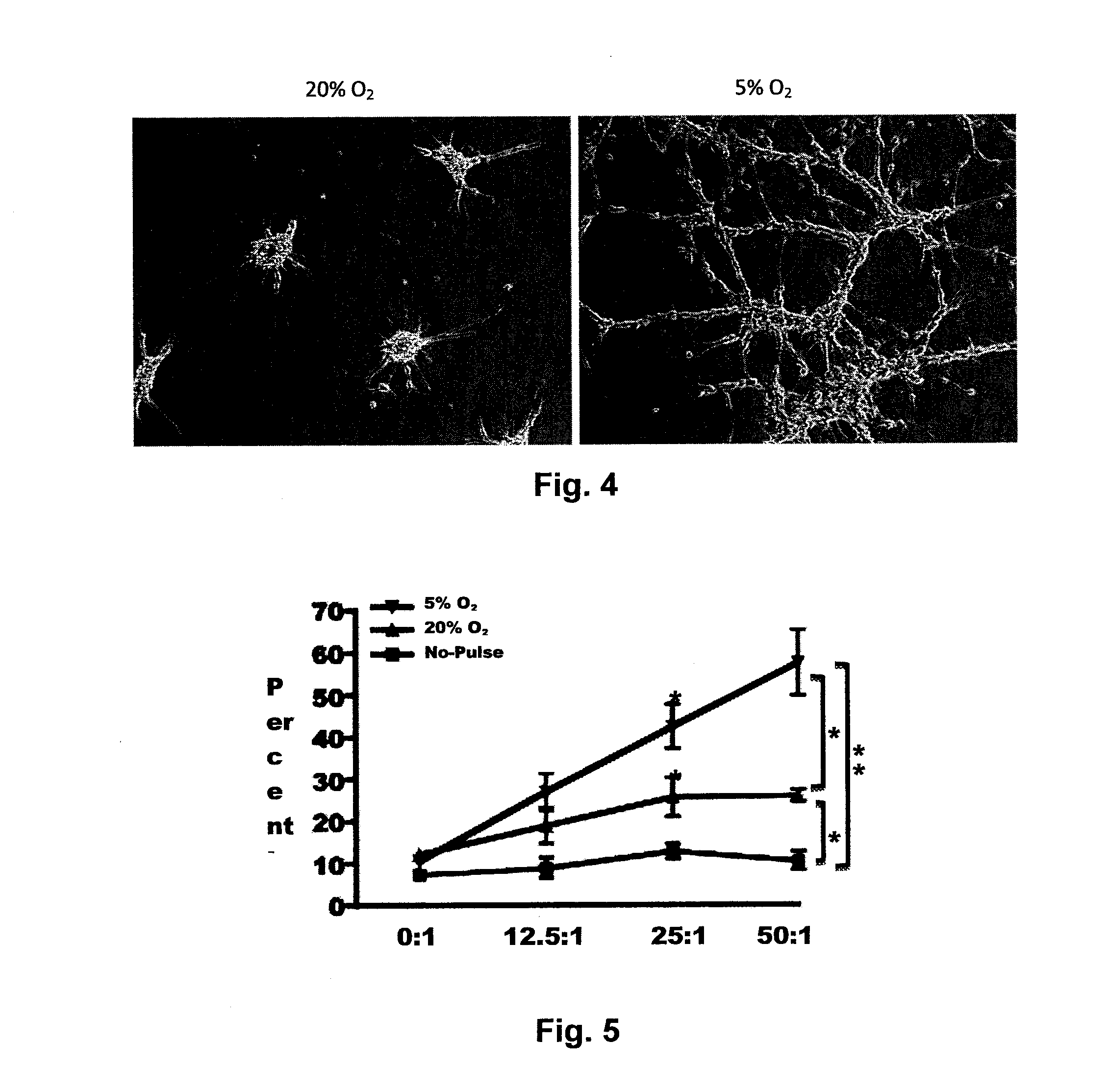
[0138]Dendritic cell (DC) vaccination is a powerful approach for cancer immunotherapy that has recently obtained regulatory approval, and tumor lysate-pulsed DC vaccines are being tested in clinical trials. Tumor cell lysates (TLs) are often generated from cells cultured in atmospheric oxygen (˜20% O2), which is higher than the O2 tension in the tumor in situ. It has been discovered that culturing murine tumor cells in 5% O2 profoundly enhanced the efficacy of TL vaccines. The inventors thus sought to determine if culturing primary human glioma cells in 5% O2 would enhance DC potency and tumor antigen expression. Unlike primary glioblastoma cells cultured in 20% O2, cultures in 5% O2 showed a partial reversion toward the gene expression patterns in the parental tumor in situ. Additionally, 5% O2 TLs were taken up more efficiently by DCs, which exhibited a superior ability to present antigen to CD8 T cells. CD8 T cells primed by 5% O2 lysate-pulsed DCs had significantly improved tumoricidal function relative to those primed by 20% O2 TLs. Collectively the inventors' results demonstrate that tissue culture O2 can: i) bias expression of target antigens to better reflect tumors in situ, ii) increase the potency of DCs for antigen presentation, iii) enhance effector function of CD8 T cells. These results have broad implications for improving the efficacy of TL-pulsed DC vaccines. Based on these data a clinical trial has been initiated using DCs pulsed with glioma cells expanded in 5% O2 in serum-free conditions.
[0139]Therapeutic vaccination utilizing dendritic cells (DCs) pulsed with tumor-associated antigens is a promising approach for cancer immunotherapy (Ridgway D. The first 1000 dendritic cell vaccines. Cancer Invest 2003; 21: 873-86). The United States Food and Drug Administration recently approved a peptide-pulsed DC vaccine for the treatment of castration-resistant prostate cancer (DeFrancesco L. Landmark approval for Dendreon's cancer vaccine. Nat Biotechnol; 28: 531-2; Kantoff P W, Higano C S, Shore N D, et al. Sipuleucel-T immunotherapy for castration-resistant prostate cancer. N Engl J Med; 363: 411-22). Tumor cells are frequently used as a source of antigen for DC vaccines. Vaccines using tumor cells and tumor cell lysates (TLs) have several advantages including targeting multiple, patient-specific tumor antigens. Vaccination with TL-pulsed DCs has demonstrated encouraging results in early stage clinical trials in numerous malignancies, but there is clearly a need for additional improvements (Le D T, Pardoll D M, Jaffee E M. Cellular vaccine approaches. Cancer J; 16: 304-10).
[0140]It remains unclear how tissue culture might affect anti-tumor immune responses evoked by tumor cell vaccines. Primary glioblastoma cells cultured in serum-containing media were genetically and phenotypically very different from the primary tumor, whereas culture of the same cells in serum-free conditions more closely reflected the primary tumor and enriched for a cancer stem cell phenotype (Lee J, Kotliarova S, Kotliarov Y, et al. Tumor stem cells derived from glioblastomas cultured in bFGF and EGF more closely mirror the phenotype and genotype of primary tumors than do serum-cultured cell lines. Cancer Cell 2006; 9: 391-403). Glioma cell lysates generated in serum-free conditions were more effective than those derived from serum-containing media when employed for TL-pulsed DC vaccines in a murine model (Pellegatta S, Poliani P L, Como D, et al. Neurospheres enriched in cancer stem-like cells are highly effective in eliciting a dendritic cell-mediated immune response against malignant gliomas. Cancer Res 2006; 66: 10247-52). Traditionally, tumor cell vaccines are derived from cultures maintained at atmospheric oxygen (˜20.95%; hereafter 20% O2), far from the average oxygen tension of less than 6% O2 measured in glioblastomas in situ (Evans S M, Judy K D, Dunphy I, et al. Hypoxia is important in the biology and aggression of human glial brain tumors. Clin Cancer Res 2004; 10: 8177-84). It is established that oxygen influences gene expression, cell metabolism, proliferation, survival (van den Brenk H A, Moore V, Sharpington C, Orton C. Production of metastases by a primary tumour irradiated under aerobic and anaerobic conditions in vivo. Br J Cancer 1972;
[0141]26: 402-12; Sciandra J J, Subjeck J R, Hughes C S. Induction of glucose-regulated proteins during anaerobic exposure and of heat-shock proteins after reoxygenation. Proc Natl Acad Sci USA 1984; 81: 4843-7; Pouyssegur J, Dayan F, Mazure N M. Hypoxia signalling in cancer and approaches to enforce tumour regression. Nature 2006; 441: 437-43; Gordan J D, Simon M C. Hypoxia-inducible factors: central regulators of the tumor phenotype. Curr Opin Genet Dev 2007; 17: 71-7), and hypoxia increases the expression of tumor stem cell markers CD133, Nestin, and SOX2 (Platet N, Liu S Y, Atifi M E, et al Influence of oxygen tension on CD133 phenotype in human glioma cell cultures. Cancer Lett 2007; 258: 286-90; McCord A M, Jamal M, Shankavaram U T, Lang F F, Camphausen K, Tofilon P J. Physiologic oxygen concentration enhances the stem-like properties of CD133+ human glioblastoma cells in vitro. Mol Cancer Res 2009; 7: 489-97; Li Z, Bao S, Wu Q, et al. Hypoxia-inducible factors regulate tumorigenic capacity of glioma stem cells. Cancer Cell 2009; 15: 501-13). However, the effect of oxygen on tumor cell immunogenicity is poorly understood.
[0142]The present results indicated that culturing primary glioma cells in 20% O2 caused a global shift in gene expression away from the primary tumor, which was partially reversed by culturing cells at 5% O2. Notably, expression of several glioma antigens known to elicit T cell responses was increased in 5% O2. Primary human glioma cells grown in 5% O2 exhibited intrinsic adjuvant properties as assessed by superior DC uptake, antigen presentation, and priming of tumoricidal CD8 T cells. Thus, primary human glioma cells grown in serum-free media at 5% O2 emerge as the most recent improvement in the evolution of tumor cell vaccines.
[0143]Materials and Methods
[0144]Tumor cell culture: Surgically resected gliomas (Table 1) were enzymatically dissociated and cultured in neural stem cell media (Wu A, Oh S, Wiesner S M, et al. Persistence of CD133+ cells in human and mouse glioma cell lines: detailed characterization of GL261 glioma cells with cancer stem cell-like properties. Stem Cells Dev 2008; 17: 173-8).
TABLE 1 Table of the tumor identification, patient information, and the experiment in which the tumor was used, and passage number of the cells. Patient number Id Tumor Experiment Passage Patient 1 *BT130 GBM Microarray 12 Patient 2 *BT132 GBM Microarray 12 Patient 3 GBM6 GBM mRNA and Fc, CTL 8 Patient 4 MNBT110 GBM mRNA, Fc, 14 Westerns, CTL Patient 5 MNBT112 Epd CTL 12 Patient 6 MNBT113 GBM mRNA, Fc 14 Western Patient 7 MNBT 124 GBM mRNA, Fc 15 *indicates that the tumor were resected at the Mayo Clinic, Rochester MN; others were resected University of Minnesota Medical Center, Fairview. GBM = Glioblastoma multiforme, Epd = Ependymoma, FC = Flow Cytometry, CTL = Cytotoxcity assay.
[0145]Unless explicitly stated, all tissue cultures were maintained at 20% O2. Cultures labeled as “5% O2” were converted from 20% O2 cultures by maintenance at 5% O2 for at least 30 days prior to use as described (Olin M R, Andersen B M, Zellmer D M, et al. Superior efficacy of tumor cell vaccines grown in physiologic oxygen. Clin Cancer Res; 16: 4800-8).
[0146]Microarray: Total RNA was isolated from snap frozen tissue at resection, or cultured cells from either 5% or 20% O2. Expression levels of 18,401 genes were analyzed using a Whole-Genome Gene Expression DASL Assay (Illumina, San Diego, Calif.).
[0147]Total RNA was isolated from snap frozen tissue at time of resection, and from cells grown in 5% and 20% O2 using the RNEasy® Plus Mini Kit (Qiagen®, Valencia, Calif.) according to the manufacturer's instructions. Quality control was performed with an Agilent 2100 Bioanalyzer (Santa Clara, Calif.). Whole-Genome Gene Expression DASL Assay (Illumina, San Diego, Calif.) was used to analyze expression levels of 18,401 genes. Briefly, biotinylated cDNA was synthesized from 100 ng of total RNA from each sample using mixed poly-T and random nonamer primers. Biotinylated cDNA was hybridized to assay oligonucleotides, which were subsequently bound to streptavidin-conjugated paramagnetic particles. Oligos were extended and ligated, creating templates that were amplified with random, fluorescent primers. The fluorescently labeled products were bound to 24,631 oligonucleotide probes contained on each microarray of a Illumina HumanRef-8_V1 Expression BeadChip, and analyzed with laser confocal microscopy. Three technical replicates were analyzed for each condition for patient 2 and tissue from patient 1. Two technical replicates were performed for cell lines derived from patient 1.
[0148]Array data were analyzed for quality control as previously described (Sarver A L. Toward understanding the informatics and statistical aspects of micro-RNA profiling. J Cardiovasc Transl Res; 3: 204-11), quantile normalized, and multiple probes for each gene were averaged. For each set of experiments (tumor, 20% and 5% oxygen) each experimental value was divided by the average of the 20% O2 culture to determine the differences between states independent of the differences between the different tumors. Two group t-tests as well as the average fold change were used to determine differentially expressed genes.
[0149]Quantitative RT-PCR: Extracted RNA was analyzed by real time PCR using SYBR Green one step PCR master mix (Qiagen®). The following conditions were used for PCR in an ABI PRISM 7500 thermocycler: 95° C. for 15 min; 94° C. for 30s, 55° C. for 30s, 68° C. for 30s in a total of 40 cycles; 72° C. for 10 min; and 10° C. until collected. Relative quantification of gene expression was calculated and normalized to GAPDH expression levels using the 2−ΔΔCt method (Livak K J, Schmittgen T D. Analysis of relative gene expression data using real-time quantitative PCR and the 2(-Delta Delta C(T)) Method. Methods 2001; 25: 402-8). Primers are listed in Table 2.
TABLE 2 List of primers used to determine transcription levels. Antigen Primer Sequence CD133 Forward-5-TCG TAC TCG GCT CCC TGT TG-3 (SEQ ID NO: 1) Reverse 5-ATT CAC GCG GCT GTA CCA CA-3 (SEQ ID NO: 2) SOX2 Forward 5-CCC CCG GCG GCA ATA GCA-3 (SEQ ID NO: 3) Reverse 5-TCG GCG CCG GGG AGA TAC AT-3 (SEQ ID NO: 4) NESTIN Forward 5-AGG TAG AGG AGC TGG CAA GGC GAC-3 (SEQ ID NO: 5) Reverse 5-TTT TCA GTA GCC CGC AGC CG-3 (SEQ ID NO: 6) EPHA2 Forward 5-CTG GCC TTC CAG GAT ATC GG-3 (SEQ ID NO: 7) Reverse 5-TGC ACA GTG CAT ACG GGG CT-3 (SEQ ID NO: 8) IL13Ra2 Forward 5-CTG ATA AGC ACA ACA TTT GGC TCT-3 (SEQ ID NO: 9) Reverse 5-TGA TGG TCT TCC ATG TTT CAC TAC C-3 (SEQ ID NO: 10) HER2/Neu Forward 5-CTG CAG CTT CGA AGC CTC ACA A-3 (SEQ ID NO: 11) Reverse 5-ATG GCA GCA GTC AGT GGG CAG T-3 (SEQ ID NO: 12) GAPDH Forward 5-GTC GGA GTC AAC GGA TTT GGT-3 (SEQ ID NO: 13) Reverse 5-GGG ATT TCC ATT GAT GAC AAG CT-3 (SEQ ID NO: 14)
[0150]Flow cytometry: 5×10⁵ cells were stained with: anti-CD133/2 (Miltenyi Biotec), EphA2, SOX2, Nestin, HER-2/neu (R&D Systems), and IL13Rα2 (SantaCruz Biotechnology). Following three washes, CD133/2 was stained with a secondary anti-mouse-647; IL13Rα2 and EphA2 were stained with secondary anti-mouse-PE along with their respective isotype and analyzed using a FACS Canto II. For DC phenotyping, 5×10⁵ DCs were stained with CD80-FITC, CD86-PE, CD83-PE, HLA-DR-APC, CCR7-FITC (eBioscience) and HLA-ABC-FITC (BD Biosciences), incubated at 4° C. for 30 min, washed, fixed, then analyzed on a FACS Caliber. Intracellular staining was performed according to the manufacturer's protocol (BD Biosciences).
[0151]Dendritic Cells: Monocytes were purified using CD14 magnetic beads (Miltenyi Biotech) from the peripheral blood of healthy HLA-A2+ donors, plated at a concentration of 5×10⁵ in 24-well plates and matured as previously described (Mailliard R B, Wankowicz-Kalinska A, Cai Q, et al. alpha-type-1 polarized dendritic cells: a novel immunization tool with optimized CTL-inducing activity. Cancer Res 2004; 64: 5934-7); on day 6, DCs were pulsed with 100 μg of TLs derived in either 5% or 20% O2, and maturated prior to experiments.
[0152]CTL Assays: 1×10⁶ HLA-A2+ PBMCs from normal donors were added to matured (day 8) lysate-pulsed DCs with 50 U IL-2 and incubated seven days. For re-stimulation, a second set of pulsed DCs was added to the co-culture for four days. On day 11, primed PBMCs were co-cultured with 2×10⁴ CFSE-labeled HLA-A2+ glioma cells for 6 h, then analyzed by flow cytometry to determine cytotoxicity as described (Olin M R, Hwa Choi K, Lee J, Molitor T W. Gammadelta T-lymphocyte cytotoxic activity against Mycobacterium bovis analyzed by flow cytometry. J Immunol Methods 2005; 297: 1-11). For the blocking assay, anti-HLA-ABC was added to target cells for 25 min, washed, and added to effector cells.
[0153]CMV assay: 5 X 10⁵ HLA-A2+ iDCs were pulsed with 100 μg of tumor lysates derived in 5% or 20% O2+/−10 μg of pp65 HLA-A2-restricted CMV antigen
[0154](NLVPMVATV) and matured as described in the methods section. Following maturation, DCs were washed 3 times, and 5×10⁵ PBMCs from CMV sera-positive donors were added to DCs. CMV sera-negative PBMCs were used as a negative control. Cells were incubated for 48 h, and supernatant was analyzed by cytometric bead array (BD Biosciences) for IFNγ. Cells were cultured for an additional 24 h, stained with anti-CD8, pp65 pentamer (ProImmune), and intracellularly stained for IFNγ production.
[0155]Increase in tumoricidal activity is not due to an increase in co-stimulatory molecules. To investigate if lysates derived in 5% O2 culture altered co-stimulatory molecule expression, DCs were pulsed with tumor lysates derived in either 5% or 20% O2 conditions, matured, and analyzed for CD83, CD80/86, HLA-DR, HLA-ABC, and CCR7 expression. In three separate experiments, no significant differences in co-stimulation markers were observed between the two oxygen conditions.
[0156]Statistical analysis: Statistical comparisons were made by ANOVA, followed by post hoc comparisons using a 2-tailed t-test. All tests were performed with Prism 4 software (Graph Pad Software, Inc). P values <0.05 were considered significant.
[0157]Results and Discussion
[0158]In order to compare tissue cultures to the primary tumor in situ, mRNA expression levels were profiled from two glioblastomas and cultured cells derived from the same tumor grown in 5% and 20% O2. As a trend, the gene expression signature shifted towards the levels observed in situ when the cells were cultured in 5% O2 relative to 20% O2. There was a significant difference in the expression of 3,333 genes between the 20% O2 culture and the in situ tumor in both patients. Of these, 77 genes were differentially expressed between the 5% and 20% O2 cultures.
[0159]Selected cancer stem cell markers (CD133, Nestin, Sox2) and TAA known to elicit T cell responses (EphA2, IL-13Ra2, HER-2/neu) were analyzed by PCR and flow cytometry. Relative to 20% O2, cells cultured in 5% O2 expressed significantly more mRNA encoding for CD133, EphA2, IL-13Rα2 and HER-2/neu (FIG. 6A). SOX2 and Nestin demonstrated the same trend but failed to reach statistical significance. With the exception of HER-2/neu, protein expression followed the same trend with significant increases in CD 133, EphA2, and IL13Rα2 (FIG. 6B). Moreover, western blot for CD133 and IL13Rα2 revealed similar changes in expression (FIG. 6C). Collectively, these experiments demonstrated that cells cultured in 5% O2 better reflect the antigen expression on the tumor in situ and are enriched for markers of “sternness” and immunogenic TAA.
[0160]It was next asked if oxygen tension would alter the ability of TLs to prime tumoricidal T cells in culture. HLA-A2+ PBMCs from normal donors were primed by autologous DCs pulsed with TLs derived from three different glioma patients. Primed PBMCs were assessed for their ability to lyse the same HLA-A2+ glioma cells that were used for priming. PBMCs primed by 5% O2 TLs demonstrated superior tumoricidal activity against target cells from 3/3 patients (FIG. 7A-C). To elucidate whether tumor cell lysis required priming CD8 T cells, an anti-HLA-ABC blocking antibody was added to tumor cells prior to effector cells. Blockade of MHC I-TCR interactions prevented a tumoricidal response, implicating cytotoxic T lymphocytes (CTLs) as the main effectors in this assay (FIG. 7D).
[0161]A plausible explanation for the enhancement in CTL priming would be improvement of DC maturation by 5% O2 TLs. The expression of co-stimulatory molecules, MHC molecules, and CCR7 on mature DCs was measured after pulsing with TLs from 20% or 5% O2. There were no significant differences in expression of CD83, CD80, CD86, or HLA-ABC, HLA-DR, or CCR7. Therefore, the adjuvant activity of 5% O2 TLs was not likely due to enhancing DC maturation, suggesting other mechanisms. Experiments were conducted to determine whether the superior CTL priming achieved with 5% O2 TLs could be due to changes in lysate uptake. Immature DCs were pulsed with TLs from CFSE-labeled glioma cells grown in 5% or 20% O2. Flow cytometry-based detection of CFSE+HLA-DR+ cells (marking DCs loaded with lysate) revealed that 5% O2 TLs were uptaken by twice the number of DCs compared to TLs derived in 20% O2 (FIG. 8A). Thus, unidentified factor(s) in the 5% O2 TLs increased the fraction of DCs that engulfed tumor-associated proteins in tissue culture.
[0162]The inventors established an assay to determine if TLs altered DC-mediated CD8 T cell priming from a defined antigen not present in the TLs. To accomplish this, TLs were mixed with pp65 peptide, a specific marker antigen with well-defined CD8 T cell responses. Cytomegalovirus (CMV)-derived pp65 is an HLA-A2-restricted immunodominant epitope to which CMV sera-positive patients typically have CD8 T cell memory responses (reviewed in (Moss P, Khan N. CD8(+) T-cell immunity to cytomegalovirus. Hum Immunol 2004; 65: 456-64)). PBMCs from sera-positive donors were co-cultured with DCs that had been pulsed with TLs with or without pp65. PBMCs from sera-negative donors were used to control for CD8 T cell activation independent of pp65. Soluble IFNγ in the tissue culture supernatant was quantified as a measure of CD8 T cell activation. There was a background of 50-200 pg/ml of IFNγ detected in the supernatant when TLs were pulsed in the absence of pp65, possibly due to activation of T cells or natural killer (NK) cells irrespective of pp65 (FIG. 8B). There was no significant difference in elaborated IFNγ between the non-pulsed and lysate-pulsed PBMCs from sera-positive donors, ruling out reactivity to CMV antigens in the lysate, demonstrating negligible expression of CMV antigens in TLs (Cobbs C S, Harkins L, Samanta M, et al. Human cytomegalovirus infection and expression in human malignant glioma. Cancer Res 2002; 62: 3347-50). As expected, PBMCs from sera-positive donors primed with pp65 alone (no TLs) elaborated IFNγ at levels 2-4 fold above background, whereas sera-negative donor PBMCs did not respond to pp65. Thus, sera-positive donor PBMCs displayed a phenotype consistent with immunological memory to CMV, allowing the inventors to measure how TLs could change the response to a defined CD8 T cell epitope. PBMCs primed with pp65 mixed with 5% O2 TLs elaborated twice the amount of IFNγ compared to pp65 alone. In marked contrast, 20% O2 TLs significantly suppressed pp65-dependent IFNγ secretion (FIG. 8B). In order to confirm that pp65-specifc CD8 T cells were the main source of IFNγ, flow cytometry was conducted to assess IFNγ expression specifically in CD8+pp65-pentamer+ cells (FIG. 8C). Consistent with measured soluble IFNγ, CD8+pp65-pentamer+ cells primed by 5% O2 TLs plus pp65 produced more IFNγ on a per-cell basis relative to all other groups. Taken together, these data demonstrate that 5% O2 TLs have intrinsic adjuvant activity, independent of the amount of TAA expressed. It is noteworthy that these findings paralleled what the inventors recently reported using more precise mouse immunology reagents and established glioma cell lines; namely that 5% O2 TLs increased the presentation of exogenous ovalbumin on MHC I and enhanced antigen-specific CD8 T cell activation, whereas 20% O2 TLs were suppressive to CD8 T cell priming and alternatively promoted antibody responses (Olin M R, Andersen B M, Zellmer D M, et al. Superior efficacy of tumor cell vaccines grown in physiologic oxygen. Clin Cancer Res; 16: 4800-8).
[0163]In summary, the present data demonstrate that tissue culture oxygen functions as a master “immunologic switch” by simultaneously regulating the expression of TAA and factors that modulate DC-mediated priming of CD8 T cells. By selecting for expression of TAA that are more abundant on the tumor in situ, the inventors show that oxygen can be exploited to prime CD8 T cells with greater specificity to targets abundant in the tumor in situ. A separate and equally important finding is that tumor cells grown in physiologic oxygen are inherently more immunogenic to CD8 T cells through a mechanism that is independent of co-stimulation but involves enhancement of CD8 T cell priming A third consideration is that growing primary glioma cells in 1-7% O2 increased cancer stem cell markers in several previous studies (Platet N, Liu S Y, Atifi M E, et al Influence of oxygen tension on CD133 phenotype in human glioma cell cultures. Cancer Lett 2007; 258: 286-90; McCord A M, Jamal M, Shankavaram U T, Lang F F, Camphausen K, Tofilon P J. Physiologic oxygen concentration enhances the stem-like properties of CD133+ human glioblastoma cells in vitro. Mol Cancer Res 2009; 7: 489-97; Li Z, Bao S, Wu Q, et al. Hypoxia-inducible factors regulate tumorigenic capacity of glioma stem cells. Cancer Cell 2009; 15: 501-13), showing that the T cells primed are better suited to eradicate cancer stem cells in situ. Vaccines made by expansion of primary tumor cells in physiologic oxygen serve as a powerful system to induce clinically useful anti-tumor immune responses.
Example
EXAMPLE 3
[0164]GBM6-AD cells can be grown to generate apoptotic bodies to pulse onto autologous dendritic cells to make a vaccine.
[0165]Production of Dendritic Cells. Peripheral blood mononuclear cells (MNCs) are collected with an FDA-licensed apheresis instrument using established, standard collection procedures. Sterile technique is followed to prevent contamination of the collection. The cells are seeded in a tissue culture flask or cell factory at a viable seeding density of 1.0E+05-1.5E—05 and incubated at 37° C., 5% CO2, On day +3, 20% total volume of culture medium supplements with IL-4 and GM-CSF (1,000 IU/mL) is added to the cells. On day +6 the cells are matured by adding GM-CSF (1,000 IU/mL), ILlbeta (25 ng/mL) TNF-alpha (50 ng/mL), IFN-alpha (10,000 IU/mL), IFN-gamma ((1,000 IU/mL), and Poly-IC (20 μg/mL) and incubating (37° C., 5% CO2) until day +8. A brain tumor apoptotic body (1 apoptotic cell per iDC) is added on day 6 as well. Mature, pulsed alpha-type 1 polarized DCs are harvested from culture using TrypLE Select on day +8 and cryopreserved in Plasmlyte-A, 5% human serum albumin (HSA), and DMSO (10% final concentration).
[0166]Brain Tumor Stem Cell Processing. A single cell line derived from an adult patient with glioblastoma multiforme was tested prior to acceptance into the MCT facility. Testing included mycoplasma (PTC) and sterility; results were satisfactory. Cells were then expanded in serum-free neural stem cell medium (DMEM/F12, B-27 Supplement, N-2 Supplement) with EGF (20 ng/mL final concentration) and bFGF (2 ng/mL final concentration) at 37° C., 5% CO2, and 5% O2. The culture was split as cells formed spheres at approximately 3-5 days using TrypLE Select and mechanical dissociation. Cells were re-suspended in serum-free neural stem cell medium with EGF (20 ng/mL final concentration)/bFGF (2 ng/mL final concentration) and continued through culture until a sufficient amount of cells were expanded for the MCB. The cells were then washed in DPBS and cryopreserved in cryopreservation medium (Plasmlyte-A, 5% human serum albumin (HSA), and DMSO (10% final concentration)).
[0167]Samples of the MCB are expanded in a manner consistent with the MCB to establish a working cell mass which is frozen using a controlled-rate freezer. A portion of the mass is then be thawed and subjected to nebulization, and the resultant bulk whole cell lysate is irradiated (200 Gy) and adjusted to approximately 2 mg/mL. It is then be aseptically transferred into vials at approximately 1.2 mg in 600 μL. The vials containing lysate are frozen using a controlled-rate freezer and then stored at ≦−150° C. until distribution for administration.
PUM
Property | Measurement | Unit |
Time |
Description & Claims & Application Information
We can also present the details of the Description, Claims and Application information to help users get a comprehensive understanding of the technical details of the patent, such as background art, summary of invention, brief description of drawings, description of embodiments, and other original content. On the other hand, users can also determine the specific scope of protection of the technology through the list of claims; as well as understand the changes in the life cycle of the technology with the presentation of the patent timeline. Login to view more.