
Method for detecting cellular DNA damage and antibody against cell surface antigen responsive to DNA strand breaks

a cell surface antigen and dna strand break technology, applied in the field of detecting cellular dna damage, can solve the problems of inducing strong adverse reactions, difficult to select marker molecules that can be applied in practice, and most difficult for organisms such as cancer cells to repair damage, etc., to inhibit the survival or growth of cancer cells, reduce the antigenicity of antibodies, and high levels

Active Publication Date: 2014-01-02
FUJITA HEALTH UNIVERSITY +1
View PDF0 Cites 0 Cited by
  • Summary
  • Abstract
  • Description
  • Claims
  • Application Information

AI Technical Summary

Benefits of technology

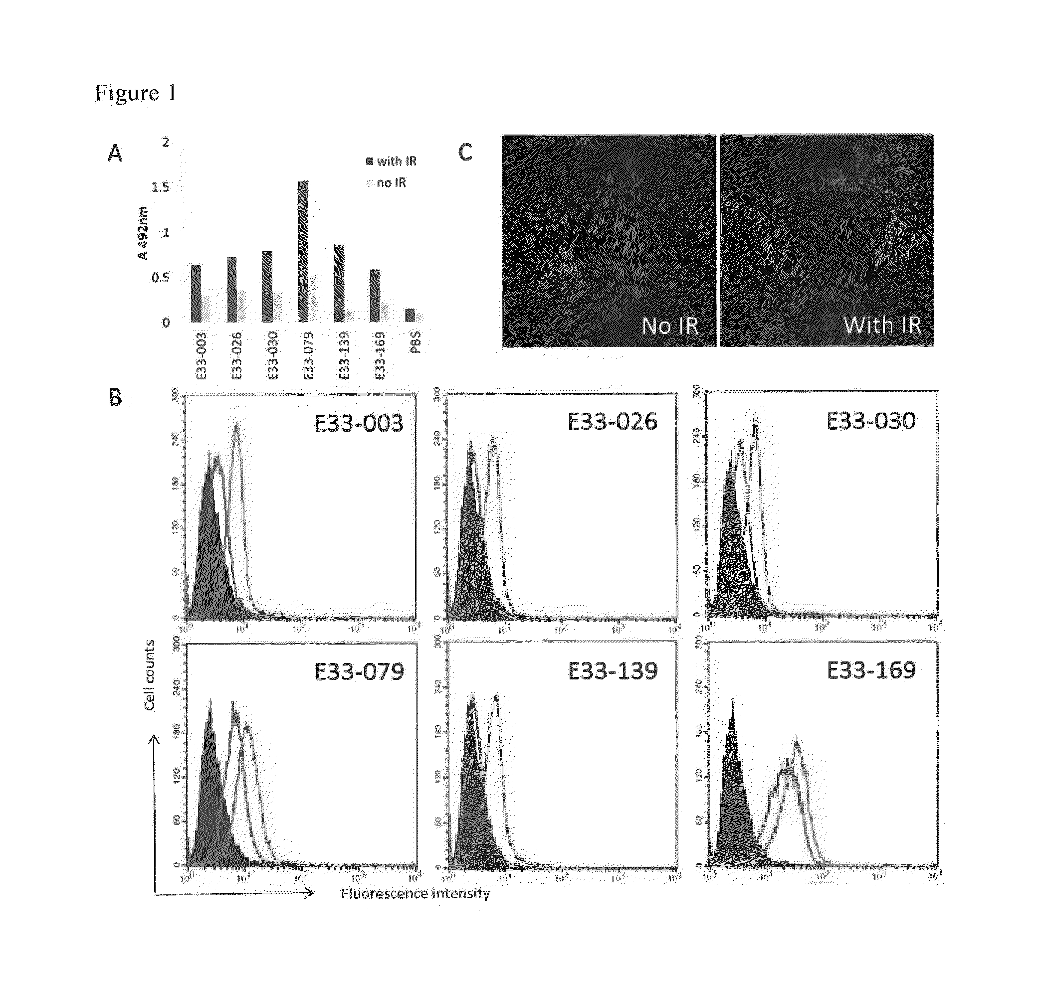
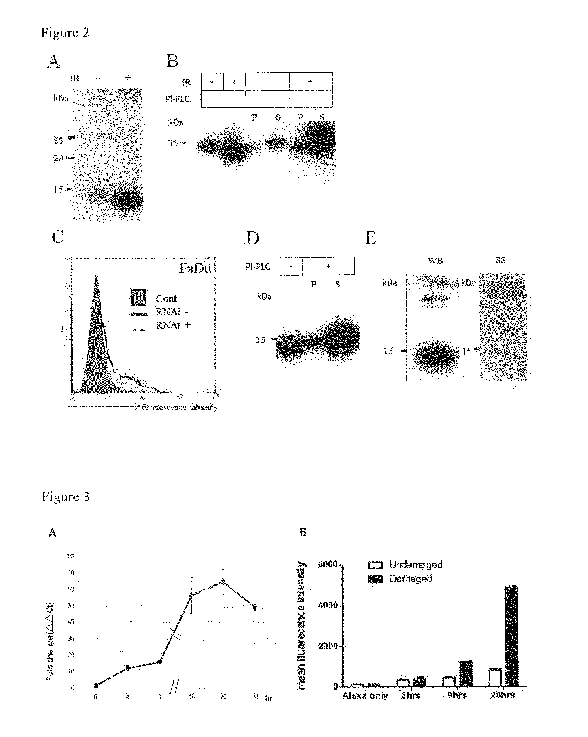
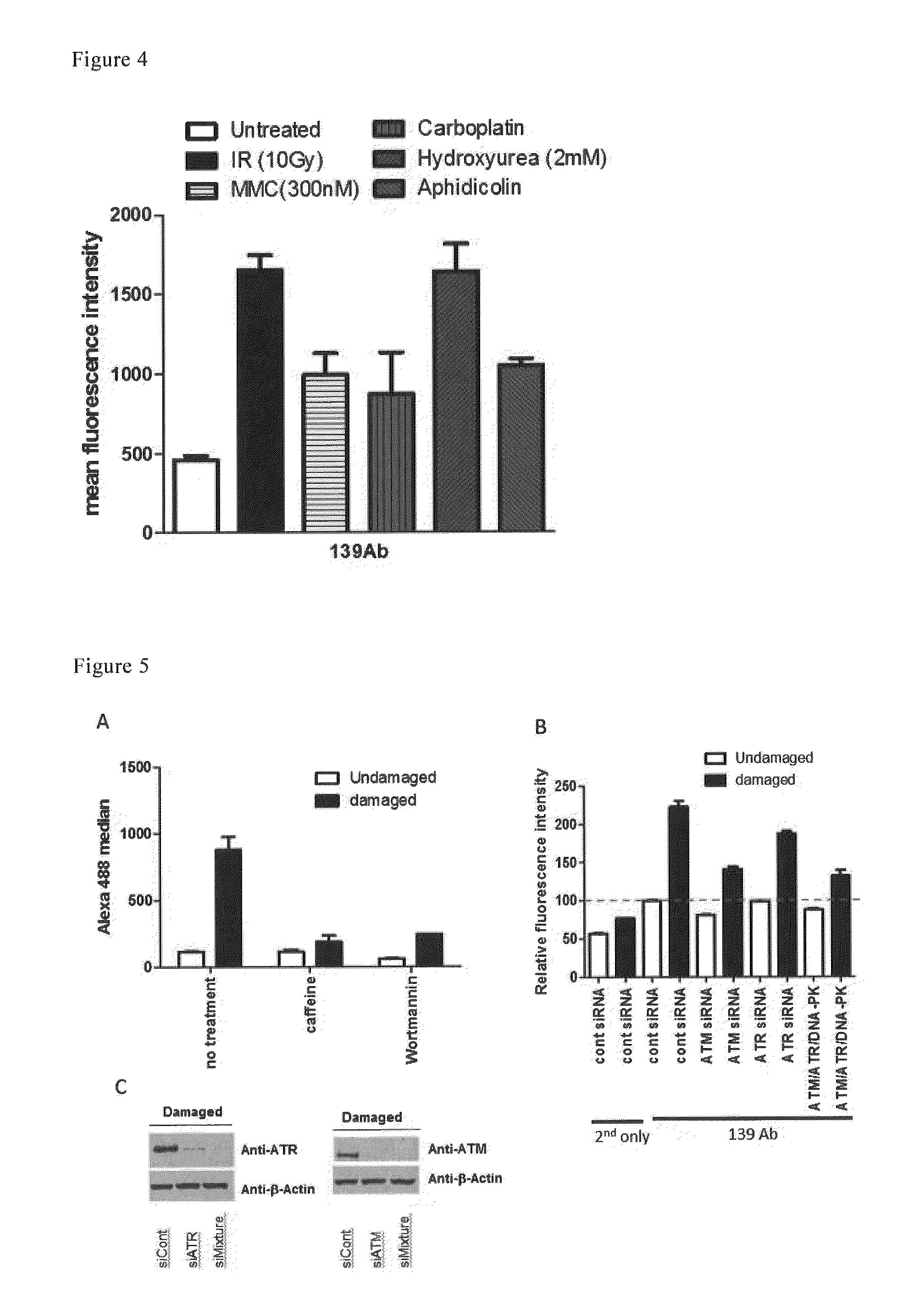
The present invention provides an antibody that specifically recognizes cells that have undergone radiation and resulted in DNA double-strand breaks. By administering this antibody to cells and analyzing their expression, DNA damage can be detected. The antibody can also be used as a reagent to detect changes in living cells and as a diagnostic and therapeutic agent for cancer patients undergoing radiation. Additionally, the invention provides a human antibody that specifically targets a cancer-related protein called Ly6D and inhibits its survival or growth. This antibody is less immunogenic and can be used as an anti-cancer agent.

Problems solved by technology

It is most difficult for organisms such as cancer cells to repair the damage.
However, it was difficult to select marker molecules that can be actually applied in practice due to a disadvantage of mismatch of the mRNA expression level and the protein expression level seen in many aspects (Non-Patent Document 5).
Conventional therapies such as chemotherapy and radiation are problematic because they can kill cancer cells, but at the same time, they cause damage to normal cells, resulting in induction of strong adverse reactions.
However, since target cancer antigens are membrane proteins, it has been difficult to obtain antibodies even against known cancer antigens.

Method used


Image

  • Method for detecting cellular DNA damage and antibody against  cell surface antigen responsive to DNA strand breaks
  • Method for detecting cellular DNA damage and antibody against  cell surface antigen responsive to DNA strand breaks
  • Method for detecting cellular DNA damage and antibody against  cell surface antigen responsive to DNA strand breaks

Examples


examples

Experimental Materials and Methods

(1) Cells, Antibodies, and an Antibody Library

[0131]The MCF10A cell line was provided by Dr. T. Ohta, St. Marianna University, Japan, and the FaDu and A431 cell lines were purchased from ATCC. Mouse anti-human LY6DAb and Mouse anti-human GML were purchased from Abnova Corp. The AIMS5 antibody library has been previously reported (Morino et al., 2001 J. Immunol. Meth. 257: 175-184).

(2) Cell Culture

[0132]MCF10A cells were cultured in Dulbecco's modified Eagle's medium (DMEM) / F12 supplemented with 2.5% FBS, 1% penicillin-streptomycin, 0.1 μg / ml cholera toxin, 0.5 μg / ml hydrocortisone, 20 ng / ml epidermal growth factor (EGF), and 10 μg / ml serum insulin. A431 cells were grown in DMEM supplemented with 10% FBS and 1% penicillin-streptomycin. FaDu cells were cultured in Eagle's minimum essential medium (EMEM) supplemented with 10% FBS and 1% streptomycin-penicillin.

(3) X-Ray Irradiation and Drugs

[0133]After the growth to confluence, the cells were irradiate...


PUM

PropertyMeasurementUnit
Surfaceaaaaaaaaaa
Cytotoxicityaaaaaaaaaa

Abstract

To provide a method for detecting cellular DNA damage by specifically recognizing molecules responsive to DNA strand breaks. A method for detecting DNA damage in cells, which comprises administering in vivo, to cells, an antibody against a cell membrane surface antigen that is expressed in cells undergoing DNA strand breaks to a greater extent than in cells not undergoing DNA strand breaks in an environment that causes DNA damage, and analyzing the expression status of the cell membrane surface antigen.

Description

TECHNICAL FIELD[0001]The present invention relates to a method for detecting cellular DNA damage, an antibody against a cell surface antigen responsive to DNA strand breaks, and a reagent for detecting DNA damage and a pharmaceutical composition comprising the antibody.BACKGROUND ART[0002]It is known that DNA damage causes DNA strand breaks, although it depends on the situation. The term “DNA strand breaks” refers to a state in which hydroxyl and phosphate groups that constitute DNA have dissociated. DNA double-strand breaks are caused by external factors such as radiation and internal factors such as DNA digestion. DNA damage is known to cause various reactions, including cell cycle arrest, DNA repair, and apoptosis (Non-Patent Document 1). All of these reactions are induced by ATR and ATM kinases belonging to the PIKK family that is important for controlling the DNA damage checkpoint mechanism and DNA repair (Non-Patent Document 2). Since DNA damage leads to destruction of well-or...

Claims


Application Information

Patent Timeline
IPC IPC(8): C07K16/30G01N33/68
CPCC07K16/30G01N33/6893A61P35/00A61P35/02C07K16/28C07K2317/622
Inventor KUROSAWA, YOSHIKAZUKUROSAWA, MAIKOKUROSAWA, GENE
Owner FUJITA HEALTH UNIVERSITY
Learn More