

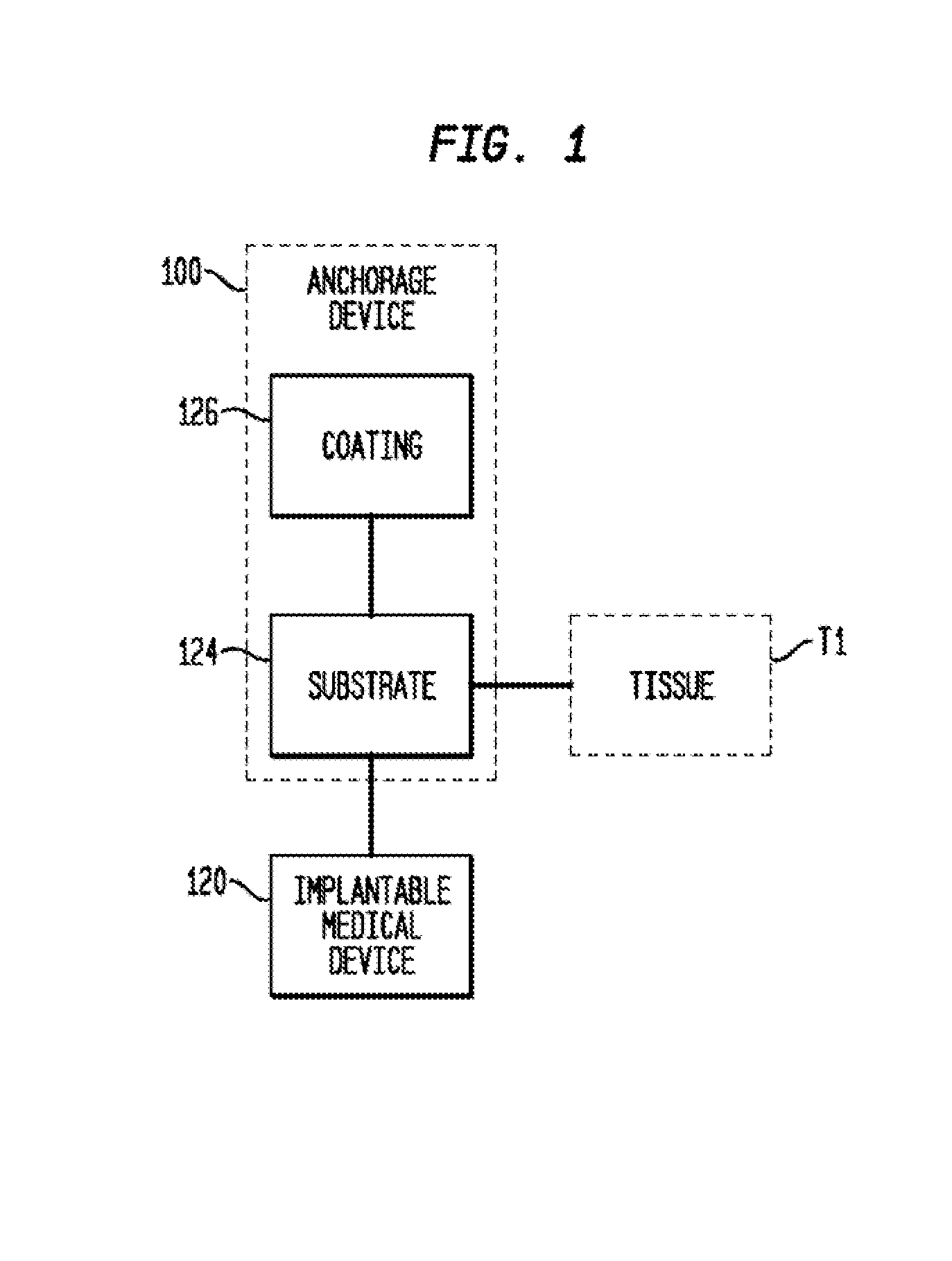
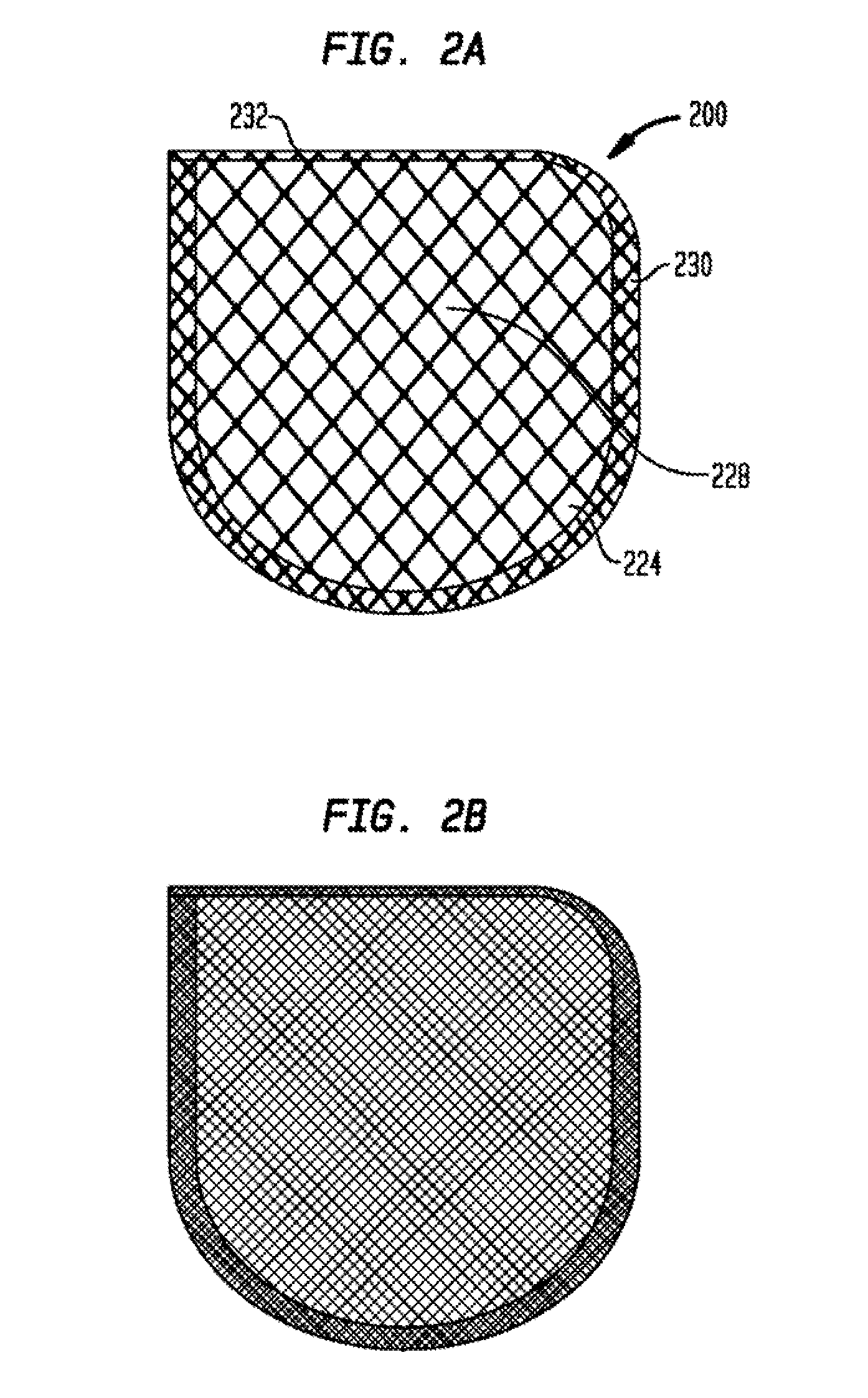
Anchorage Devices Comprising an Active Pharmaceutical Ingredient
an anchorage device and pharmaceutical technology, applied in the field of anchorage devices comprising active pharmaceutical ingredients, can solve the problems of difficult removal or explantation of the anchorage device, undesirable material portion, and difficult removal of the anchoring device without damaging the surrounding, so as to reduce the incidence of microbial infections, prevent tissue contact, and reduce the surface area
- Summary
- Abstract
- Description
- Claims
- Application Information
AI Technical Summary
Benefits of technology
Problems solved by technology
Method used
Image

Examples
example 1
[0127]FIG. 9, and Table 3 below, shows the cumulative release of rifampin and minocycline from three formulations (Formulation A, Formulation B, and Formulation C). The mesh substrate used here is a fully resorbable terpolymer of glycolic acid 6-hydroxycaproic acid and 1-3 propanediol. The total weight of the drug would range from about 5 mg to about 50 mg for Rifampin and about 5 mg to about 20 mg for Minocycline HCl. This is to keep, it is believed, the maximum drug that can be released in 1 day to a maximum of 1 / 10 of the oral daily dose. This low dose is sufficient for the product to be efficacious, since the drug is delivered locally at the site of action. This results in high tissue concentrations, which are above the minimum inhibitory concentrations of common pathogens. In some embodiments, the amount of each of rifampin and minocyclin in the devices of FIG. 9 comprise from about 0.85 to about 1.20 micrograms / cm2.
TABLE 3TimeCumulative Release (%)(h)FORMULATION AFORMULATION B...
example 2
[0128]Standard in vitro studies were conducted to demonstrate effectiveness against several pathogenic organisms.
Minimum Inhibitory Concentrations
[0129]Establishing the MIC of antimicrobials is a necessary step in the process of establishing effective use concentrations. Approved standards for this activity with antibiotics are published by NCCLS. These standards are primarily intended for use in clinical settings with patient isolates. However, they represent a consensus methodology for best practice in determining MICS that are reproducible and defensible. The principles upon which they are based provide a sound framework for determining the MIC for the test.
Materials:
[0130]The broth dilution method. was used to measure quantitatively the in vitro activity of an antimicrobial agent against a given microbial isolate. To perform the test, a series of tubes were prepared with a broth to which various concentrations of the antimicrobial agent were added. The tubes were then. inoculate...
example 4
Tissue Concentration
[0151]In some embodiments, the antibiotic is minocycline and a tissue concentration of the minocycline is between about 0.65 μg / mL and 0.8 μg / mL after about 2 hours; where the tissue concentration of the minocycline is between about 2.55 μg / mL and about 2.75 μg / mL after about 6 hours; and where the tissue concentration of the minocycline is between about 1.2 μg / mL and about 1.9 μg / mL after about 24 hours. In other embodiments, the antibiotic is rifampin and a tissue concentration of the rifampin is between about 0.6 μg / mL and 1.4 μg / mL after about 2 hours; where the tissue concentration of the rifampin is between about 1.9 μg / mL and about 2.3 μg / mL after about 6 hours; and where the tissue concentration of the rifampin is between about 2.6 μg / mL and about 4.2 μg / mL after about 24 hours. (See, e.g., Table 7).
PUM
| Property | Measurement | Unit |
|---|---|---|
| concentration | aaaaa | aaaaa |
| concentration | aaaaa | aaaaa |
| concentration | aaaaa | aaaaa |
Abstract
Description
Claims
Application Information