Large oncosomes in human tumors and in circulation in patients with cancer

Inactive Publication Date: 2014-02-27
CEDARS SINAI MEDICAL CENT
View PDF2 Cites 12 Cited by
  • Summary
  • Abstract
  • Description
  • Claims
  • Application Information

AI Technical Summary

Benefits of technology

[0098]The “oncosomes” described in Di Vizio et al. (Cancer Res 2009 Vol 69(13) pages 5601-5609), are a mixture exosomes and large oncosomes. Specifically, as shown in FIGS. 10 and 11(a), the oncosome purification method described in DiVizio et al. results in a mixture of exosomes (FIG. 11(a) middle panel black bar) and large oncosomes (FIG. 11(a), middle panel white bars), compared to the filtration method described herein (FIG. 11(a) right panel) which yields an isolated population of large oncosomes.
[0099]As described herein, presence of large oncosomes in a subject is indicative of increased likelihood of cancer metastasis. The isolation methods described herein, such as (i) the filtration method described in FIG. 11(a), optionally followed by fluorescence-activated cell sorting (FACS) with size beads of 1 μm to 10 μm, and (ii) differential centrifugation followed by FACS with size beads of 1 μm to 10 μm, rapidly and selectively enrich for large oncosomes. The purified large oncosomes are selected away from other microvesicles, including exosomes to yield essentially purified large oncosomes (FIG. 1 and (FIG. 11(a) right panel)). Since presence and/or an increase in the number of large oncosomes are ind

Problems solved by technology

Existing imaging techniques have their limitat

Method used


Image

  • Large oncosomes in human tumors and in circulation in patients with cancer
  • Large oncosomes in human tumors and in circulation in patients with cancer
  • Large oncosomes in human tumors and in circulation in patients with cancer

Examples


example 1

Methods of the Invention

Cell Culture

[0101]LNCaP, DU145, PC3 and WPMY-1 were from the American Type Culture Collection (ATCC) (Di Vizio D et al., Oncosome formation in prostate cancer: association with a region of frequent chromosomal deletion in metastatic disease, Cancer Res 2009, 69:5601-5609; Adam R M et al., Cholesterol sensitivity of endogenous and myristoylated Akt, Cancer Res 2007, 67:6238-6246; Di Vizio D et al., Caveolin-1 interacts with a lipid raft-associated population of fatty acid synthase, Cell Cycle 2008, 7:2257-2267). LNCaP / LacZ and LNCaP / MyrAkt1 lines were described (Adam R M et al., Cholesterol sensitivity of endogenous and myristoylated Akt, Cancer Res 2007, 67:6238-6246). For the generation of LNCaP / Vo and LNCaP / Cav-1, parental LNCaP were plated at 70-80% confluence and transfected using Lipofectamine™ 2000 Tranfection Reagent (Invitrogen). Stable populations were isolated following selection with Gentamicin G418 0.5 mg / ml. LNCaP were cultured in RPMI-1640 mediu...

example 2

Filtration-Based Isolation Protocol

[0117]Serum-free conditioned media or human platelet-poor plasma are centrifuged at low speed (2,800 g / 10 min) to eliminate cells and cell-debris. In comparison with the ultracentrifugation based protocol, in which extracellular vesicles (EV) are recovered from the supernatant after centrifugation at 10,000 g / 30 min or 100,000 g / 60 min, in our new protocol supernatant is filtered at 8,000 g / 30 sec using Vivaspin ultrafiltration spin columns. Large oncosomes are recovered form the top of the filter membrane (upper chamber), whereas small EV, are eluted (lower chamber) (FIGS. 10 and 11).

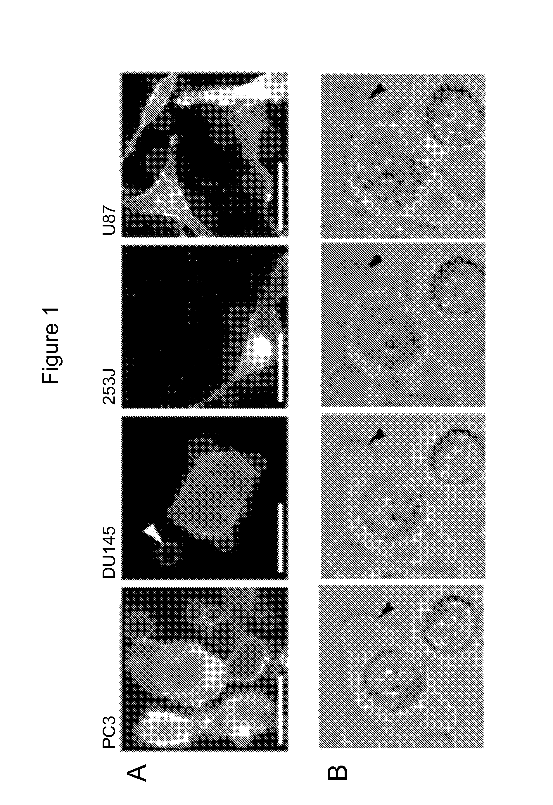
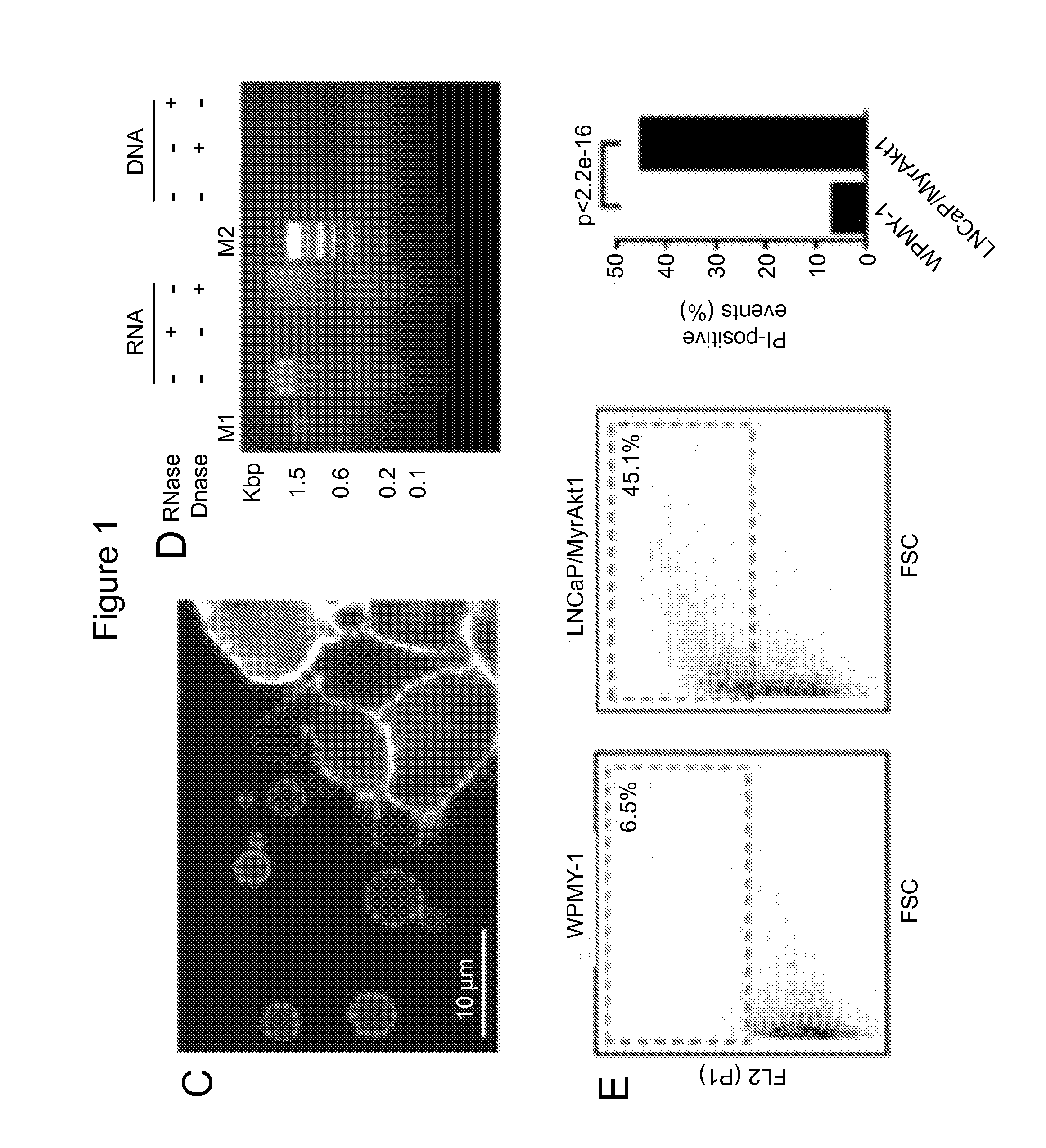
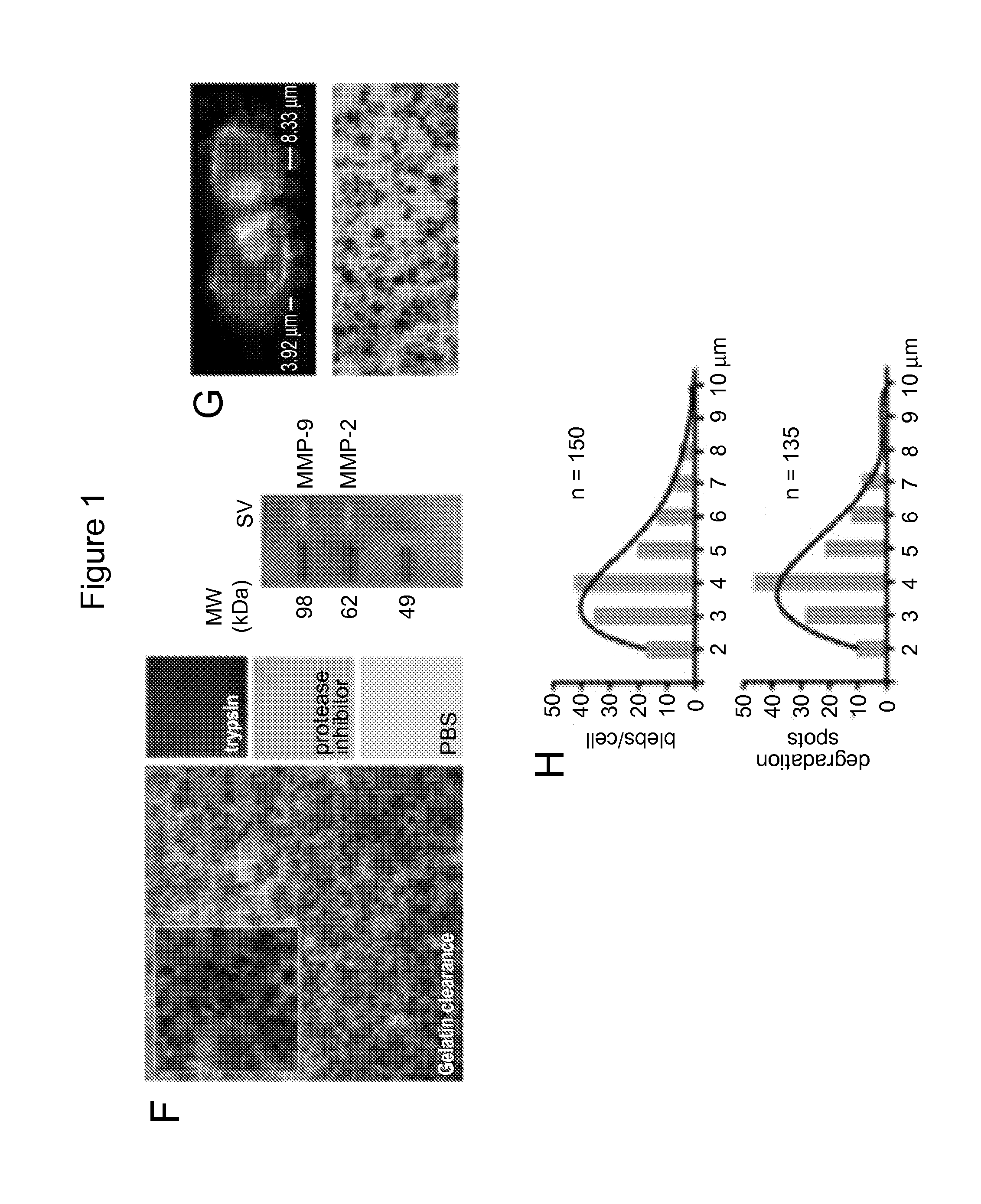
[0118]Tumor cells that exhibit the amoeboid feature of membrane blebbing can release large, plasma membrane-derived vesicles into the extracellular space (Di Vizio D et al., Oncosome formation in prostate cancer: association with a region of frequent chromosomal deletion in metastatic disease, Cancer Res 2009, 69:5601-5609) (FIGS. 1A, 1B, 2A). To determine whether the...

example 3

Large Oncosomes in the Circulation and In Situ

[0122]Microvesicles range from about 20 nm to about 10 μm. Exosomes (a type of microvesicles) range from about 20 nm to about 100 nm. Large oncosomes (another type of microvesicles) range in size from about 1 μm to about 10 μm. To distinguish large oncosomes from smaller TMV unequivocally, ultracentrifugation and immuno-flow cytometry were used in combination with sizing beads to create a calibrated domain of analysis based on size. Following biochemical purification, shed vesicles were stained with DAPI to verify the absence of cellular contamination and subsequently labeled with an HA antibody, for detection of membrane-localized MyrAkt1. MyrAkt1-negative and -positive populations were then distinguished on a cell sorter, with gates set using 1 and 10 μm beads (FIG. 3A). Because aggregates may potentially be a significant component of the vesicle preparation, we excluded them from the analysis. To do this, forward scatter signal (FSS) ...


PUM

PropertyMeasurementUnit
Pore sizeaaaaaaaaaa
Lengthaaaaaaaaaa
Lengthaaaaaaaaaa

Abstract

The invention provides methods for isolating large oncosomes and determining cancer metastasis based on the presence of large oncosomes in a subject in need thereof.

Description

GOVERNMENT RIGHTS[0001]The invention was made with government support under Grant Nos. CA131472, CA131471 and CA143777 awarded by the National Institutes of Health. The government has certain rights to the invention.CROSS-REFERENCE TO RELATED APPLICATIONS[0002]This application claims priority from U.S. Provisional Patent Application No. 61 / 692,591, filed on Aug. 23, 2012, which is incorporated herein by reference in its entiretyFIELD OF INVENTION[0003]The invention provides methods for isolating large oncosomes, methods for identifying large oncosomes, methods for determining the likelihood of cancer metastasis in a subject in need thereof by detecting large oncosomes and / or its molecular contents (proteins, nucleic acids and / or lipids) and methods for treating cancer in a subject in need thereof.BACKGROUND[0004]All publications herein are incorporated by reference to the same extent as if each individual publication or patent application was specifically and individually indicated ...

Claims


Application Information

Patent Timeline
IPC IPC(8): G01N1/34
CPCG01N1/34G01N33/5076G01N33/57488
Inventor DI VIZIO, DOLORESFREEMAN, MICHAEL R.MORELLO, MATTEO
Owner CEDARS SINAI MEDICAL CENT
Why Eureka
Try Eureka