
High numerical aperture telemicroscopy apparatus

a telemicroscopy and numerical aperture technology, applied in the field oftelemicroscopy, can solve the problems of low numerical aperture optics, insufficient magnification and resolution for most microscopy applications, and inability to meet the needs of developing world countries, and achieve high resolution, and high numerical aperture optics

Active Publication Date: 2014-05-01
RGT UNIV OF CALIFORNIA
View PDF0 Cites 23 Cited by
  • Summary
  • Abstract
  • Description
  • Claims
  • Application Information

AI Technical Summary

Benefits of technology

This patent is about a device that combines high-resolution optics with a cell phone or other communication device to create a microscope. This would allow for quick disease diagnosis or other microscopy applications. The device has a high numerical aperture, which means it can capture high-quality images. It also includes a system for reducing image noise in biological assays. Overall, this invention would make it easy to use high-quality microscopy in a portable and accessible way.

Problems solved by technology

The only device coupled directly to a cellular phone provides low numerical aperture optics and inadequate magnification and resolution for most microscopy applications.
Much of this capability is unavailable in the developing world, for reasons including: cost; maintenance difficulty for delicate instruments; lack of supply chain for frequent replacement parts (arc lamps have lifetimes of only ˜200 hrs); lack of electricity to power lamps, etc.
; lack of associated computer and / or internet resources; lack of portability to remote villages; etc.
Even in the developed world, expense, complexity, and portability issues prevent widespread use of microscopy for medical purposes in non-hospital settings such as at-home monitoring of patient blood counts during chemotherapy.
As a result, sick, immunocompromised patients must risk infection to travel to central medical facilities for testing.
However, developing countries often lack access to both clinical-quality microscopes to gather patient data and qualified medical personnel to provide diagnoses and treatment.
Even in the U.S. and other developed countries, it is difficult to use microscopy for medical purposes in a non-hospital setting such as in the home.
Camera-equipped cell phones (“camera phones”) have optical systems that, due to the requirement of imaging large objects and fields of view (e.g. faces, people, or landscapes) onto small (˜5 mm on a side) sensors arrays (e.g. CCD or CMOS imaging array sensors), using a small (˜5 mm diameter, ˜5 mm focal length lens systems) must: have low object-side numerical aperture (NA), typically ≦0.01; non-telecentric design, with limitations known to those with ordinary skill in the art and including non-uniform collection efficiency across the field of view, and changes in the apparent magnification of an image with defocus; optical magnifications (M) (defined as from the object to the image on the sensor array) of <1, typically <0.03.
Thus, there is not presently available a telemicroscopy apparatus based on a cell phone or similar device designed to be used for telemedicine applications.

Method used


Image

  • High numerical aperture telemicroscopy apparatus
  • High numerical aperture telemicroscopy apparatus
  • High numerical aperture telemicroscopy apparatus

Examples


Embodiment Construction

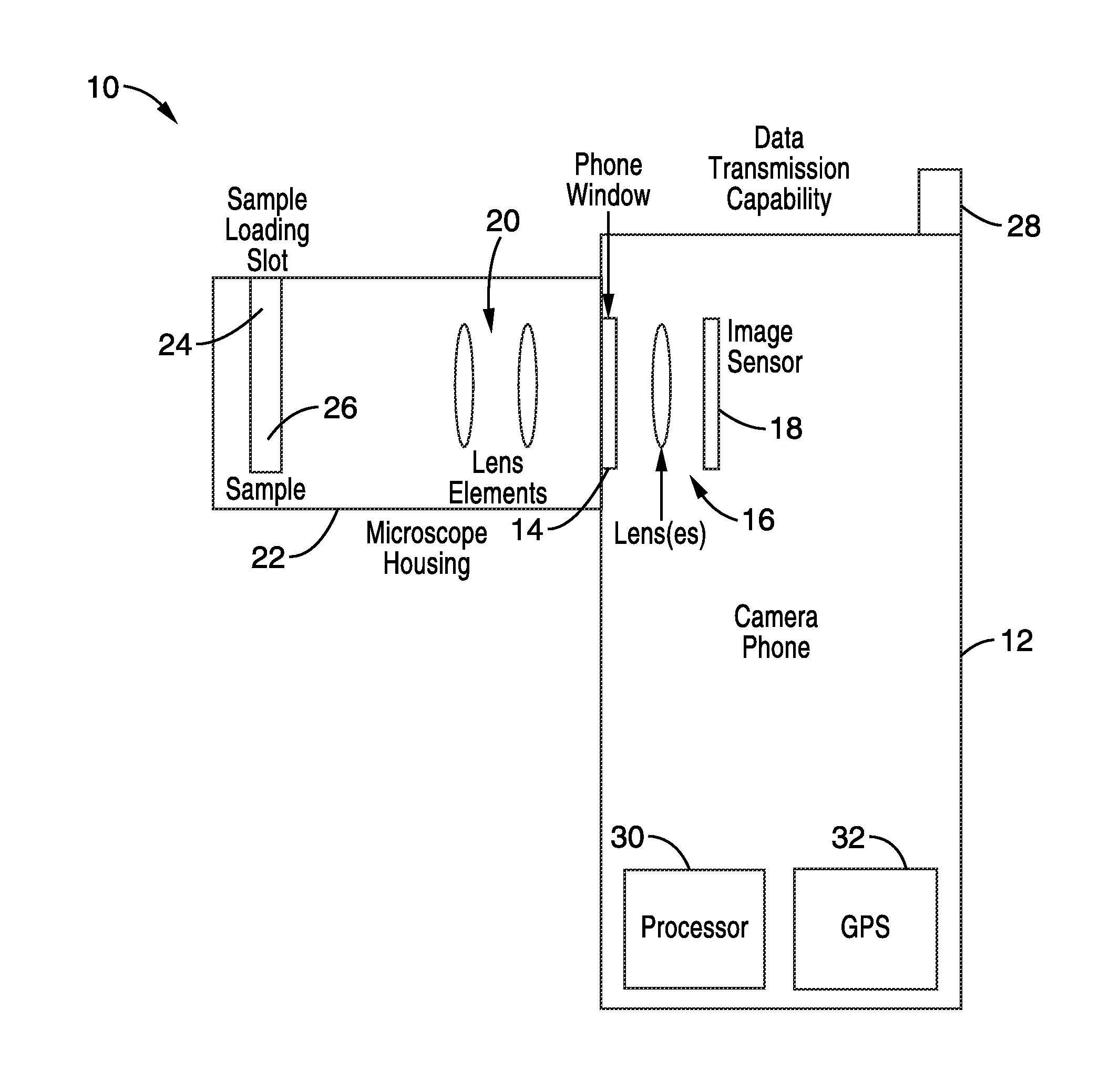
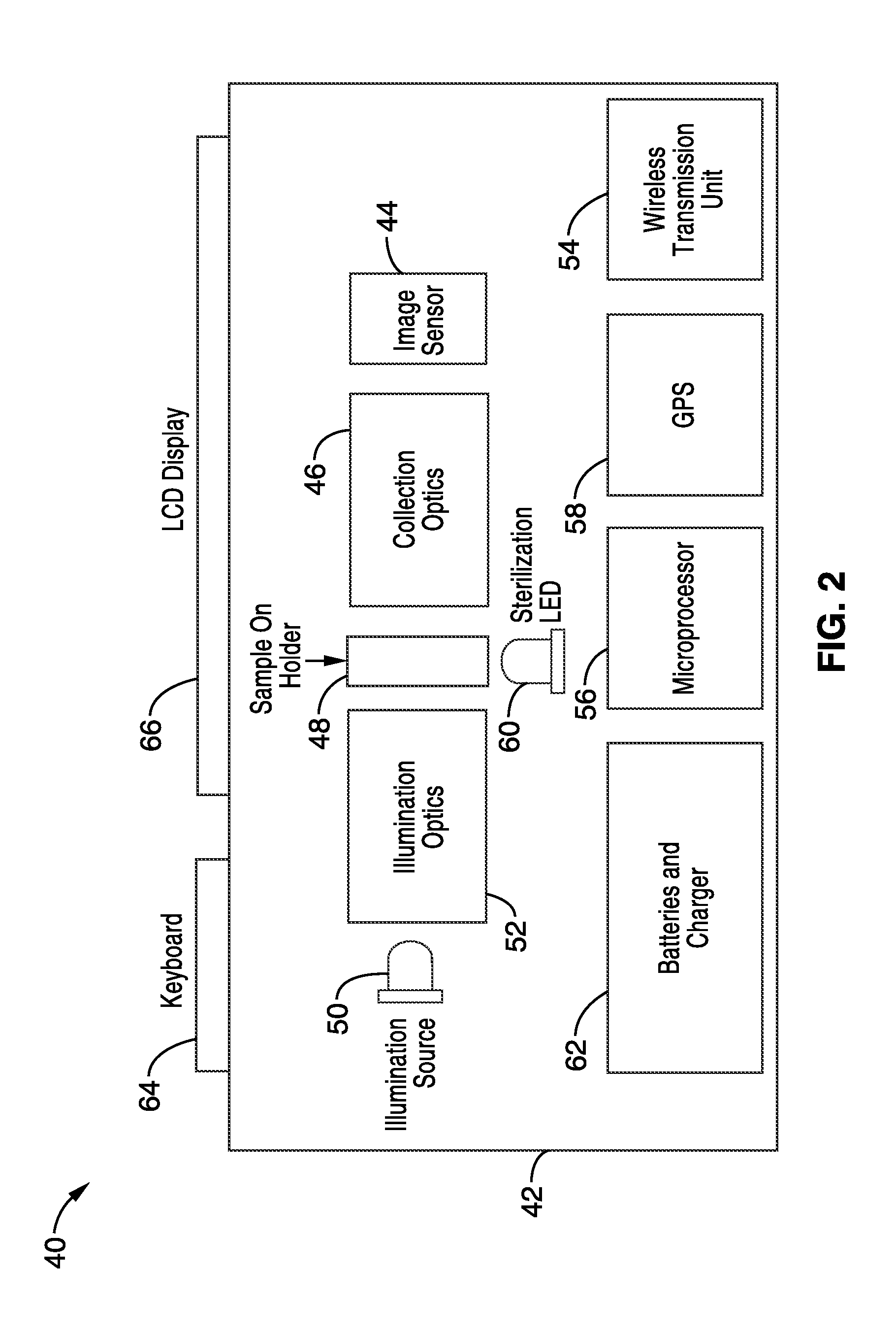
[0086]The present invention is a device which combines many of the benefits of the medical instruments with portability, low power consumption, cellular data transfer capability, and lower cost. In the simplest implementation, additional optics are attached to a standard camera-phone (cell phone with built-in camera). Such a system can also have software that assists in image acquisition, image processing (e.g. counting of the bacilli in a tuberculosis-positive sputum smear), appending patient information to the image or data derived from the image, and uploading of the information to a server or other computer system (possibly including another phone) for diagnosis, medical consultation, general record keeping (e.g. for reimbursement purposes), epidemic tracking or general epidemiology, etc. Furthermore, information can be downloaded to the phone in use, e.g. to provide patient record data for patients to be seen the next day, synchronize records, provide updated results from other...


PUM


Abstract

An imaging system consisting of a cell-phone with camera as the detection part of an optical train which includes other components. Optionally, an illumination system to create controlled contrast in the sample. Uses include but are not limited to disease diagnosis, symptom analysis, and post-procedure monitoring, and other applications to humans, animals, and plants.

Description

CROSS-REFERENCE TO RELATED APPLICATIONS[0001]This application is a continuation of U.S. patent application Ser. No. 12 / 826,375 filed on Jun. 29, 2010, incorporated herein by reference in its entirety, which is a 35 U.S.C. §111(a) continuation of, PCT international application number PCT / US2008 / 088646 filed on Dec. 31, 2008, incorporated herein by reference in its entirety, which claims priority from U.S. provisional application Ser. No. 61 / 018,537 filed on Jan. 2, 2008, incorporated herein by reference in its entirety.[0002]This application is also related to PCT International Publication No. WO 2009 / 088930 published on Jul. 16, 2009, incorporated herein by reference in its entirety.STATEMENT REGARDING FEDERALLY SPONSORED RESEARCH OR DEVELOPMENT[0003]Not ApplicableINCORPORATION-BY-REFERENCE OF MATERIAL SUBMITTED IN A COMPUTER PROGRAM APPENDIX[0004]Not ApplicableNOTICE OF MATERIAL SUBJECT TO COPYRIGHT PROTECTION[0005]A portion of the material in this patent document is subject to cop...

Claims


Application Information

Patent Timeline
Patent Type & Authority Applications(United States)
IPC IPC(8): H04M1/02G02B21/36G02B21/00
CPCH04M1/0264G02B21/0008G02B21/361G02B21/0092G02B21/16G02B21/36G01N21/8483G02B21/365G02B21/02H04N5/44G02B21/025G02B21/06G02B21/26
Inventor FLETCHER, DANIELHANSEN, WENDYSWITZ, NEILBRESLAUER, DAVID N.DOUGLAS, ERIKMAAMARI, ROBILAM, WILBURDILL, JESSE
Owner RGT UNIV OF CALIFORNIA
Learn More