

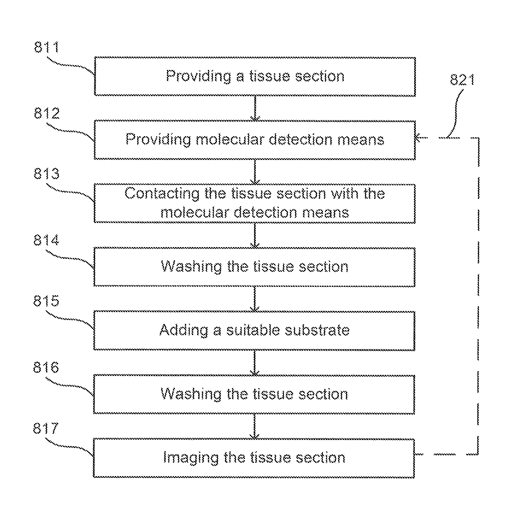
Method for providing images of a tissue section
a tissue section and image technology, applied in the field of immunohistochemistry and computer-based analysis of images, can solve the problems of not providing detailed information about the spatial relationship (physical relationship) between the analysed cell types, and 2 cell types can be simultaneously detected
- Summary
- Abstract
- Description
- Claims
- Application Information
AI Technical Summary
Benefits of technology
Problems solved by technology
Method used
Image

Examples
example 1
Tissue Handling, Sample Preparation, and Generation of Sections
[0205]The following human tissues were included:[0206]Human distal colon: surgical resection due to chronic inflammation and suspected non-specific colitis.[0207]Human lung tissue from patients with Chronic Obstructive Pulmonary Disease, COPD and Cystic Fibrosis: lung resection due to suspected lung cancer, the analyzed tissue was not affected by cancer and obtained as far away from the tumour as possible.[0208]Human lymph nodes: Large draining lymph nodes collected in association with lung transplantation due to severe COPD or cystic fibrosis.[0209]Human tonsils: collected as part of routine tonsillectomy due to repeated episodes of tonsillitis.
[0210]Samples (i.e. blocks of tissue) from all tissue types were immersed subjected to routine fixation by immersion into routine fixative (4% buffered formaldehyde, pH 7.6). After fixation overnight, the samples were dehydrated in a series of solutions with increasing concentrat...
example 2
Multiple Immunohistochemical Staining and Generation of Serial Digital Images
[0211]Immunohistochemical staining was performed using an automated immunohistochemistry robot (Autostainer CL-classic; Dako Cytomation, Glostrup, Denmark) with the DAKO REAL EnVision detection system), a sensitive standard method intended for detection of primary mouse or rabbit antibodies (IHC kit Code K5007, Dako Cytomation, Denmark; for details see www.dako.com). The primary antibodies used to detect the cell-specific antigens (earlier referred to as “cell markers”) are listed in Table 2 and applied onto the sections at the dilution recommended by the commercial producers for immunohistochemical staining of human tissues prepared for routine pathological examination (i.e. sections from formalin-fixed and paraffin-embedded samples). Examples of series of markers that were used in the evaluation of ESMS are listed in Table 3.
TABLE 2Examples of Antibodies Used for ExperimentalValidation of the ESMS Techniq...
example 3
Computerized Image Analysis and Decoding of Marker-Specific Staining Patterns
[0228]Digitalized sections from Aperio's slide scanner, corresponding to a series of primary images according to the disclosed invention, were inspected manually using a viewing software (Aperio ImageScope, version 10.0.35.1798, Aperio Technologies Inc). In the initial evaluation regions of interest in each section were selected for further detailed analysis. Using the extract and export image function in the ImageScope software, raw images, i.e. primary digital images, were exported as TIFF or JPEG files; one image for each staining cycle. Together, the images formed one series of images per region of interest.
[0229]For some images the distribution pattern of the brown DAB precipitation was outlined already before the image export by the colour segmentation features (“positive pixel” algorithm) included in the ImageScope software after selecting RGB and Hue values characteristic of the brown DAB precipitat...
PUM
 Abstract
Description
Claims
Application Information