

Fluorescent label for biological substance detection method
a fluorescent label and biological substance technology, applied in the field of biological substance detection methods, can solve the problems of affecting the effect of the staining concentration, the difficulty of simultaneous observation, and the inability to estimate the actual amount of an antibody or the like from the staining concentration
- Summary
- Abstract
- Description
- Claims
- Application Information
AI Technical Summary
Benefits of technology
Problems solved by technology
Method used
Image

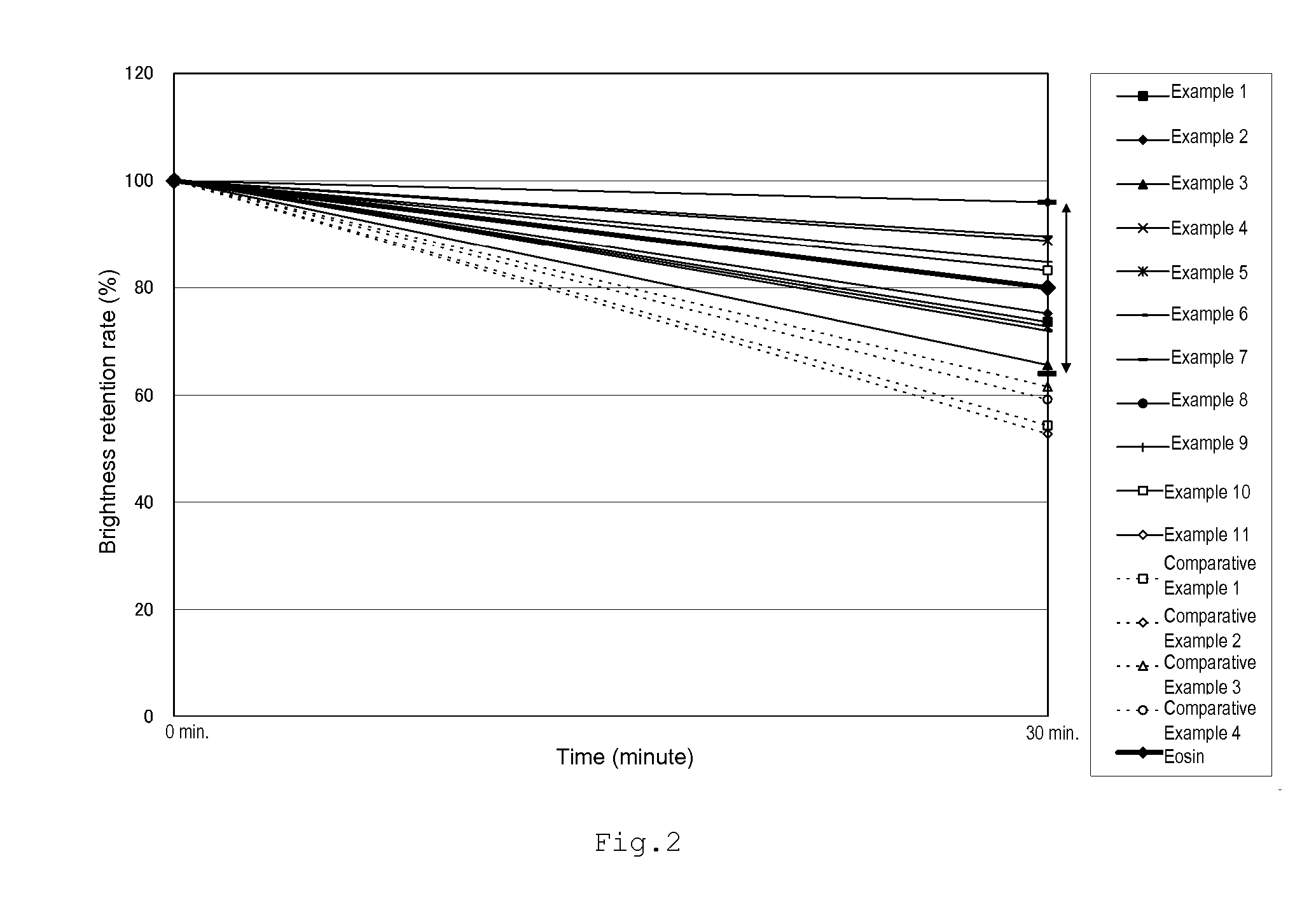
Examples
examples
[0076]The present invention will now be described in detail by way of examples thereof; however, the present invention is not restricted to the following examples.
Sample 1-1
Texas Red-Containing Silica Particle
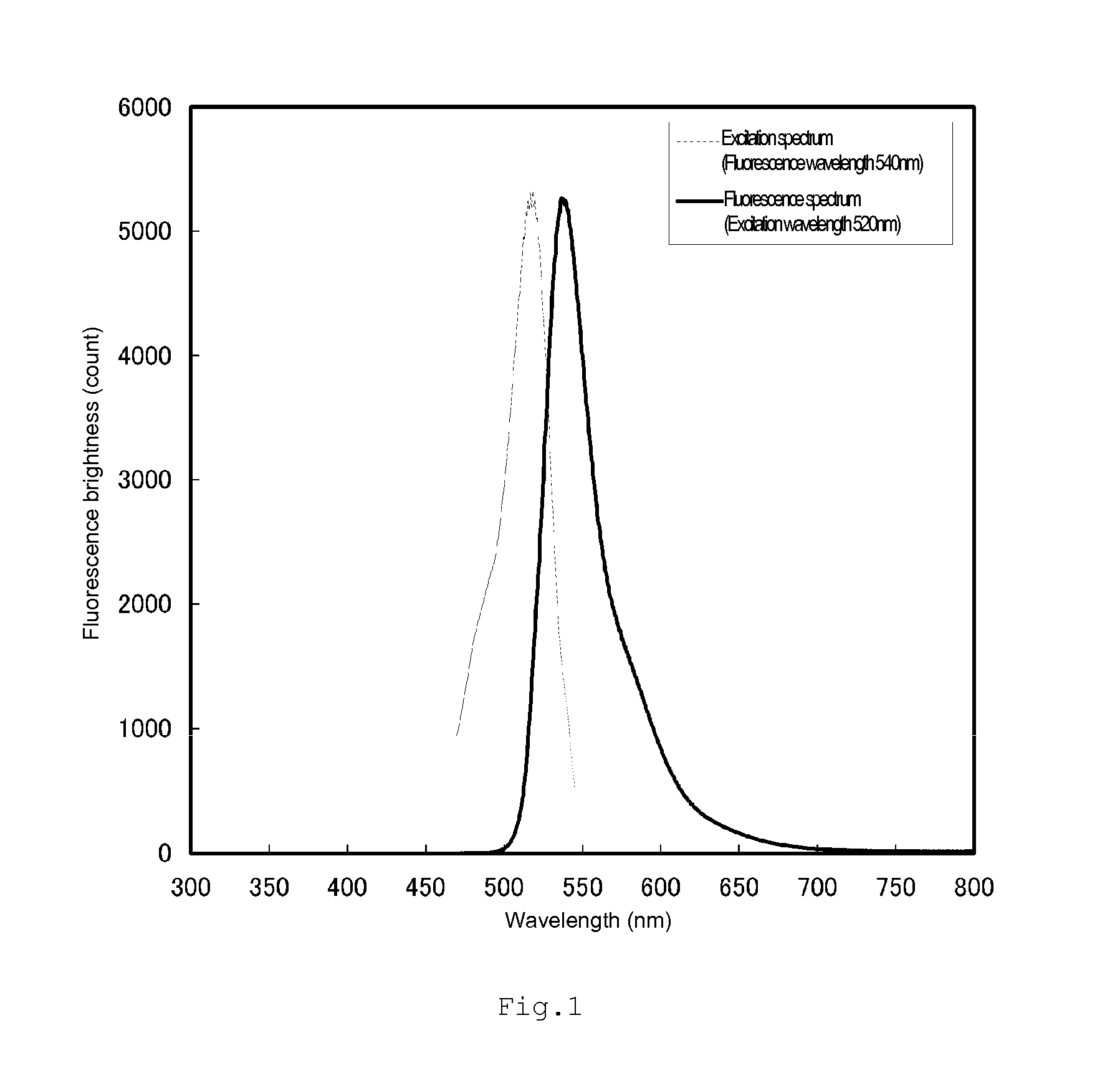
[0077]In DMF, 3.4 mg of a fluorescent dye, Sulforhodamine 101 acid chloride (manufactured by Dojinsha Co., Ltd., Texas Red dye), and 3 μL of 3-aminopropyltrimethoxysilane (manufactured by Shin-Etsu Chemical Co., Ltd., KBM903) were mixed to obtain an organoalkoxysilane compound. Then, 0.6 mL of the thus obtained organoalkoxysilane compound was mixed for 3 hours with 48 mL of ethanol, 0.6 mL of TEOS (tetraethoxysilane), 2 mL of water and 2 mL of 28% aqueous ammonia. The mixture produced in the above-described step was centrifuged at 10,000 G for 20 minutes and the resulting supernatant was removed. Then, ethanol was added to disperse the precipitates and the resultant was centrifuged again. The precipitates were washed twice with each of ethanol and pure water b...
PUM
| Property | Measurement | Unit |
|---|---|---|
| wavelength range | aaaaa | aaaaa |
| emission wavelength | aaaaa | aaaaa |
| particle diameter | aaaaa | aaaaa |
Abstract
Description
Claims
Application Information