Eureka AIR delivers breakthrough ideas for toughest innovation challenges, trusted by R&D personnel around the world.

Equine Amniotic Membrane-Derived Mesenchymal Stem Cells

Active Publication Date: 2014-11-13
KANGSTEM BIOTECH
View PDF1 Cites 2 Cited by
  • Summary
  • Abstract
  • Description
  • Claims
  • Application Information

AI Technical Summary

Benefits of technology

The present invention is about using the equine amniotic membrane as a source of mesenchymal stem cells. These stem cells have the ability to grow and differentiate, making them useful for treating injuries and diseases in horses. This invention provides an effective solution for veterinary medicine to treat bone, tendon, and muscle injuries and loss diseases in racehorses and other equine animals.

Problems solved by technology

Formerly, organ transplantation, gene therapy, etc., were proposed for the treatment of incurable human diseases, but their use has not been achieved efficiently due to immunorejection, a small supply of organs, and insufficient knowledge of genes.
These cells are also called embryonic stem cells and can differentiate into various other tissue cells but cannot form new living organisms.
However, stem cells in adult tissues, such as the bone marrow, are very rarely present, and such cells are difficult to culture without inducing differentiation, and thus difficult to culture in the absence of specifically screened media.
When such racehorses with a good pedigree are injured, the healing of the racehorses is problematic.
Considering the size of horses, it is a major technical issue to reliably supply a large number of cells.
Therefore, isolation and characterization of stem cells derived from various equine tissues have become important issues in the stem cell field.
However, studies on cell therapeutic agents for the treatment of bone, cartilage, tendon or muscle of equine animals, in particular racehorses, have not yet been sufficient, even though the need for the cell therapeutic agents is great.
However, equine animals do not have much adipose tissue, unlike other mammals, and thus it is very difficult to obtain adipose tissue from equine animals.
In addition, a method of obtaining adipose from individuals is invasive and causes pain.
Also, due to the nature of racehorses, there is a distinct limitation in the isolation of stem cells from adipose tissue or bone marrow.
Particularly, a system of sorting cells according to the expression level of the markers is expensive.
Due to such shortcomings, the use of the cell sorting method has been greatly limited.
Under culture conditions for such mixed cells, the distribution of nutrients cannot be uniform, thereby causing heterogeneity in differentiation of cells.
Conclusively, the problem that the cells cannot be produced as a homogeneous cell population serves as a fatal disadvantage, as when they are used as the therapeutic agent the actual effect may be different from the intended effect.

Method used


Image

  • Equine Amniotic Membrane-Derived Mesenchymal Stem Cells
  • Equine Amniotic Membrane-Derived Mesenchymal Stem Cells
  • Equine Amniotic Membrane-Derived Mesenchymal Stem Cells

Examples


example 1

Collection of Equine Amniotic Membrane

[0056]Amniotic membranes, which were normally disposed of after separation by Cesarean section delivery, were used (the College of Veterinary Medicine, Seoul National University). These membranes were for research purposes only and were provided without cost. The separated membranes were used only for the isolation and characterization of stem cells from the tissue.

[0057]All placental samples (n=4) were obtained from pure-bred female horses after delivery in Korean private horse farms. In order to reduce the contamination and damage of the tissue, all the samples were collected immediately after delivery using sterile surgical tools. The collected placental samples were stored at 4° C. and transferred to the laboratory as soon as possible in order to avoid possible contamination from a dusty environment. The amniotic membrane was physically separated from the chorion.

example 2

Isolation and Culture of Stem Cells

[0058]Cell isolation and culture were performed by a slight modification of the previously described method [S. Diaz-Prado et al., Tissue Eng. Part C Methods, 2010; C. M. Mihu et al., Rom. J. Morpho. Embryol, 50: 73-77, 2009]. All the placental samples were collected from equine animals through Cesarean section delivery by the method of Example 1. To separate the amniotic membrane from the whole placenta, the amniotic membrane was physically separated from the chorion. Under sterile conditions, the collected amniotic membrane was washed 3-4 times with physiological saline (0.9%). To remove epithelial cells, the collected amniotic membrane was treated with trypsin-EDTA (0.25%) at 37° C. for 30 minutes and washed 3-4 times with physiological saline. Then, the amniotic membrane from which epithelial cells were removed was cut into small pieces with a surgical knife and treated with collagenase type I (2 mg / ml; Worthington biochemical, Freehold, N.J.) ...

example 3

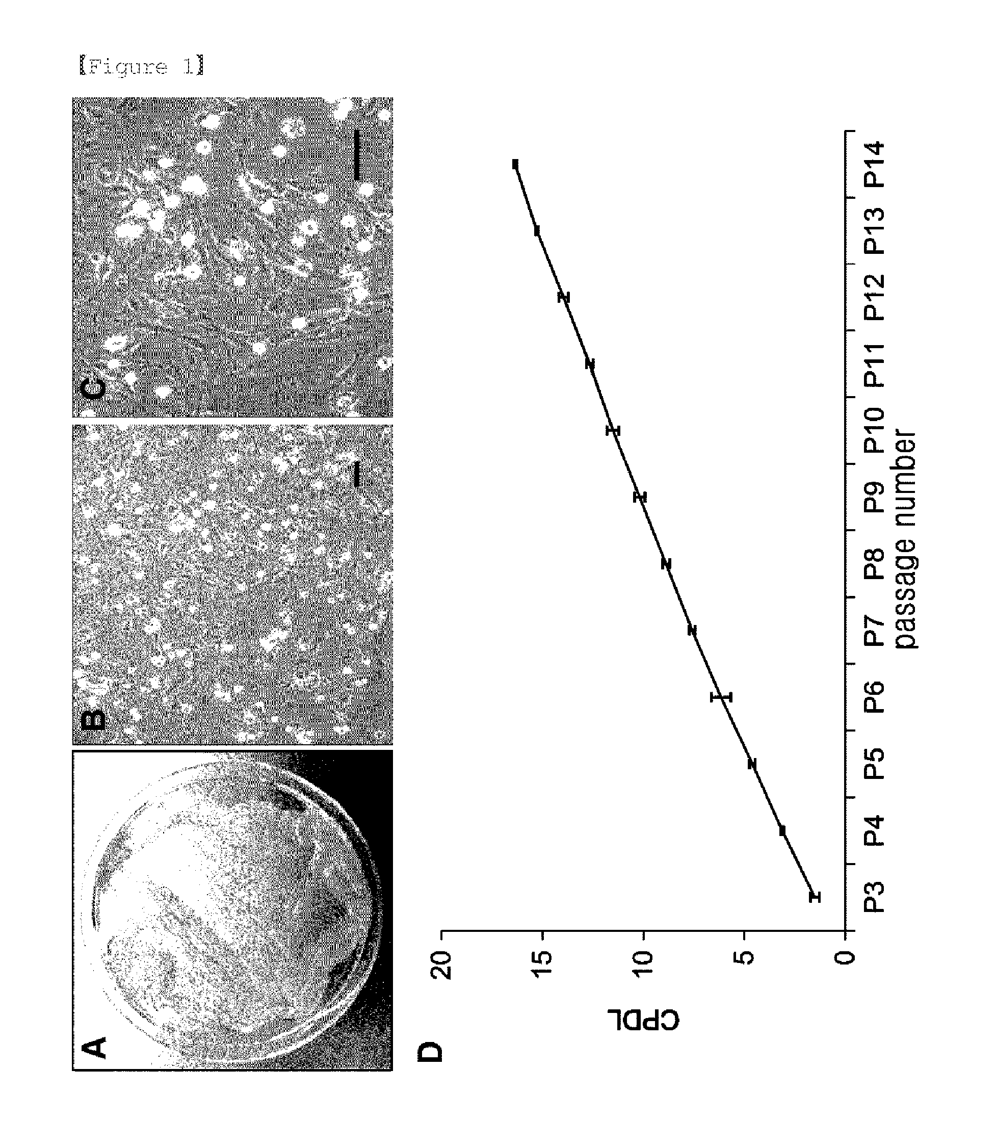
Cumulative Population Doubling Level Analysis

[0060]The analysis of cell proliferation was performed by a slight modification of the previously described method [S. B. Park et al., Cytotherapy, 13: 1431-43, 2011]. Stem cells, including multipotent stem cells, have self-renewal capacity which is associated with continuous and steady proliferation rate [Reya T. et al., Nature, 414 (6859): 105-11, 2001]. Therefore, the estimated growth efficiency and proliferation potential of the eAM-MSCs obtained in Example 2 were determined based on the total cumulative population doubling level using the formula CPDL=ln (Nf / Ni) ln 2, wherein Ni is the initial seeding cell number, Nf is the final harvesting cell number, and ln is the natural log. The cells (5×10⁴) were seeded into three 6-well culture plates, and after 5-7 days, subcultured. The number of final cells was counted, and 5×10⁴ cells were re-seeded. To determine the cumulative population doubling level, the population doubling level of ea...


PUM


Abstract

The present invention relates to equine amniotic membrane-derived mesenchymal stem cells (eAM-MSCs) and a preparation method thereof. More specifically, the present invention relates to equine amniotic membrane-derived mesenchymal stem cells, which show negative immunological responses to all of the human markers CD19, CD20, CD28, CD31, CD34, CD38, CD41a, CD62L, CD62P and CD200, and positive immunological responses to all of the human markers CD44, CD90 and CD105, and have the ability to be maintained in an undifferentiated state for 14 passages or more and the ability to differentiate into ectoderm, mesoderm and endoderm-derived cells.

Description

TECHNICAL FIELD [0001]The present invention relates to equine amniotic membrane-derived mesenchymal stem cells (eAM-MSCs) and a preparation method thereof. More specifically, the present invention relates to equine amniotic membrane-derived mesenchymal stem cells, which show negative immunological responses to all of the human markers CD19, CD20, CD28, CD31, CD34, CD38, CD41a, CD62L, CD62P and CD200, and positive immunological responses to all of the human markers CD44, CD90 and CD105, and have the ability to be maintained in an undifferentiated state for 14 passages or more and the ability to differentiate into ectoderm, mesoderm and endoderm-derived cells.BACKGROUND ART [0002]Biotechnology in the 21st century presents the possibility of new solutions to food, environment and health problems, with the ultimate object of promoting human prosperity. In recent years, the technology of using stem cells has been considered as a new way to treat incurable diseases. Formerly, organ transp...

Claims


Application Information

Patent Timeline
IPC IPC(8): C12N5/074A61K35/50
CPCC12N5/0607C12N2500/32C12N2500/34C12N2500/38C12N2500/24C12N2501/00C12N2501/33C12N2506/03A61K35/50C12N5/0668A61K35/56C12N1/38C12N5/0662
Inventor KANG, KYUNG SUNSEO, MIN SOOPARK, SANG BUM
Owner KANGSTEM BIOTECH
Why Eureka
Eureka Blog
Learn More