Method of deep tissue imaging using multi-photon excitation of a fluorophore

a fluorophore and deep tissue imaging technology, applied in the field of biological tissues, can solve the problems of limited penetration depth of current imaging techniques, limited imaging depth, and no method has been entirely satisfactory, and achieves the effect of increasing energy

Active Publication Date: 2014-12-18
ALFANO ROBERT R
View PDF2 Cites 12 Cited by
  • Summary
  • Abstract
  • Description
  • Claims
  • Application Information

AI Technical Summary

Benefits of technology

[0006]In a first embodiment, a method for imaging tissue using a multi-photon process is provided In the method, both excitation and emission wavelengths fall within an optical window. The method comprises sequential steps of irradiating a fluorescent agent with a multi-photon excitation wavelength, the step of irradiating being performed with a laser with at least picosecond or faster pulses. The fluorescent agent is present within a biological tissue and has a first singlet energy gap (S0 to S1) corresponding to an emission wavelength between about 650 nm and about 1100 nm and an excited singlet energy gap (S0 to Sn where n is greater than 1) corresponding to the multi-photon excitation wavelength that is within an optical window. The step of irradiating produces an excited singlet state (Sn) that is higher in energy than a first singlet state (S1) of the fluorescent agent. The excited singlet state (Sn) is permitted to undergo non-radiative relaxation to the first singlet state (S1). The first singlet state (S1) is permitted to undergo fluorescence to a ground state (S0) to produce the emission wavelength, the emission wavelength corresponding to the first singlet energy gap (S0 to S1). The biological tissue is imaged using the emission wavelength to produce an image of the biological tissue.
[0007]In a second embodiment, a method for imaging tissue using a multi-photon process is provided. In the method, both excitation and emission wavelengths fall within an optical window. The method comprises sequential steps of providing indocyanine green (ICG) to a biological tissue and irradiating the ICG with a multi-photon excitation wavelength, the step of irradiating being performed with a laser with at least picosecond or faster pulses. The ICG is present within a biological tissue and has a first singlet energy gap (S0 to S1) corresponding to an emission wavelength between about 650 nm and about 1100 nm and an excited singlet energy gap (S0 to Sn where n is greater than 1) corresponding to the multi-photon excitation wavelength that is within an optical window, the step of irradiating producing an excited singlet state (Sn) that is higher in energy than a first singlet state (S1) of the ICG. The excited singlet state (Sn) is permitted to undergo non-radiative relaxation to the first singlet state (S1). The first singlet state (S1) is permitted to undergo fluorescence to a ground state (S0) to produce the emission wavelength, the emission wavelength corresponding to the first singlet energy gap (S0 to S1). The biological tissue is imaged using the emission wavelength to produce an image of the biological tissue.

Problems solved by technology

One of today's main challenges of the biomedical optical community is to image deeper into a layer of biological tissue.
In the ultraviolet to visible, the limitation of imaging depth in tissue is due to the scattering owing to extracellular and nuclear structures and attenuation of blood, e.g. oxyhemoglobin (HbO2) and deoxyhemoglobin (Hb) and water.
The penetration depth of current imaging techniques are still limited by the scattering of the operating light in the visible range and re-absorption of the emitted light.
To date, no method has been entirely satisfactory.

Method used


Image

  • Method of deep tissue imaging using multi-photon excitation of a fluorophore
  • Method of deep tissue imaging using multi-photon excitation of a fluorophore
  • Method of deep tissue imaging using multi-photon excitation of a fluorophore

Examples


examples

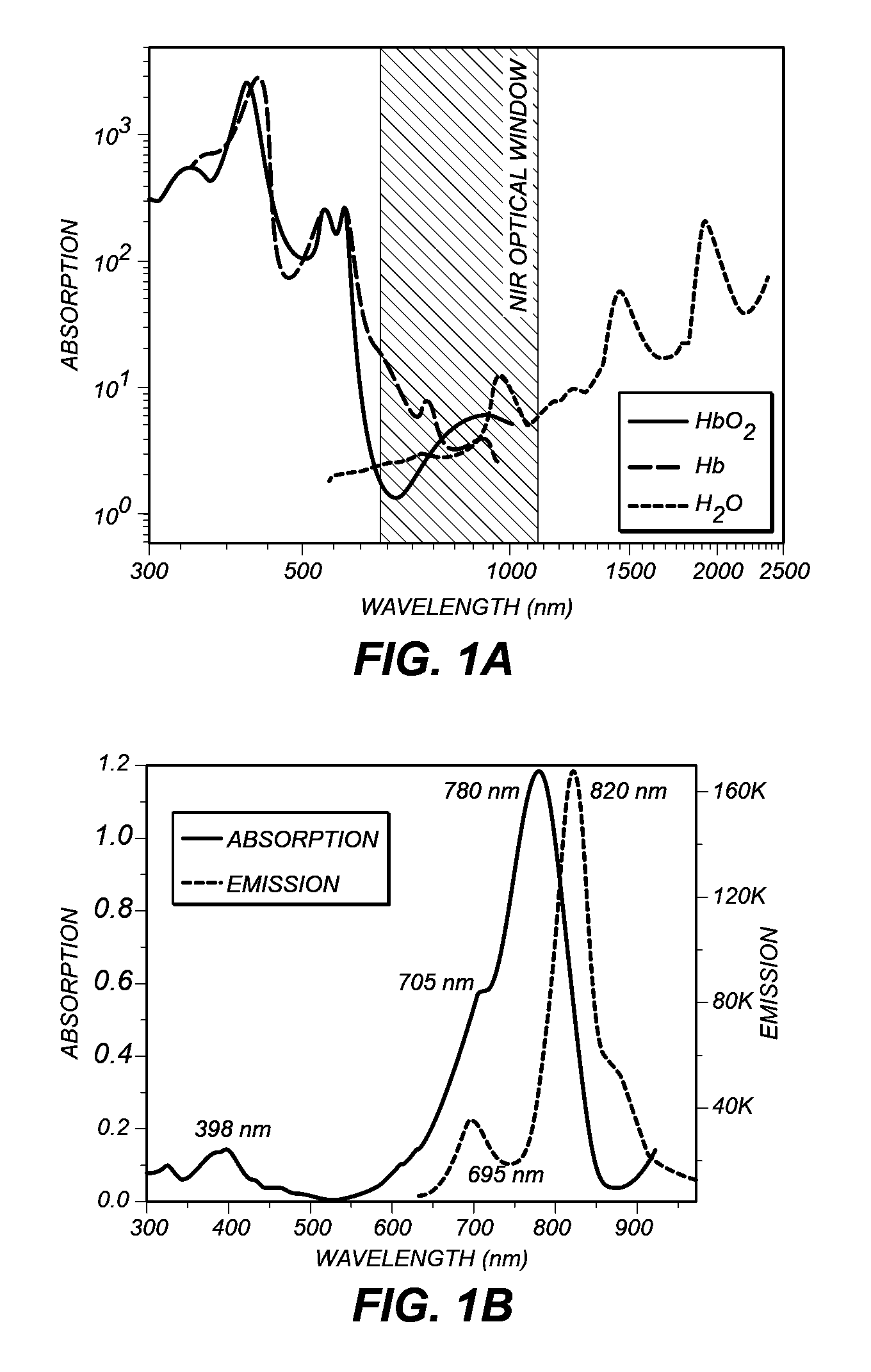
[0030]The specimens to be imaged were Chl α- and ICG-stained uncoated pore glass beads (37 micrometer with pore diameter of about 24 nm). The beads were respectively soaked in a Chl α and ICG solution overnight. All the sample preparations and measurements were performed at room temperature. The size of beads was selected to approximate microvessels in a human brain.

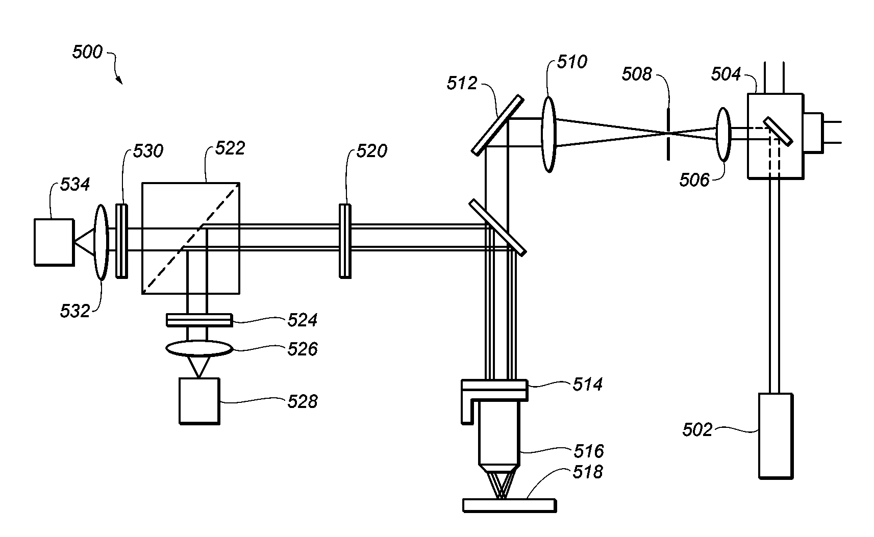
[0031]FIG. 5 is a schematic diagram of a system 500 for imaging. A picosecond or faster laser 502 (e.g. a Ti:Sa laser) provides a multi-photon wavelength to galvanometer-drive X-Y mirror 504. The multi-photon wavelength is passed through a scan lens 506, a field aperture plane 508 and through a tube lens 510. A mirror 512 directs the multi-photon wavelength to a piezoelectric translator 514 which is in communication with objective lens 516 and specimen 518. An emission wavelength is produced by specimen 518 which is subsequently directed to IR-blocking filter 520. IR-blocking filter 520 selects wavelengths between 400 an...


PUM


Abstract

A method is provided for deep tissue imaging using multi-photon excitation of a fluorescent agent. The fluorescent agent is irradiated with an ultrafast laser to produce an excited singlet state (Sn) which subsequently undergoes non-radiative relaxation to a first singlet state (S1). The S1 state undergoes fluorescence to the ground state S0 to produce an emission wavelength. Both the excitation and emission wavelengths are within the near infrared optical window, thereby permitting deep tissue penetration for both the incoming and outgoing signals.

Description

CROSS-REFERENCE TO RELATED APPLICATIONS[0001]This application claims priority to and the benefit of U.S. Provisional Patent Application Ser. No. 61 / 956,620 (filed Jun. 13, 2013) the entirety of which is incorporated herein by reference.STATEMENT OF FEDERALLY SPONSORED RESEARCH OR DEVELOPMENT[0002]This invention was made with Government support under contract number W81 XWI-I-11-1-0335 awarded by the U.S. Army Medical Research and Material Command (USAMRMC), contract number 5SC1HD068129 awarded by the Eunice Kennedy Shriver National Institute of Child Health & Human Development, contract number 2G12RR03060-26A1 awarded by the National Center for Research Resources and contract number 8G12MD007603-27 awarded by the National Institute on Minority Health Disparities from the National Institutes of Health.BACKGROUND OF THE INVENTION[0003]The subject matter disclosed herein relates to imaging of biological tissues using fluorescence from intrinsic and / or extrinsic agents. One of today's m...

Claims


Application Information

Patent Timeline
Patent Type & Authority Applications(United States)
IPC IPC(8): A61B5/00
CPCA61B5/0071
Inventor ALFANO, ROBERT R.PU, YANGSHI, LINGYANPRATAVIEIRA, SEBASTIAO
Owner ALFANO ROBERT R
Why Eureka
Try Eureka