Cardiac imaging processing for interventions

a technology for intervention and cardiac imaging, applied in the field of intervention cardiac imaging processing, can solve the problems of inability to reproduce electromechanical mapping and easy errors, and achieve the effect of facilitating the understanding of the target region

Inactive Publication Date: 2015-03-05
UMC UTRECHT HLDG BV
View PDF5 Cites 4 Cited by
  • Summary
  • Abstract
  • Description
  • Claims
  • Application Information

AI Technical Summary

Benefits of technology

[0005]Another aspect of the present invention is to provide a system for facilitating real-time image guided (pre-) clinical stem cell injection. This system provides the possibility to guide intramyocardial injections with high accuracy to locations with distinct infarct, ischemia (perfusion / viability), or myocardial deformation characteristics. In addition, this system can be used to specify the definition of border zone of the infarct.
[0010]The system allows the target region for the treatment to be clearly defined in the interventional image modality, so that during an intervention, the target region can be reached more reliably.
[0016]The target region may correspond to a region comprising an isoline where the local disease severity is equal to the reference value and a margin on both sides of the isoline. It has been found that such a margin around an isoline provides relatively good results.
[0018]The method may comprise determining a no-go zone for intervention corresponding to an area with a high local disease measure according to a set of predetermined constraints. Clear identification of such a no-go zone provides additional safety for the patient, because an injection in the highly diseased area, for example a core infarction area, may impose a risk to the patient.
[0023]The method may comprise displaying the registered target region fused with an image of the interventional image modality during an intervention. The image of the interventional image modality during the intervention may, for example, be indicative of a location of an interventional instrument, and / or real-time anatomical information related to an interventional instrument, and the fused image containing both the target region and the image of the interventional image modality may help to locate the target region with the instrument and / or in respect of the real-time anatomical information.
[0024]The method may comprise displaying the registered target region in a bull's eye plot format or a polar coordinate system. Such a format may facilitate the understanding of the target region by an observer.

Problems solved by technology

Since the measures of electromechanical mapping are often not distinctive in non-transmural infarctions, and measurements are interpolated in regions where no measurements are taken, the results obtained by electromechanical mapping are not reproducible, and prone to errors.

Method used


Image

  • Cardiac imaging processing for interventions
  • Cardiac imaging processing for interventions
  • Cardiac imaging processing for interventions

Examples


Embodiment Construction

[0046]In the following description, a number of embodiments of the invention will be described in more detail. However, it should be noted that the detailed description presents example embodiments, which do not limit the invention.

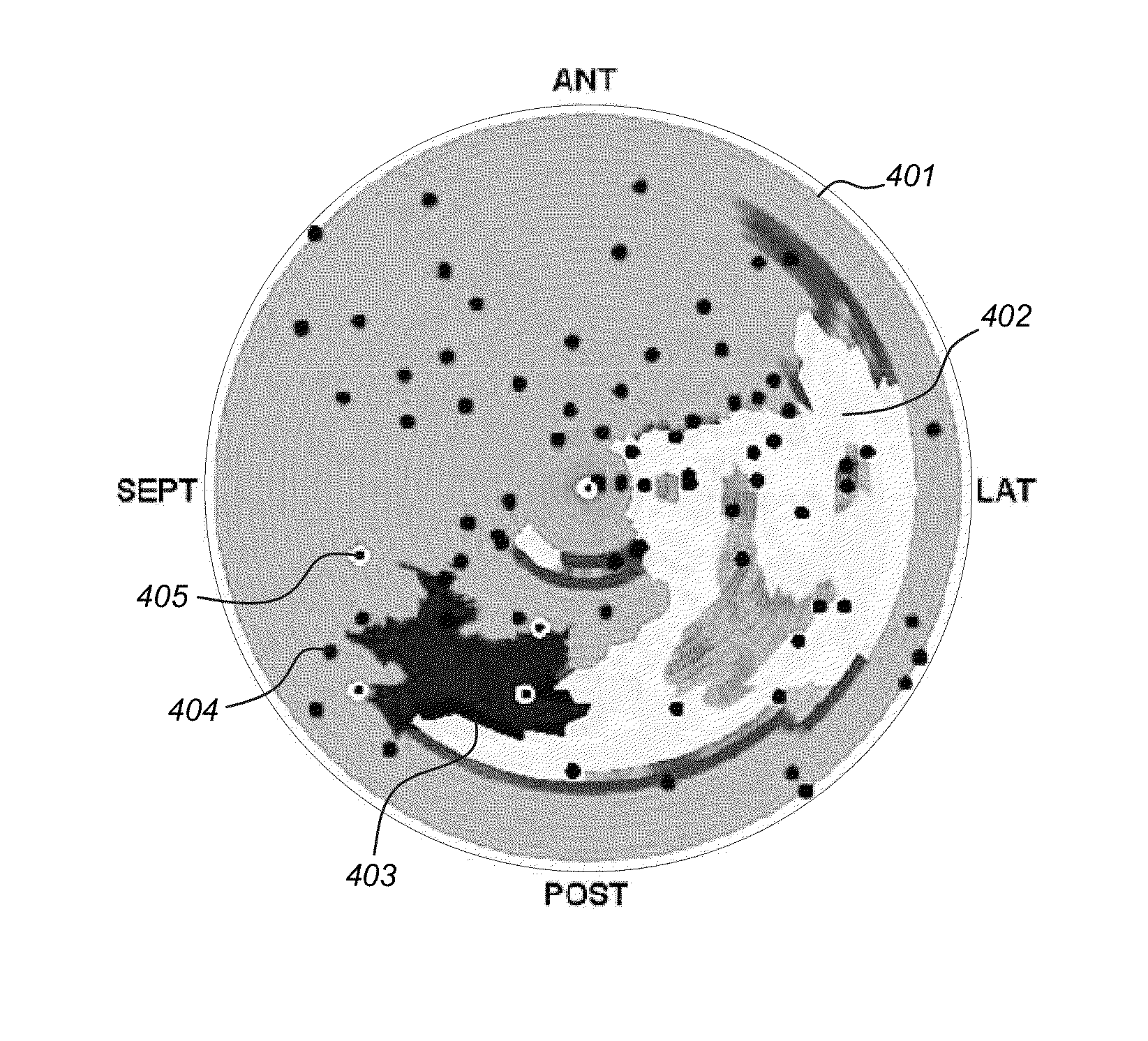
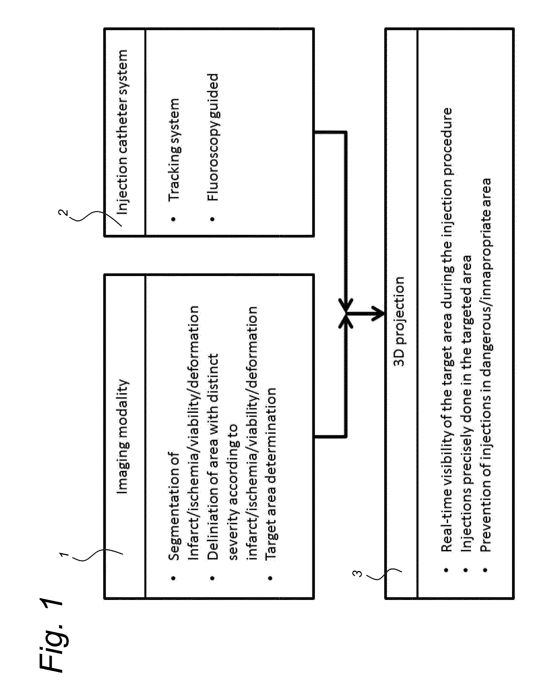
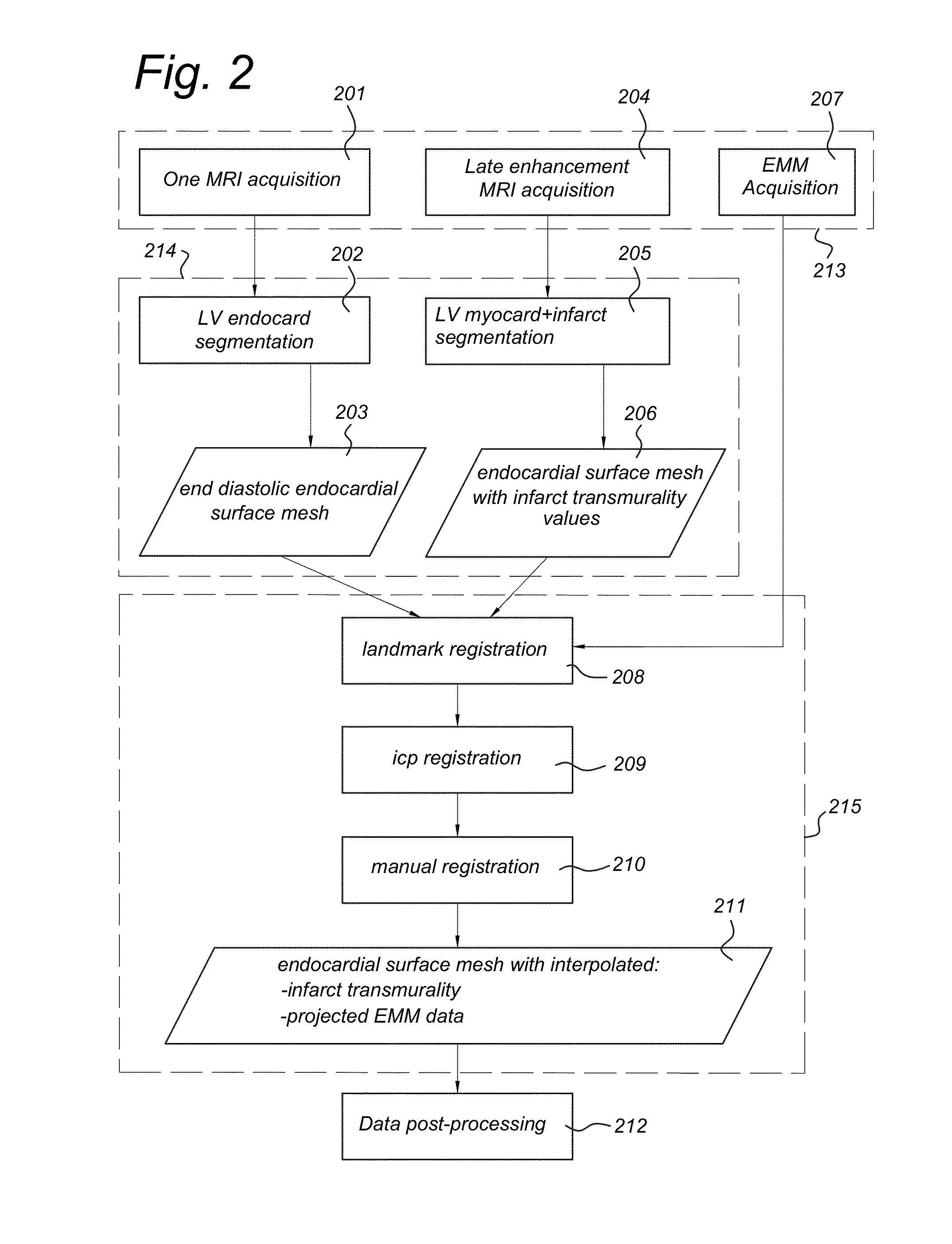
[0047]According to a method of treatment planning in cardiac stem cell therapy, prior to the injection procedure, gold standard diagnostic imaging techniques are used to assess the severity of the disease. Using MRI, infarct transmurality may be assessed using late gadolinium enhancement. Perfusion of the myocardium may be assessed. Deformation of the myocardium may be assessed, for example using MRI tagging. Using SPECT / CT, perfusion and / or viability of the myocardium may be assessed. These are examples of local disease severity measures. The diagnostic imaging data may be converted into a target area by an automatic algorithm using user defined thresholds for the local disease severity measure. For an image guided injection, the target area may be proje...


PUM


Abstract

A method of processing an image includes determining a local disease severity measure based on a 3D dataset. The method further includes determining a target region for a treatment, based on the local disease severity measure, wherein the target region corresponds to a transition region between a first region and a second region, wherein the first region has values of the local disease severity measure which are substantially distinct from the values of the local disease severity measure in the second region. The method further includes registering at least the target region with an interventional image modality. For example, the local disease severity measure is indicative of a measure of a local disease severity at a location along a surface of a myocardium.

Description

FIELD OF THE INVENTION[0001]The invention relates to cardiac image processing for interventions.BACKGROUND OF THE INVENTION[0002]Cardiac regenerative therapy for ischemic heart disease (IHD) aims to provide local cardiac protection and regeneration by means of vasculogenesis, cardiomyogenesis, and matrix support. Studies have shown that injection of stem / progenitor cells into the border zone of the infarcted area helped to stimulate cardiac repair via cell-to-cell contact and secretion of paracrine factors. Moreover, therapeutic effects may rely on the delivery and retention of the regenerative therapeutics on a location where oxygen and nutrients are available to enable survival. Via intramyocardial catheters stem cells, stem cell derived factors, progenitor cells, and / or biomaterials can be injected with minimal invasiveness into the myocardium. Injection locations can be chosen based on tissue viability measures obtained from electromechanical mapping (EMM). For example, NOGA®XP ...

Claims


Application Information

Patent Timeline
Patent Type & Authority Applications(United States)
IPC IPC(8): A61B5/00A61B19/00A61B10/02A61B5/055A61M25/00
CPCA61B5/0035A61B5/055A61B19/5225A61B10/0233A61M25/0084A61B18/1492A61B5/287
Inventor VAN SLOCHTEREN, FREBUS JANVAN ES, RENECHAMULEAU, STEVEN ANTON JOZEF
Owner UMC UTRECHT HLDG BV
Why Eureka
Try Eureka