Eureka AIR delivers breakthrough ideas for toughest innovation challenges, trusted by R&D personnel around the world.

Method for peptide histochemical diagnosis

a histochemical diagnosis and peptide technology, applied in the field of peptide histochemical diagnosis, can solve the problems of difficult to identify whether the peptide is present, poor result in paraffin-embedded surgical specimens, and inability to anticipate the efficacy of chemotherapy for cancer patients after surgery using this method, and achieves the effect of a small volume and more binding ability

Inactive Publication Date: 2015-04-23
NAT TAIWAN UNIV
View PDF6 Cites 2 Cited by
  • Summary
  • Abstract
  • Description
  • Claims
  • Application Information

AI Technical Summary

Benefits of technology

The present invention provides a targeted peptide chemotherapy for treating cancer patients using nanoparticles. The targeted peptides are small molecules that can enter the extracellular space, increasing the binding ability for targeting tumor cells. A peptide histochemical diagnosis can be used to identify whether the targeted peptides can bind to a paraffin section of tumor specimen of the patients, making therapy more effective. This innovation allows for a more effective and targeted treatment for cancer patients.

Problems solved by technology

However, it is difficult to identify whether the peptides can be used to bind to the peptide binding protein in cancer cells by staining the formalin-fixed paraffin-embedded surgical specimens, to confirm the peptides for the targeted chemotherapy in cancers has efficacy in patients before clinical use.
Thus, it is the key to evaluate therapy effects of the patient before peptide-targeted chemotherapy, and the main problem is to identify whether the targeted peptide can directly bind to the surgical tumor specimens from the patient.
It has been reported that biotin can be directly linked to the peptide to form a biotin-peptide, the biotin-peptide can also bind to cancer cells, but it has poor result in the paraffin-embedded surgical specimens.
It is impossible to anticipate the efficacy of chemotherapy for cancer patient after surgery using this method.
Currently, there is not yet a method of pathological diagnosis for targeted peptide to bind to formalin-fixed paraffin-embedded specimens in surgical sections.

Method used


Image

  • Method for peptide histochemical diagnosis
  • Method for peptide histochemical diagnosis
  • Method for peptide histochemical diagnosis

Examples


example 1

Synthesis and Characterization of Magnetic Nanoparticles

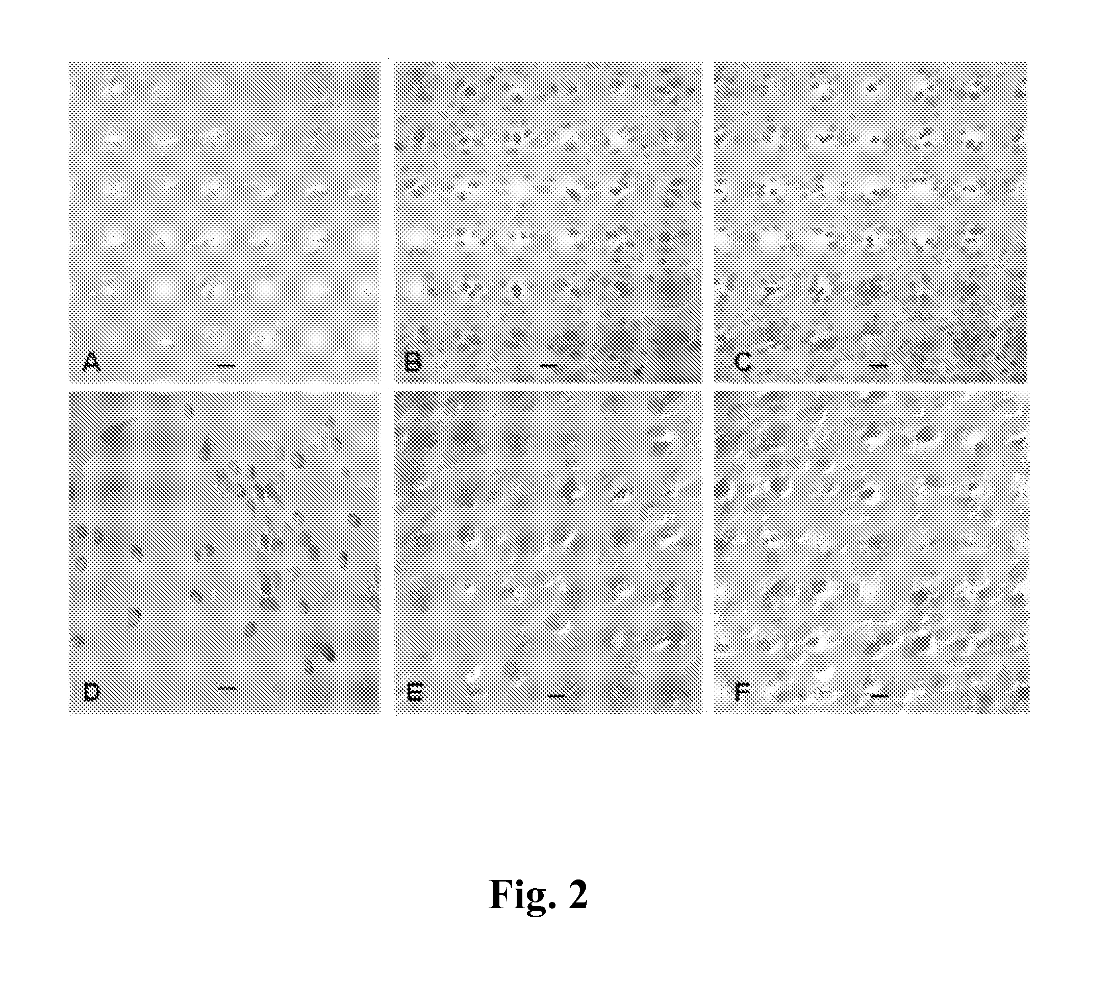
[0035]The dextran coated-Fe3O4 is linked with the targeted peptide SEQ ID NO:1 or the FITC-targeted peptide SEQ ID NO:1 by the MagQu Company. Dextran is used as the hydrophilic surfactant layer. The iron oxide fluid with the desired concentration is available by diluting the highly concentrated iron oxide fluid with a pH 7.4 phosphate buffered saline (PBS) solution. For iron oxide labeling, the NPC-TW01 cells are cultured for 24 hr, incubated with or without the targeted peptide SEQ ID NO:1-linked dextran-coated iron oxide nanoparticle (P-Dex-Fe3O4) at a concentration of 10 μg Fe3O4 / mL in the incubation media for 1 hr at 4° C. and incubated for 4 hr at 37° C. in a 10% CO2 incubator. Cells are washed in a 2% fetal bovine serum in PBS. After fixation, the binding ability between the targeted peptide SEQ ID NO: 1 and tumor cells can be observed by Prussian blue reagent; each iron oxide nanoparticle is bound by approximately above ...

example 2

Flow Cytometry Analysis of the Targeted Peptide SEQ ID NO: 1 Binding Cells in Different Cell Lines

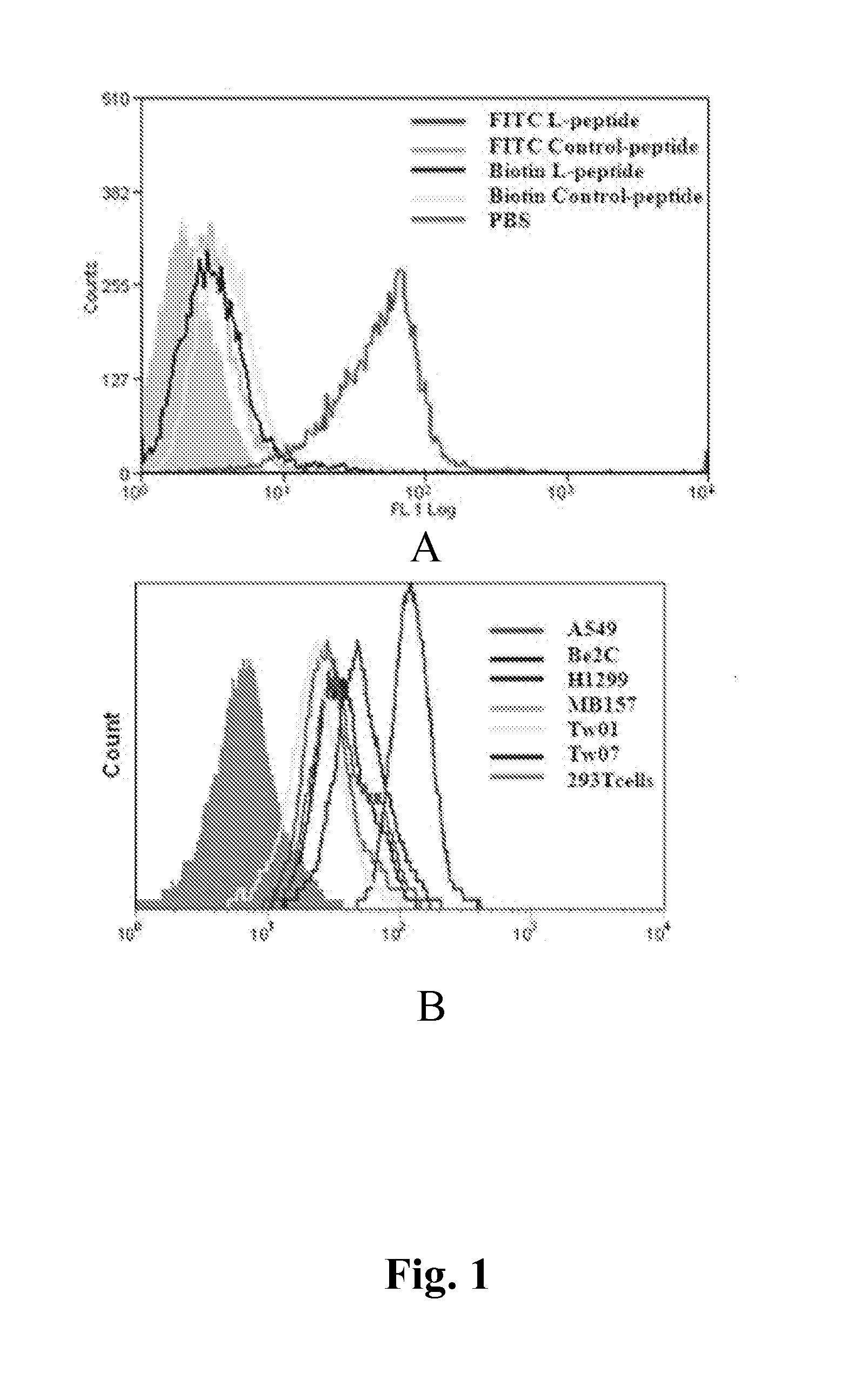
[0036]The cultured cells, including nasopharyngeal carcinoma (NPC-TW01 and NPC-TW07), lung cancer (H1299 and A549), neuroblastoma (Be2C), breast cancer (MB157) and immortalized embryonic renal epithelia (TEKID) (239T) are subjected to flow cytometer (FACSCAN, BD Co., U.S.A.) after the FITC (Fluorescein isothiocyanate)-targeted peptide SEQ ID NO:1 treatment. Simultaneously, FITC-control peptide, FITC-biotin-targeted peptide SEQ ID NO: 1 (B-P) and FITC-biotin-control peptide (B-C-P) are used for flow cytometry comparison.

[0037]When NPC-TW07 cells are incubated with FITC-targeted peptide SEQ ID NO: 1, a clear peak is found in the histogram (FIG. 1A). When the targeted peptide SEQ ID NO: 1 is linked with biotin, very weak or no binding is seen. When other NPC cell line, such as NPC-TW01, and other cancer lines including lung cancer (A549 and H1299), neuroblastoma (Be2C) and breast cancer (M...

example 3

Localization of the Targeted Peptide SEQ ID NO: 1 in Nasopharyngeal Carcinoma

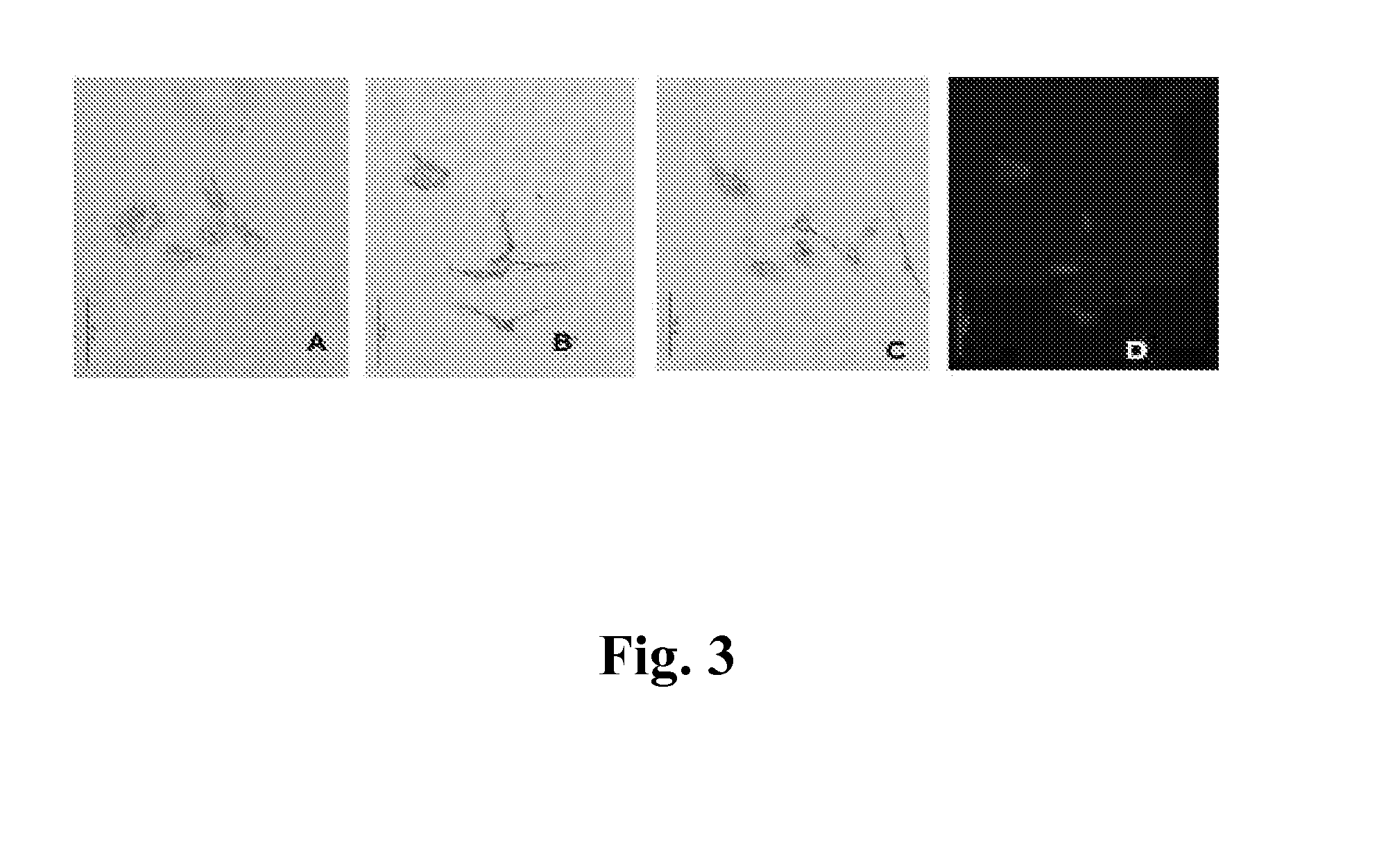
[0038]Because the targeted peptide SEQ ID NO:1 binds weakly to the cancer cells after conjugating to biotin, the sequence of this peptide was modified as Biotin-modified-peptide (B-m-P). B-m-P is modified by adding 5 amino acids spacer SEQ ID NO: 4 to N-terminal and linking with biotin. B-m-P can bind to the small surgical NPC specimens, but the result is not good enough to observe a clear reaction product. For the surgical specimens with more than 1 cm, B-m-P still can not bind to the surgical specimens, there is no binding phenomenon can be observed, while the targeted peptide SEQ ID NO:1-linked dextran-coated iron oxide nanoparticles (P-Dex-Fe3O4) of the present invention can bind to tumor cells and be stained with Prussian blue reagent. The result is excellent so as to name “peptide histochemical diagnosis”.

[0039]In the present invention, P-Dex-Fe3O4 has to be prepared at first. The nanoparticle (MW 60,...


PUM

PropertyMeasurementUnit
Pressureaaaaaaaaaa
Coloraaaaaaaaaa

Abstract

The present invention provides a method of peptide histochemical diagnosis to detect the peptide binding protein in the cancer tissue. This peptide binding specifically to tumor cells is linked to the dextran coated iron oxide nanoparticle. The peptide linked dextran coated iron oxide nanoparticle can be used to bind to the formalin-fixed and paraffin-embedded tumor surgical specimens, and the method of present invention can be used to evaluate the efficacy of peptide-targeted chemotherapy for treatment of cancer patients.

Description

CROSS-REFERENCE TO RELATED APPLICATIONS[0001]This application claims the priority benefit of Taiwan application serial no. 102137765, filed on 18 Oct. 2013. The disclosure of the Taiwan application is incorporated by reference herein.BACKGROUND OF THE INVENTION[0002]1. Field of the Invention[0003]The invention relates to a method for peptide histochemical diagnosis, and in particular, to a method for peptide histochemical diagnosis of the formalin-fixed paraffin-embedded surgical specimens.[0004]2. The Prior Arts[0005]Currently, some targeted peptides for the targeted chemotherapy in cancers have been reported. However, it is difficult to identify whether the peptides can be used to bind to the peptide binding protein in cancer cells by staining the formalin-fixed paraffin-embedded surgical specimens, to confirm the peptides for the targeted chemotherapy in cancers has efficacy in patients before clinical use. Although the patients suffer the same type of cancer, it is still unknown...

Claims


Application Information

Patent Timeline
IPC IPC(8): G01N33/574
CPCG01N33/57492G01N33/57407G01N33/57438G01N33/57423G01N33/57415C07K7/08
Inventor LIN, CHIN-TARNGWU, HAN-CHUNG
Owner NAT TAIWAN UNIV
Why Eureka
Eureka Blog
Learn More