Mesenchymal-like stem cells derived from human embryonic stem cells, methods and uses thereof

a technology of embryonic stem cells and stem cells, applied in the field of embryonic stem cell derived stem cells, can solve the problems of wasting resources, wasting resources, and current methods that cannot conduct such differentiation in an efficient manner, and achieves the effects of high yield, high yield, and efficient generation of mscs

Active Publication Date: 2015-07-09
UNIV OF CONNECTICUT +1
View PDF3 Cites 18 Cited by
  • Summary
  • Abstract
  • Description
  • Claims
  • Application Information

AI Technical Summary

Benefits of technology

[0013]Disclosed herein is a method to efficiently produce T-MSC in high purity and high yield. The method has the features of relatively few steps and fewer required differentiation factors than previously reported.
[0069]It is an object of the disclosed method to provide a method that is high in yield. The differentiation method described herein can produce 1-5×10¹⁰ T-MSC cells within 30 days from 1×10⁵ of hESC, whereas other method can only produce up to 1×10⁸ MSC cells within 30 days.

Problems solved by technology

However, current methods are not able to conduct such differentiation in an efficient manner to produce a high yield of high purity MSCs.
However, the currently available adult tissue-derived MSCs have several pitfalls.
First, the limited sources and varying quality of the donor tissues such as the bone marrow restrict the study and application of the MSCs and prevent the standardization of the MSCs as a medical product for large-scale clinical use.
To obtain enough cell numbers for clinical use, in vitro expansion is necessary, which can decrease the immunosuppressive and homing abilities of MSCs (Javazon et al., 2004).
Third, there are safety issues regarding to the use of adult-derived MSCs including malignant transformation (Wong, 2011) and potential transmission of infectious pathogens from donors.
In 2010, the inventors and Advanced Cell Technology developed another method to derive MSC from hemangioblast, which involved the use of many expensive cytokines and methylcellulose medium, but the derivation efficiency is also low using this method.
Currently known methods for differentiation of hESCs into MSCs are each characterized as having one or more serious shortcomings and weaknesses: Differentiation of MSCs from hESCs co-cultured with the OP9 stromal cells has the disadvantages of being time consuming, producing cells of low yield, low purity, and using animal feeder cells and undefined culture conditions (Barbed et al., 2005).
Differentiation from outgrowing cells around replated embryoid bodies formed by hESCs has the disadvantages of being time consuming, producing cells in low yield, using undefined culture condition, and being an expensive method (Olivier et al., 2006).
Differentiation from hESCs cultured on collagen-coated plates has the disadvantages of very low yield, undefined culture conditions, and being time consuming (Liu et al., 2012).
Differentiation with hESCs treated with inhibitors of TGFβ signaling has the disadvantages including low purity of cells (per our tests), low cell yield, time consuming method, and low immunosuppressive effect of the cells that are produced (Chen et al., 2012; Sanchez et al., 2011).
However, these offer only palliative relief and are associated with serious adverse effects including increased infection, heart attack, stroke, progressive multifocal leukoencephalopathy, arrhythmia, pain, depression, fatigue, macula edema, and erectile dysfunction (Johnston and So (2012); Weber et al.
The efficacy of BM-MSC on treatment of the disease is questionable.
There is very little cell loss when hESCs are differentiated through the trophoblast stage, whereas, other methods usually have over 90% loss of the starting cells during the initial differentiation step, resulting in much lower cell yields than the method disclosed herein.

Method used


Image

  • Mesenchymal-like stem cells derived from human embryonic stem cells, methods and uses thereof
  • Mesenchymal-like stem cells derived from human embryonic stem cells, methods and uses thereof
  • Mesenchymal-like stem cells derived from human embryonic stem cells, methods and uses thereof

Examples


example 1

Derivation of T-MSC Material and Methods

[0343]The following reagents and materials were obtained from the below-described sources:[0344]Customed mTeSR1 Medium: Stem Cell Technology, Inc.[0345]BMP4: Stemgent or other vendors[0346]SB431542: Cayman Chemical or other vendors[0347]A83-01: Stemgent or other vendors.[0348]ALK5 inhibitor: Stemgent or other Vendors[0349]DMEM / F12: GIBCO Life Technologies[0350]alpha-MEM: GIBCO Life Technologies[0351]Fetal Bovine Serum: GIBCO Life Technologies or other vendors

[0352]CT2 hESC line derived at the University of Connecticut Stem Cell Core was cultured for two passages on irradiated mouse embryonic fibroblast (MEF) as feeders. The hESCs were then split on plates coated with Matrigel (BD Biosciences, San Jose, Calif.) and cultured in mTeSR1 (Ludwig et al., 2006) (Stem Cell Technologies, Vancouver, Canada). ESI-017, ESI-051, ESI-053, ESI-049, and ESI-35 human embryonic stem cells were purchased from BioTime, Inc. (CA).

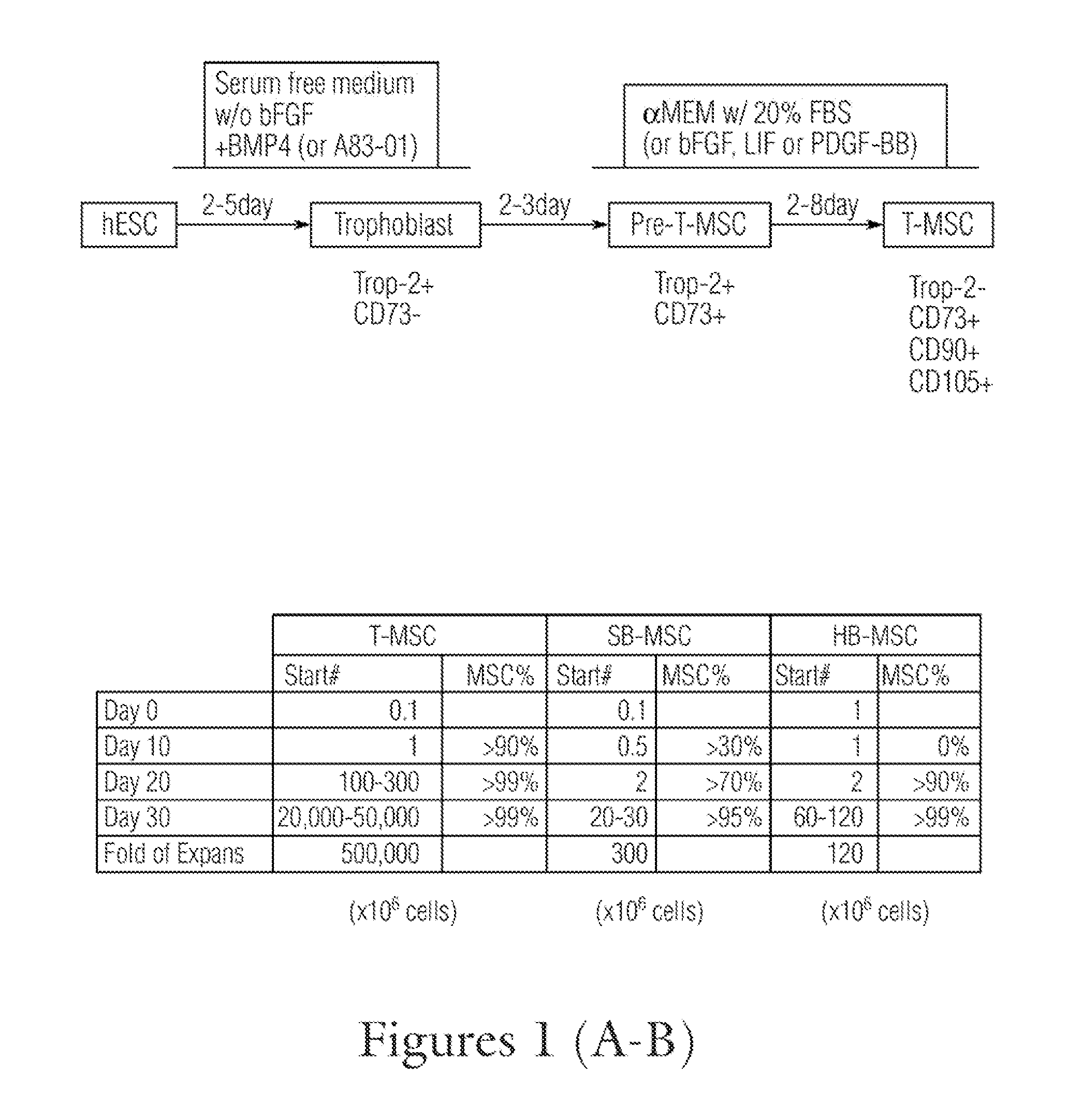
[0353]Derivation of T-MSC

[0354]As ...

example 2

Characterization of T-MSC cells

[0365]The T-MSC cells obtained in Example 1 were further analyzed using flow cytometry immunofluorescence staining.

[0366]Materials and Methods

[0367]Flow cytometry staining was used to characterize the T-MSCs. Cells were washed and blocked with 2% BSA in PBS, and stained with antibodies for various cell surface markers Trop-2 (Trp-2, eBioscience), CD31. CD34, CD29. CD73, CD90, CD105, CD44, CD45, CD146, CD166, HLA-ABC, HLA-DR, HLA-G (BD Bioscience or eBioscience) by following the manufacturers' instructions. Data were collected on FACS LSR II Flow Cytometer using FACSDiva software (BD Bioscience). Post-acquisition analysis was performed with the FlowJo software (Treestar).

[0368]Results

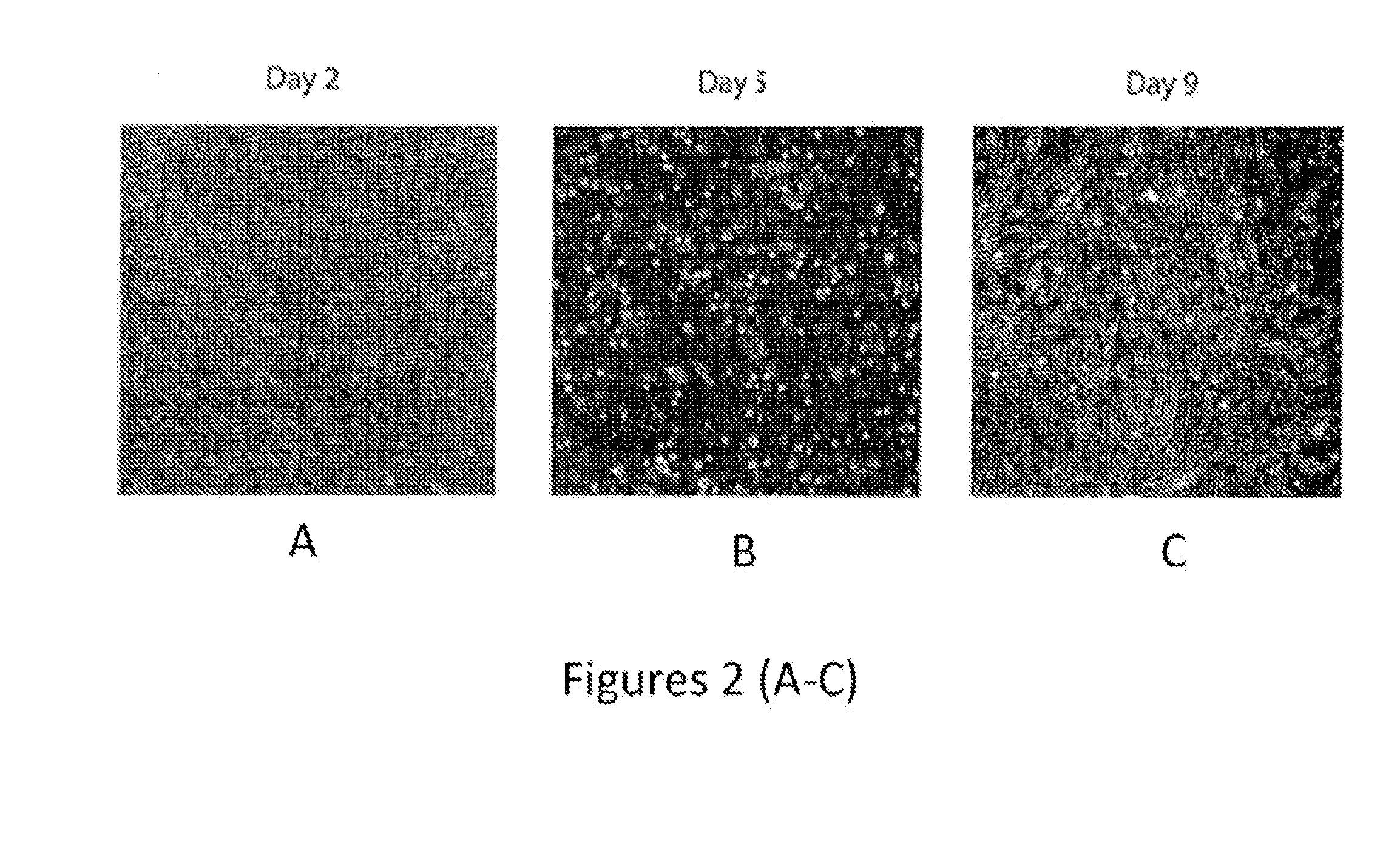
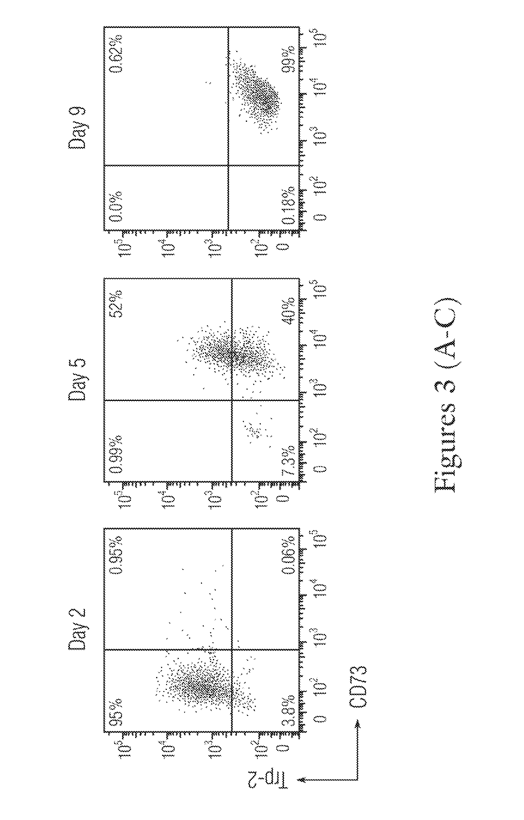
[0369]The attached cells obtained from Day 2 trophoblast, Day 5 pre-T-MSC and Day 9 T-MSC were stained with CD73 and Trop-2. The trophoblast cells only expressed high levels of Trop-2 (greater than 95%), but less than 1% of CD73 (FIG. 3A); the pre-T-MSC at day 5 has more th...

example 3

T-MSCs have a Stronger Inhibition on T Cell Functions In Vitro than BM-MSC

[0371]hEs-MSCs and BM-MSCs were compared for their ability to inhibit T cell proliferation in vitro following antigen stimulation.

[0372]Materials and Methods

[0373]Culture of BM-MSCs

[0374]BM-MSCs were derived from BM mononuclear cells (BMMNCs) or obtained from AllCells, Inc. (Alameda) and Lonza (Basel, Switzerland) BMMNCs. For derivation, BMMNCs were thawed and plated onto tissue culture plastic dishes in □MEM+20% FBS. Adherent cells began to appear within the first 4-5 days and fed every 3 days until day ˜10-12, when cells were harvested and replated at 3,000-5,000 cells / cm2.

[0375]The in vitro assay for T cell proliferation was performed using lymphocytes isolated from mouse peripheral lymph nodes. These lymphocytes were labeled with 5 μM of carboxyfluorescein succinimidyl ester (CFSE) to track their proliferation by monitoring CFSE dilution in their daughter cells, for 10 minutes at 37° C. 10,000 T-MSCs or BM...


PUM

PropertyMeasurementUnit
timeaaaaaaaaaa
timeaaaaaaaaaa
concentrationaaaaaaaaaa

Abstract

The disclosure provided herein relates generally to mesenchymal-like stem cells “hES-T-MiSC” or “T-MSC” and the method of producing the stem cells. The method comprises culturing embryonic stem cells under conditions that the embryonic stem cells develop through an intermediate differentiation of trophoblasts, and culturing the differentiated trophoblasts to hES-T-MSC or T-MSC, T-MSC derived cells and cell lineages “T-MSC-DL” are also described. Disclosed also herein are solutions and pharmaceutical compositions comprising the T-MSC and / or T-MSC-DL, methods of making the T-MSC and T-MSC-DL, and methods of using the T-MSC and T-MSC-DL for treatment and prevention of diseases, specifically, T-MSC and T-MSC-DL are used as immunosuppressive agents to treat multiple sclerosis and autoimmune diseases.

Description

RELATED APPLICATIONS[0001]The present application claims priority to U.S. Provisional Application Ser. No. 61 / 670,192, filed Jul. 11, 2012 and U.S. Provisional Application Ser. No. 61 / 684,509, filed Aug. 17, 2012, which are hereby incorporated by reference in their entireties.1. INTRODUCTION[0002]The disclosure provided herein relates generally to mesenchymal-like stem cells “hES-T-MSC” or “T-MSC” and the method of producing the stem cells. The method comprises culturing embryonic stem cells under conditions that the embryonic stem cells develop through an intermediate differentiation of trophoblasts, and differentiating trophoblasts into hES-T-MSC or T-MSC. Disclosed herein are the T-MSC, solutions and pharmaceutical compositions comprising the T-MSC, methods of making the T-MSC, methods of using the T-MSC for treatment and prevention of diseases, specifically, T-MSC are used as an immunosuppressive agent to treat multiple sclerosis and other autoimmune diseases, for tissue regener...

Claims


Application Information

Patent Timeline
Patent Type & Authority Applications(United States)
IPC IPC(8): C12N5/0775A61K35/28
CPCC12N5/0668A61K35/28C12N2506/02C12N2500/90C12N2501/235C12N2501/115C12N2501/135C12N2506/45C12N2501/155C12N2533/90C12N2501/145C12N2501/165C12N2501/26A61P17/00A61P17/02A61P19/02A61P19/04A61P19/08A61P21/00A61P25/00A61P35/00A61P37/02A61P43/00A61P9/00A61K35/545C12N2529/00
Inventor WANG, XIAOFANGXU, REN-HE
Owner UNIV OF CONNECTICUT
Why Eureka
Try Eureka