Method for nerve detection by raman scattering spectroscopy

a raman scattering and nerve technology, applied in the field of nerve detection, can solve the problems of unrealized comprehensive detection of nerves, difficult to use staining for observation during operations, and difficult to apply this measurement to unmyelinated nerves lacking a myelin sheath, etc., and achieve the effect of suppressing the degradation of qol after the operation caused by nervous disorder

Inactive Publication Date: 2015-10-22
KYOTO PREFECTURAL PUBLIC UNIV CORP
View PDF2 Cites 1 Cited by
  • Summary
  • Abstract
  • Description
  • Claims
  • Application Information

AI Technical Summary

Benefits of technology

[0025]The present invention provides a non-staining optical nerve detection method and device using Raman scattering spectroscopy that employs a light scattering phenomenon caused by molecular vibration. In particular, the present invention provides a non-staining method of detecting nerves containing unmyelinated nerves, which has been impossible hitherto.
[0026]The present invention enables nerves containing unmyelinated nerves to be displayed specifically. Thus, the presence and position of nerves can be grasped accurately during an operation through use of the detection method and device of the present invention, and the degradation in QOL after the operation caused by nervous disorder can be suppressed.

Problems solved by technology

However, the staining itself is harmful to humans in most cases, and hence it is difficult to use the staining for observation during operations.
Therefore, it is difficult to apply this measurement to unmyelinated nerves lacking a myelin sheath, and thus the comprehensive detection of nerves has not been realized.
However, none of Non-patent Literatures and Patent Literatures have attempted to detect nerves comprehensively.

Method used


Image

  • Method for nerve detection by raman scattering spectroscopy
  • Method for nerve detection by raman scattering spectroscopy
  • Method for nerve detection by raman scattering spectroscopy

Examples


example 1

General View of Experimental Device and Experimental Method

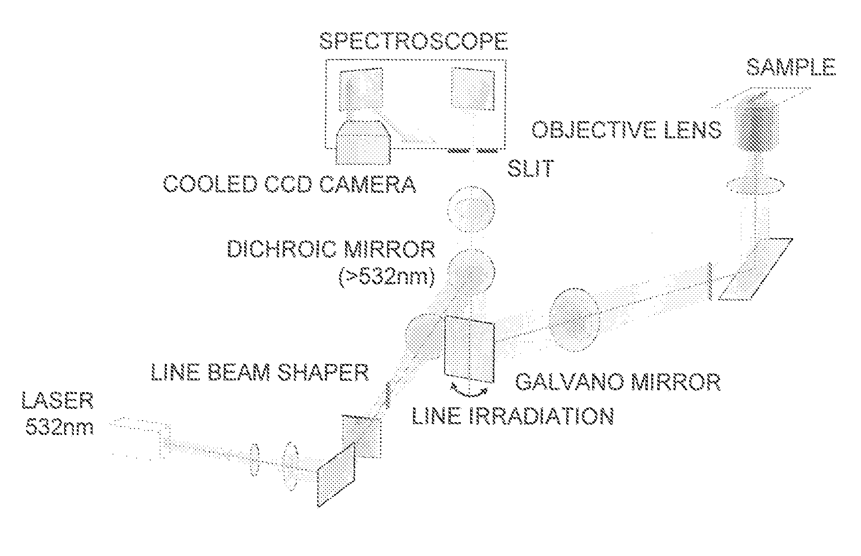
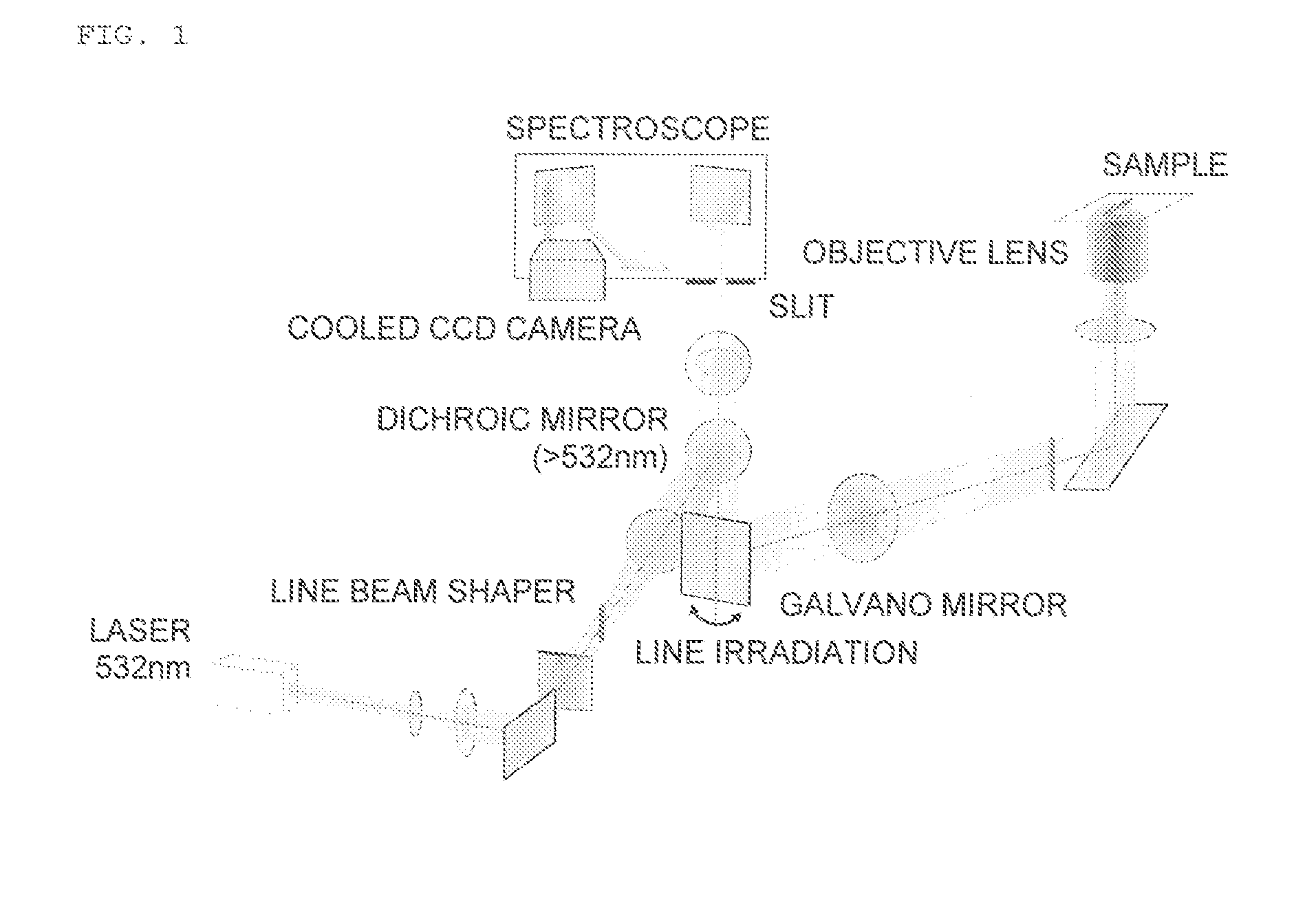
[0082]FIG. 1 illustrates a general view of an experimental device.

[0083]Slit-scanning Raman scattering microscope: RAMAN-11, manufactured by Nanophoton Corporation

[0084]Cooled CCD camera: Pixis 400 BR, manufactured by Princeton Instruments, −70° C., 1,340×400 pixels

[0085]Objective lens: UPLSAPO, manufacturedby Olympus Corporation, x60, NA=1.2

[0086]Experimental Method[0087](1) Tissue Sample

[0088]Rat Tissue

[0089]A healthy Wistar rat was euthanized by an overdose of an anesthetic, and each tissue was obtained.

[0090]Chest tissue containing intercostal nerves, tissue in the vicinity of the esophagus containing vagus nerves, femoral nerves and surrounding tissue thereof, celiac plexus, cerebellum

[0091]Type: Wister rat

[0092]Age: Young-adult (8-10 weekly age)

[0093]Human Tissue

[0094]The periprostatic tissue of a patient who has undergone radical prostatectomy was obtained. The tissue containing a vagus nerve gastric branch of a patie...

example 2

Distinguishing Nerves and Surrounding Tissue Thereof Based on Intensity Ratio

[0110]Raman spectra of myelinated nerves, unmyelinated nerves, connective tissue, fat tissue, muscle tissue (striated muscle), and blood vessels (media) were obtained, and whether or not there was a significant difference in intensity ratio in each Raman shift was investigated.

[0111]Method of calculating intensity ratio

Iratio=Iω2Iω1[Math.2]

Iratio: Intensity ratio between Iω1 and Iω2

Iω1: Raman scattering light intensity at Raman shift ω1 (left axis in the figure)

Iω2: Raman scattering light intensity at Raman shift ω2 (lower axis in the figure)

[0112]Method of Calculating Significant Difference

[0113]The intensity ratio calculated by the above-mentioned equation was measured at a plurality of points of two kinds of tissues (“connective tissue vs myelinated nerves”, etc. described in an upper part of the figure), and the two kinds of measurement groups were subjected to statistical analysis by a t-test to calcu...

example 3

Raman Spectrum by 671 nm Excitation

[0121]In order to show that nerves can be detected at various wavelengths, a Raman spectrum at an excitation optical wavelength of 671 nm was measured (FIG. 14). For reference, FIG. 15 shows the measurement result at an excitation optical wavelength of 532 nm.

[0122]A Raman spectrum that was substantially the same as that at 532 nm was also obtained at each spectrum. From this, it is considered that nerves can be detected similarly at various wavelengths.


PUM

PropertyMeasurementUnit
wavelengthaaaaaaaaaa

Abstract

The present invention provides a method of detecting nerves, including: step 1 of irradiating a sample with excitation light; step 2 of detecting Raman scattering light from the sample; step 3 of calculating an intensity ratio of a wave number within a specific range of the Raman scattering light detected in the step 2 or extracting a feature of the intensity ratio and subjecting the feature to multivariate analysis and/or statistical analysis; and step 4 of specifically displaying nerves containing unmyelinated nerves, using as an index the intensity ratio or a result from the multivariate analysis and/or the statistical analysis.

Description

TECHNICAL FIELD[0001]The present invention relates to a method of detecting nerves using Raman scattering spectra from biological tissue. The present invention further relates to a device for detecting nerves using Raman scattering spectra.BACKGROUND ART[0002]Preserving nerves in operations play important roles in the QOL (quality of life) of patients, as well as preserving organ functions. Hitherto, in order to determine the position of thin nerves, a staining technology using a dye has been improved. However, the staining itself is harmful to humans in most cases, and hence it is difficult to use the staining for observation during operations. Therefore, only thick nerves that can be observed with naked eyes of operators or with white light imaging using an image sensor have been mainly targeted for nerve preservation. There is no technology for determining the position of thin nerves, and hence the determination of the position of nerves has no choice but to depend on the anatomi...

Claims


Application Information

Patent Timeline
Patent Type & Authority Applications(United States)
IPC IPC(8): A61B5/00G01N21/65
CPCA61B5/0075G01N21/65A61B5/742A61B5/40
Inventor TAKAMATSU, TETSUROMINAMIKAWA, TAKEOHARADA, YOSHINORI
Owner KYOTO PREFECTURAL PUBLIC UNIV CORP
Why Eureka
Try Eureka