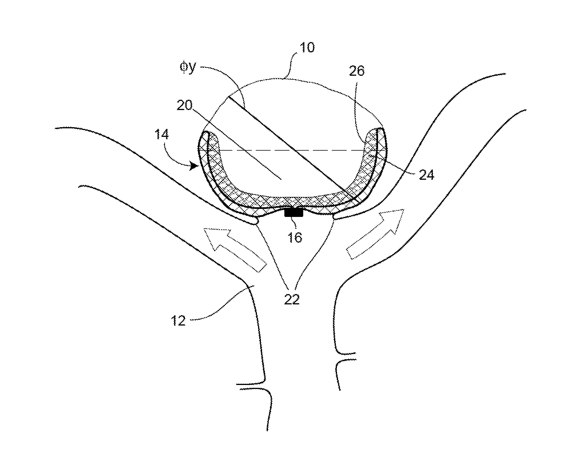
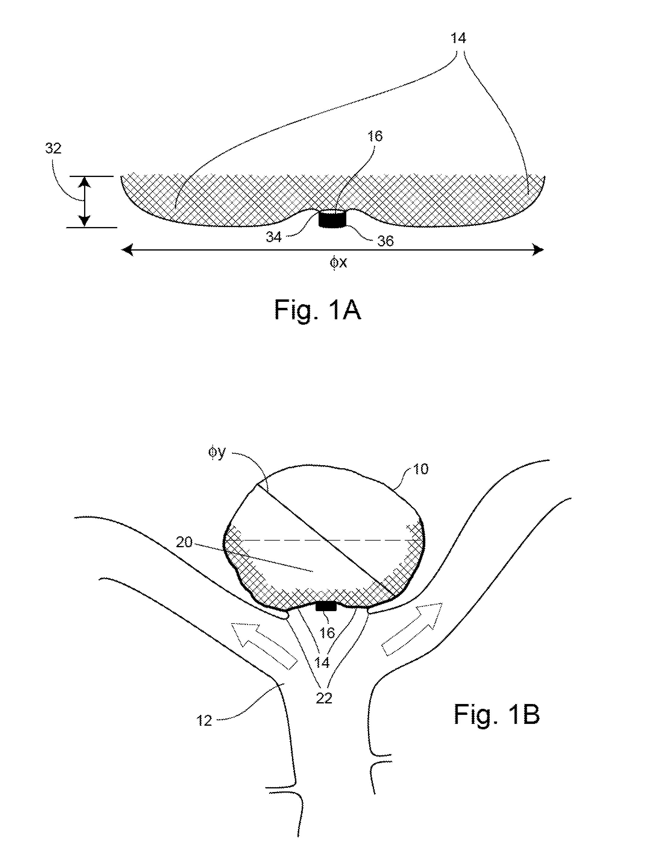
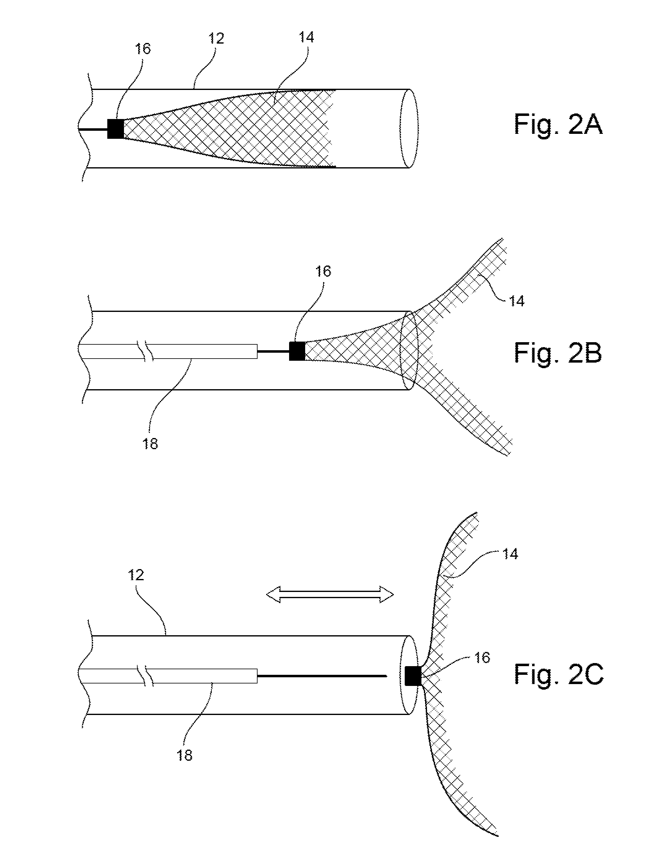
Occlusion Device

a technology of occlusion device and occlusion chamber, which is applied in the field of occlusion device, can solve the problems of unmet need for providing aneurysm treatment and/or amelioration, risk of leakage and/or rupture, hemorrhagic stroke, brain damage, etc., and achieves the effect of less material, less need for anti-coagulation therapy, and less risk of clot emboli formation

Active Publication Date: 2015-11-05
CERUS ENDOVASCULAR
View PDF10 Cites 92 Cited by
  • Summary
  • Abstract
  • Description
  • Claims
  • Application Information

AI Technical Summary

Benefits of technology

[0022]The present inventor has designed an occlusion device for providing aneurysm treatment and/or amelioration through the use of a minimum amount of fully-retrievable deployable low profile resilient mesh material which is oversized to the diameter of the aneurysm. As such, an occlusion device, having less material

Problems solved by technology

However, there still remains an unmet need for providing aneurysm treatment and/or amelioration, particularly for neurovascular aneurysms, via occlusion devices comprised of a minimum amount of fully-retrievable deployable material.
The weakened part of the artery forms a bulge, or a ballooning area, that risks leak and/or rupture.
Subarachnoid hemorrhage from a ruptured neurovascular aneurysm can lead to a hemorrhagic stroke, brain damage, and death.
Aneurysms, however, may be discovered during routine medical exams or diagnostic procedures for other health problems.
This method may not detect some aneurysms due to overlapping structures or spasm.
As such, there is a potential for the development of epilepsy in the patients due to the surgery.
The injection of these types of embolic materials suffers from disadvantages, most of which are associated with migration of the embolic material out of the aneurysm into the parent artery.
This can cause permanent and irreversible occlusion of the parent artery.
Further, it is at times difficult to gauge exactly how full the sac of the aneurysm is when detachable coils are deployed.
Therefore, there is a risk of overfilling the aneurysm in which case the detachable coils also spill out into the par

Method used


Image

  • Occlusion Device
  • Occlusion Device
  • Occlusion Device

Examples


example i

[0083]Non-detachable occlusion device lot 30680, Detachable occlusion device lot 30676, Aneurysm size 4.5 millimeters (mm) (height)×2.5 width

[0084]A 5-F (5-French) sheath placed in the femoral artery through which a 5F Cordis catheter, lumen 0.035″, length 65 centimeters (cm) and a 0.035″ Terumo guide-wire access was gained into the carotid artery.

[0085]The non-detachable occlusion device was positioned into the aneurysm with the marker at the neck of the aneurysm and contrast runs performed at timed intervals. Immediately after deployment of the device, it was observed to be in a good position in the aneurysm and there was some slowing of flow. At 5 minutes post deployment, there was some stasis observed in the aneurysm. At 10 minutes post deployment, further stagnation in the aneurysm was observed and the device was repositioned closer to the aneurysm neck. At 15 minutes post deployment, stagnation of flow in the aneurysm was observed. When the device was removed from the aneurysm...

example ii

[0087]Non-detachable occlusion device lot 30680, Detachable occlusion device lot 30676, Aneurysm size 10 mm height×4 mm wide×3 mm neck

[0088]A similar procedure to Example I was performed with the placement of the non-detachable device into the aneurysm neck. In this example, a 4F system was used to introduce the device into the 5F sheath and a “step” on the internal hub of the catheter was noted to cause the device to catch. The device was placed and timed angiographic runs obtained as before. Immediately post deployment, some flow reduction was observed in the aneurysm. At 5 minutes post deployment, a filling defect was observed in the aneurysm sac. At 10 minutes post deployment, an increase in size of the filling defect was observed.

[0089]This device was removed, angiography demonstrated that flow had returned to pre-deployment status in the aneurysm, and an implantable (detached) device was deployed using the same method as previously. The implant required some force initially to...

example iii

[0092]Detachable device lot 30676, Aneurysm size 6.5 mm×3.1 mm width×2.4 mm neck

[0093]The procedure followed the same protocol as Example II, however, on contrast agent injection it was noticed that the aorta had dissected. It was possible to deploy a device into the aneurysm neck and timed angiographic runs obtained as before.

[0094]Observations

[0095]This series of angiograms confirm the wire mesh braid configuration of the occlusion device disclosed herein is sufficiently dense to reduce blood flow in the aneurysm leading to stasis of blood and thrombosis in the aneurysm sac. The study, accounting for variability in animal morphology, allowed for the understanding and consideration of device development and its deployment technique.

[0096]All femoral punctures were carried out via femoral cut-downs with the use of a vein picker. The sheaths used were 5-F and specifically had very narrow tips allowing expansion of the femoral vessel without damaging it. The length of the catheter was...


PUM


Abstract

Provided herein is an occlusion device for intrasaccular implantation and/or vascular occlusion comprising: (a) a substantially solid marker having a proximal end, and a distal end; and (b) a low profile resilient mesh body attached to the distal end of the marker, the body having a delivery shape and a deployed shape capable of conforming to aneurysm walls; wherein the body has a diameter greater than a diameter of an aneurysm to be treated. Also provided herein is a kit comprising the occlusion device disclosed herein and a means for delivery thereof. Methods of manufacture and use of the occlusion device disclosed herein are also disclosed.

Description

RELATED APPLICATIONS[0001]This application claims priority to U.S. Provisional Application No. 61 / 986,369, filed Apr. 30, 2014, and U.S. Provisional Application No. 61 / 083,672, filed Nov. 24, 2014, the disclosures of which are hereby incorporated herein by reference in their entirety. In addition, all documents and references cited herein and in the above referenced applications, are hereby incorporated herein by reference.FIELD OF THE INVENTION[0002]The present invention relates generally to the field of occlusion devices and / or occlusion device systems and / or implantable occlusion devices and the use of the same for the treatment and / or amelioration of aneurysms.BACKGROUND OF THE DISCLOSURE[0003]There is a significant demand for the development of improved occlusion-type devices and / or systems for the treatment and / or amelioration of aneurysms. This observation is supported by the abundance and wide-range of current occlusion devices and / or systems currently in the aneurysm treatm...

Claims


Application Information

Patent Timeline
IPC IPC(8): A61B17/12
CPCA61B17/12172A61B17/12031A61B2017/1205A61B2017/12063A61B17/12113A61B17/12A61B17/12168A61B2017/00867A61B90/39A61B2090/3937
Inventor GRIFFIN, STEPHEN
Owner CERUS ENDOVASCULAR
Why Eureka
Try Eureka