Cardiac late gadolinium enhancement MRI for patients with implanted cardiac devices

a technology of cardiac implants and enhancement scars, applied in the field of clinical practice of medical imaging on patients with metal-containing devices, can solve the problems of cardiac implant device mri, icds or pacemakers mri, and non-applicability of medical imaging techniques such as magnetic resonance imaging (mri), and achieve the effects of eliminating image quality issues, high quality, and enhancement scar imaging

Active Publication Date: 2015-12-03
RGT UNIV OF CALIFORNIA
View PDF3 Cites 14 Cited by
  • Summary
  • Abstract
  • Description
  • Claims
  • Application Information

AI Technical Summary

Benefits of technology

[0011]Provided herein are methods and systems for high quality late gadolinium enhancement scar imaging even in the presence of implanted cardiac devices. The technologies disclosed can be widely used by all major manufacturers of clinical MRI systems both in the U.S. and worldwide to eliminate any image quality issues associated with these cardiac devices. In some embodiments, a custom made MRI pulse sequence will be needed, which is a piece of software that controls the MRI system and can be easily implemented.

Problems solved by technology

Currently, medical imaging techniques as such Magnetic Resonance Imaging (MRI), also known as nuclear magnetic resonance imaging (NMRI) or magnetic resonance tomography (MRT), are not applicable to patients with cardiac implant devices: partly because the myocardial scar (viability) imaging using late gadolinium enhancement MRI, which is a clinical gold standard for myocardial tissue characterization, are often nondiagnostic due to the presence of these devices.
Despite the important role of LGE MRI and the large number of patients for whom an LGE cardiac MRI could provide a non-invasive means of establishing diagnosis and guiding treatment, many of the patients do not undergo cardiac MRI due to prior implantation of cardiac devices.
Potential risks of MRI for patients with ICDs or pacemakers include heating of the tissue adjacent to lead electrodes, mechanical forces and vibration, induction of arrhythmias, and alteration of device function.
While these recent advances in MRI safety are encouraging, the diagnostic value of the LGE images for these patients, however, is often severely limited by significant imaging artifacts that arise from the device generator, a metal box that is implanted predominantly on the upper-left side of the patient's chest.
The device generator results in multi-kHz off-resonance within myocardial tissue and typically causes hyper-intensity image artifacts in LGE images, which can appear similar to the hyper-enhancement of scar tissue (see, e.g., FIG. 1).
Additional MRI signal artifacts due to off-resonance, including image distortions, signal pile-up and signal voids are also a potential concern.
These bright-signal imaging artifacts have been previously reported; however, there has been no study to date that investigated the origin of the artifacts or proposed a solution to address this important issue.

Method used


Image

  • Cardiac late gadolinium enhancement MRI for patients with implanted cardiac devices
  • Cardiac late gadolinium enhancement MRI for patients with implanted cardiac devices
  • Cardiac late gadolinium enhancement MRI for patients with implanted cardiac devices

Examples


example 1

Imaging Patients with ICDs

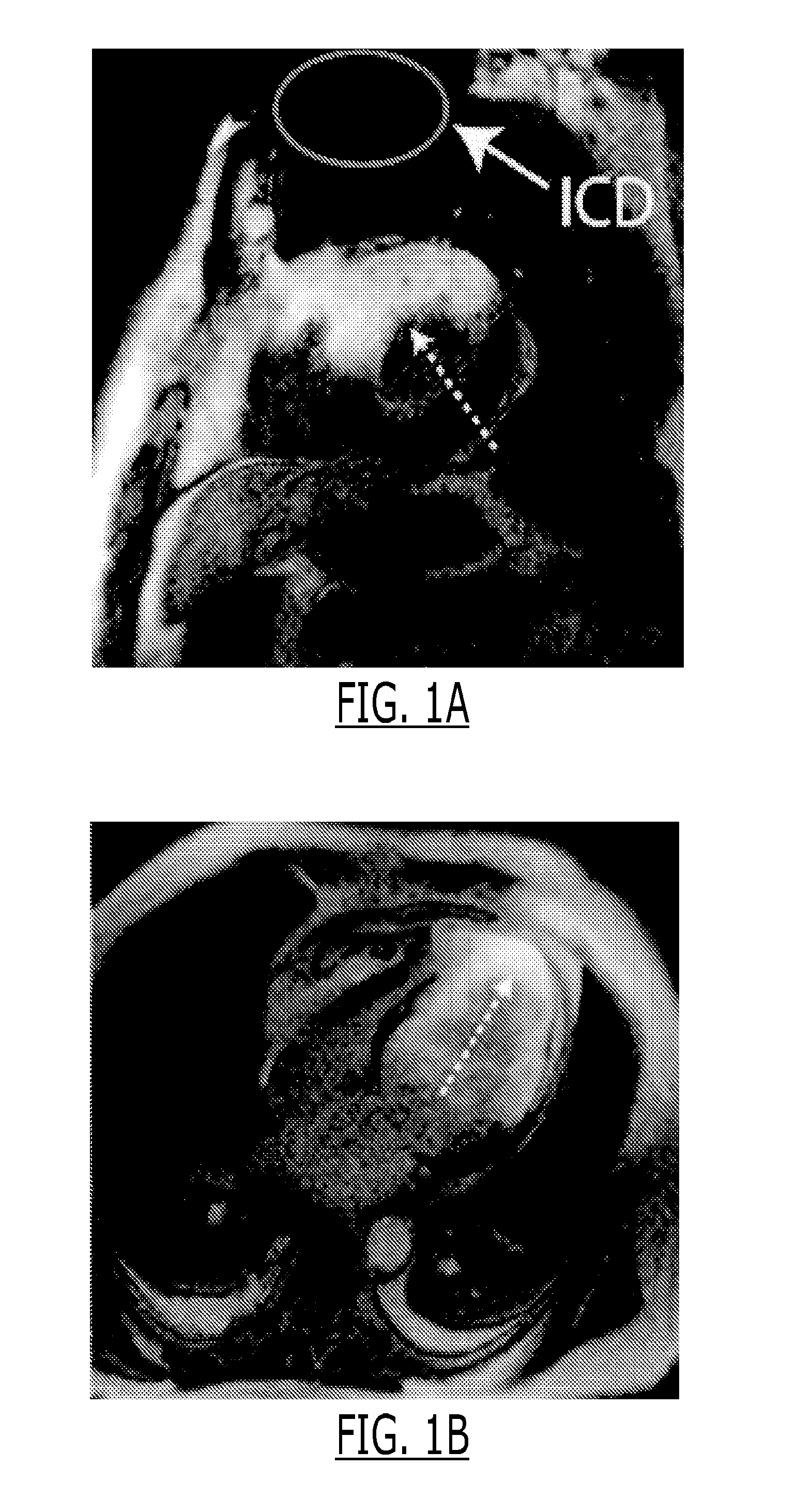
[0139]Images from 6 VT patients with ICDs who underwent cardiac MRI were examined. FIG. 1 shows representative LGE images acquired in two of these patients, where the anterior and apical regions of the left ventricle show bright signal intensity that makes it impossible to assess any myocardial scar in these regions. Although efforts were taken to reduce these artifacts at the time of scanning, the LGE images of the 6 patients were either totally non-diagnostic or the diagnostic value was severely limited by the presence of the bright artifacts. The observations of the artifacts were consistent with that of previous studies.

[0140]The conventional LGE sequence uses a non-selective adiabatic IR pulse typically with an inversion time (TI) of 200-300 ms to null the healthy myocardium. In the presence of a metallic object, the regions well beyond the physical boundary of the metallic object will be affected by strong off-resonance, often reaching up to 15-20 kHz...

example 2

Development of Strategies for Multi-spectral Imaging

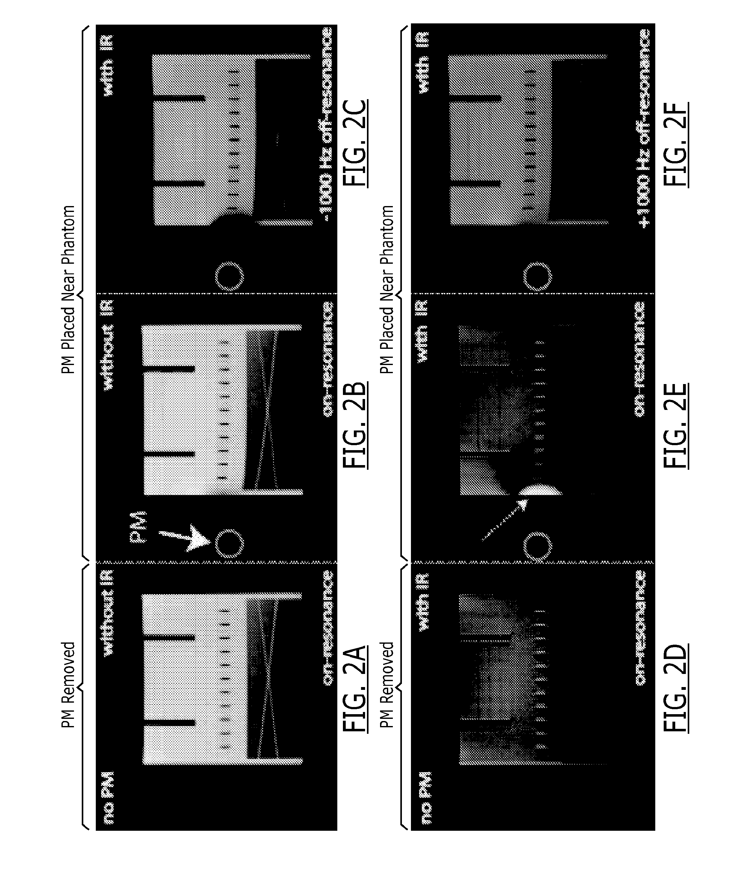
[0141]A phantom study was performed on a 1.5 T MRI system (Avanto, Siemens). A pacemaker (PM) that was previously implanted in a patient was placed approximately 7 cm to the right of a phantom (shown in red circle in FIG. 2). LGE images were acquired using the clinical 2D IR spoiled gradient echo LGE sequence with TI=200 ms.

[0142]The same LGE sequence was repeated with the exception that the modulation frequency of the inversion and excitation RF pulses was manually shifted up to ±1 kHz. On-resonance LGE MRI without the IR pulse was also performed. The same experiments were repeated after removing the pacemaker.

[0143]Results are shown in FIG. 2. In the on-resonance image with pacemaker placed, the part of the phantom affected by the off-resonance is bright (dashed arrow) and the “myocardium” is suppressed, a similar observation as in the in vivo example shown in FIG. 1. The images with −1000 Hz RF frequency shift shows the affected...

example 3

Case Study

[0144]Methods:

[0145]Recent literature shows that cardiac MRI, including LGE MRI, can be safely performed for patients with cardiac devices. However, the current LGE MRI sequence suffers from bright signal artifacts as shown in FIG. 5A. These image artifacts prevents assessment of presence or absence of myocardial scar in the affected regions, which are typically apical and anterior left ventricular wall for a device implanted on the left side of the heart. Although these image artifacts were previously reported, there has been no solution proposed for this problem. It was hypothesized that the bright signal artifact is caused by the affected myocardium not being properly inverted by the non-selective inversion pulse that is typically played in a traditional LGE MRI pulse sequence due to the limited spectral bandwidth of the inversion radiofrequency pulse.

[0146]To preliminarily test the hypothesis, a patient with an implantable cardiac defibrillator was imaged twice. Firstl...


PUM


Abstract

Disclosed herein are methods and systems for clinical practice of medical imaging on patients with metal-containing devices, such as implanted cardiac devices. In particular, Disclosed herein are methods and systems for improved late gadolinium enhancement (LGE) MRI for assessing myocardial viability for patients with implanted cardiac devices, i.e., cardiac pacemakers and implantable cardiac defibrillators.

Description

CROSS REFERENCE TO RELATED APPLICATION[0001]The application is a US national stage entry of International application No. PCT / US2013 / 068161 filed on Nov. 1, 2013, which in turn claims priority to U.S. provisional application No. 61 / 722,081 filed on Nov. 2, 2012, each of which is hereby incorporated by reference herein in its entirety.GOVERNMENT-SPONSORED RESEARCH AND DEVELOPMENT[0002]This invention was made with Government support under Grant No. R21HL118533 awarded by the National Institutes of Health. The Government has certain rights in this inventionFIELD OF THE INVENTION[0003]This invention relates to methods and systems for clinical practice of medical imaging on patients with metal-containing devices, such as implanted cardiac devices. More specifically, this invention relates to methods and systems for clinical practice of medical imaging on cardiac patients with implanted cardiac devices. Clinical practice includes, for example, device artifact reduction and electroanatomic...

Claims


Application Information

Patent Timeline
Patent Type & Authority Applications(United States)
IPC IPC(8): G01R33/56A61K49/10A61N1/05A61B5/02A61B5/044
CPCG01R33/5602A61B5/02028A61B5/044A61K49/105G01R33/5601A61K49/103A61N1/05A61B5/055G01R33/56325G01R33/5635A61B5/339
Inventor HU, PENG
Owner RGT UNIV OF CALIFORNIA
Why Eureka
Try Eureka