
Antigen-binding molecule for eliminating aggregated antigens

an antigen-binding molecule and aggregate technology, applied in the field of antigen-binding molecules for eliminating aggregated antigens from plasma, can solve the problems of inability to completely neutralize antigens with an amount of antibodies smaller than the amount of antigens, limitations in the reduction of the required antibody dose, and the development of al amyloidosis, etc., to achieve the elimination of proteins causing the disease, enhanced cytotoxicity, and increased antigen concentration

Inactive Publication Date: 2015-12-10
CHUGAI PHARMA CO LTD
View PDF6 Cites 37 Cited by
  • Summary
  • Abstract
  • Description
  • Claims
  • Application Information

AI Technical Summary

Benefits of technology

The invention relates to a system for controlling electronic devices using handheld controllers. The system allows users to control devices by tilting the controller, which triggers specific actions on the device. The technology recognizes different tilting angles and uses this information to perform various functions. The system can switch between devices and use different control methods for each device. The main technical effect of this invention is the ability to easily and intuitively control electronic devices using a handheld controller.

Problems solved by technology

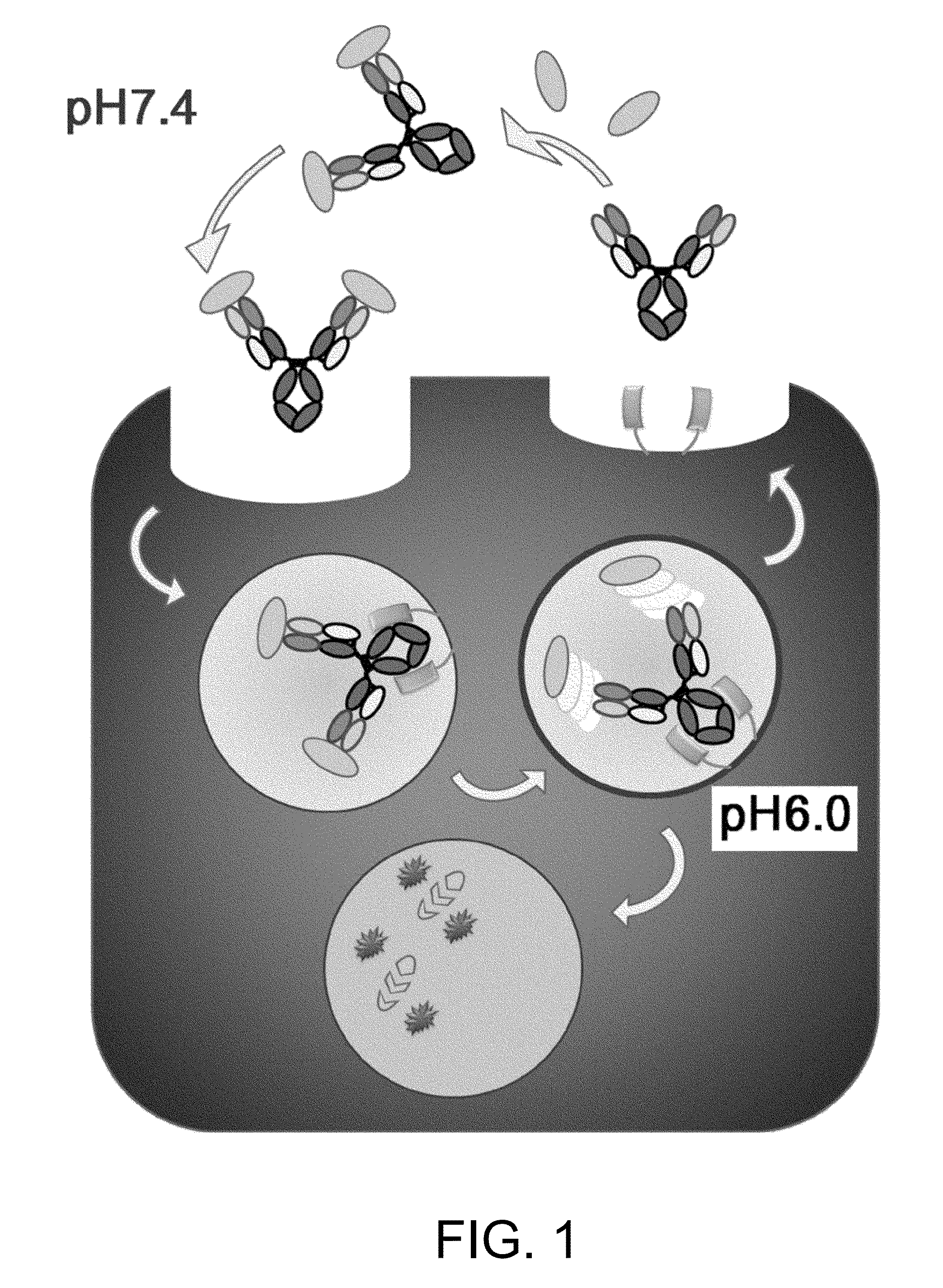
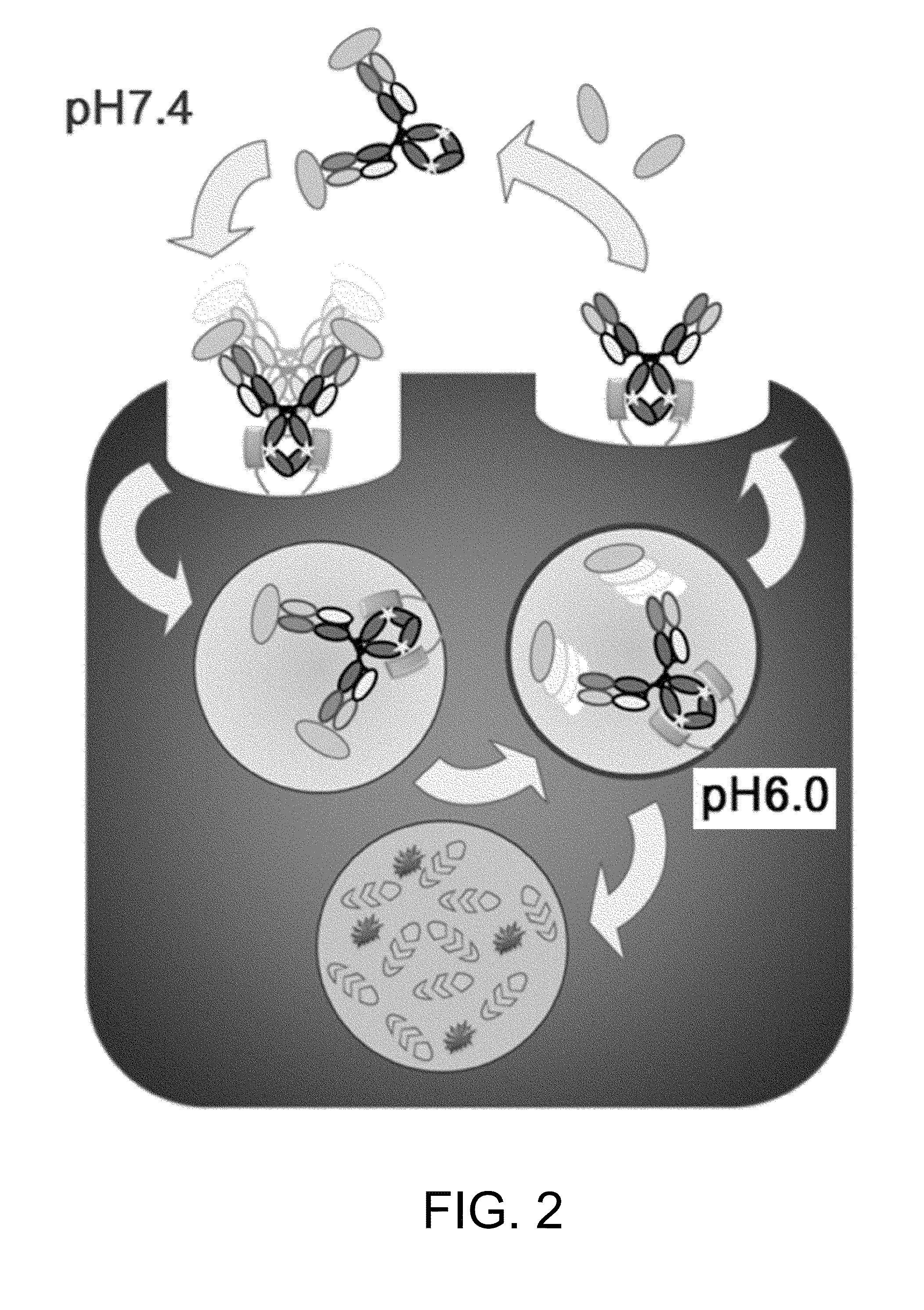
When proteins form aggregates due to various factors such as gene mutations and environmental changes, they are known to become causes of various diseases by reducing physiological functions of proteins or by posing toxic effects on cells.
Furthermore, when immunoglobulin L chain aggregates and deposits in each organ to cause organ failure, AL amyloidosis develops.
However, the stoichiometric neutralization of one antibody against one antigen (one divalent antibody against two antigens) is the limit of pre-existing methods, and thus it was impossible to completely neutralize antigen with an amount of antibody smaller than the amount of antigen.
Therefore, with just the above-described improvement of antibody pharmacokinetics or affinity maturation technology, there were limitations when it comes to reduction of the required antibody dose.
In addition, the plasma retention of an antigen is very short when compared to that of antibodies that are recycled by binding to FcRn.
Thus, when a typical antibody is administered, the antibody binds to an antigen, the plasma retention of the antigen is prolonged (becomes difficult to be eliminated from plasma) by binding to the antibody, and thus the plasma antigen concentration is increased.

Method used


Image

  • Antigen-binding molecule for eliminating aggregated antigens
  • Antigen-binding molecule for eliminating aggregated antigens
  • Antigen-binding molecule for eliminating aggregated antigens

Examples


example 1

Preparation of Antibodies that Show Calcium-Dependent Binding to Human IgA

(1-1) Preparation of MRA-hIgA, GC-hIgA-FLAG, and GC-hIgA-MYC

[0448]As human IgA, MRA-hIgA (heavy chain SEQ ID NO: 33; light chain SEQ ID NO: 36),

[0449]GC-hIgA-FLAG (heavy chain SEQ ID NO: 34; light chain SEQ ID NO: 37), and GC-hIgA-MYC (heavy chain SEQ ID NO: 35; light chain SEQ ID NO: 37) were prepared as follows.

Preparation of MRA-hIgA

[0450]MRA-hIgA which is a recombinant of human IgA (hereinafter, MRA-hIgA) was prepared as follows. A gene fragment encoding MRA-hIgA (heavy chain SEQ ID NO: 33; light chain SEQ ID NO: 36) was inserted into an animal cell expression vector. The constructed plasmid vector was transfected into FreeStyle 293 (Invitrogen) using 293Fectin (Invitrogen) along with an EBNA1-expressing gene. Then, the transfected cells were cultured at 37° C. under 8% CO2 to secrete the MRA-IgA protein into the culture supernatant. The protein was purified using ion exchange chromatography and gel filtra...

example 2

(2-1) Preparation of Aggregated hIgA

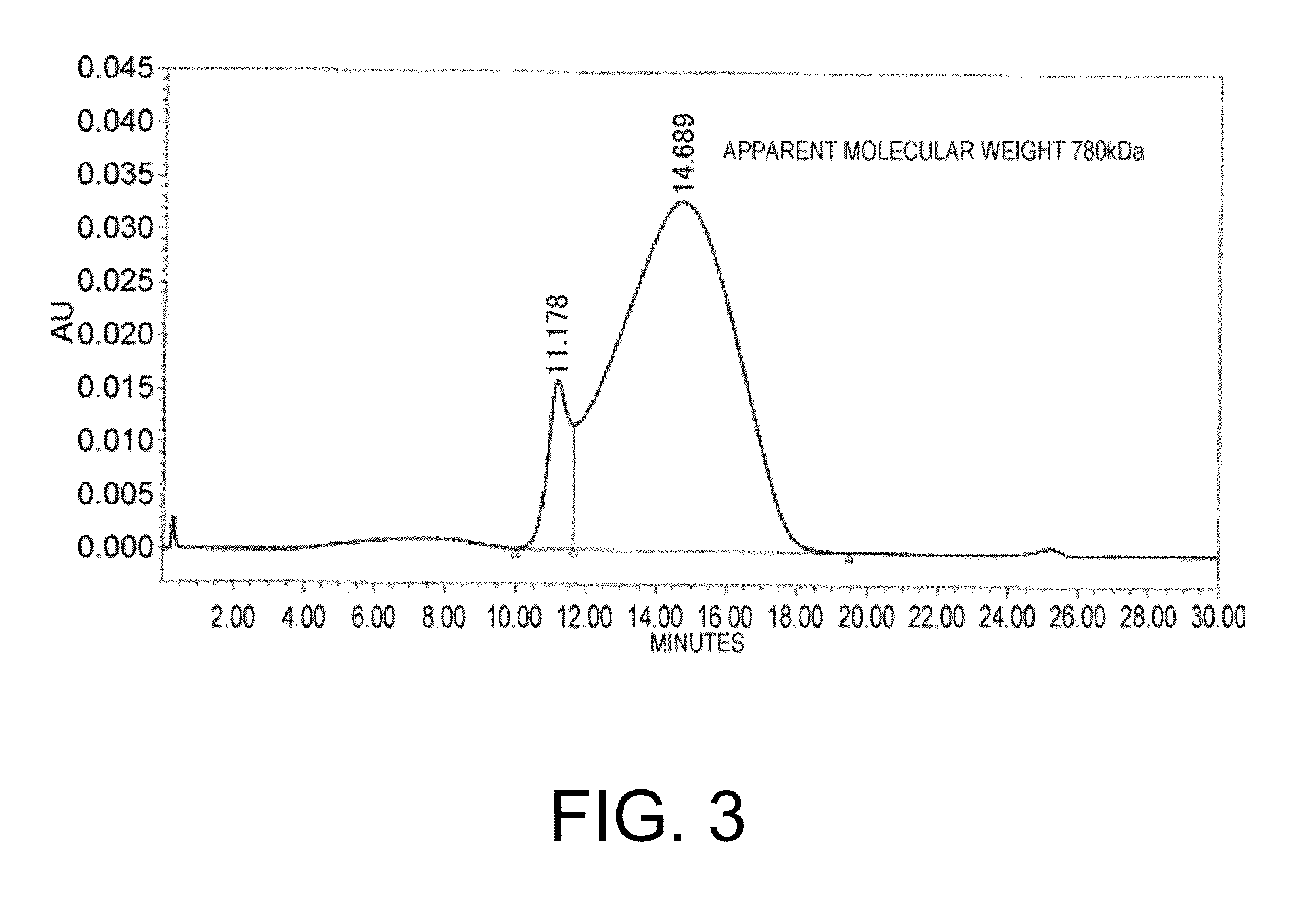
[0463]Aggregated hIgA was prepared using the crosslinking agent SPDP (N-Succinimidyl 3-(2-pyridyldithio)propionate, Thermo Scientific). GC-hIgA-MYC prepared in Example 1 was modified using SPDP, and hIgAs were cross-linked with each other by mixing a fraction that has not been treated subsequently with a fraction that has been treated under reducing conditions. After the crosslinking reaction, the macromolecular component was fractioned by gel filtration chromatography to obtain aggregated hIgA. The result of gel filtration chromatographic analysis on aggregated hIgA is shown in FIG. 3. The apparent molecular weight of the aggregated hIgA, calculated from the elution position of the molecular-weight marker, was 780 kDa. Since the elution peak is broad, aggregates of various sizes are conceivably being formed. As a comparison, GC-hIgA-FLAG that has not been treated with SPDP was used as the unaggregated hIgA.

(2-2) Assessment of the Obtained Antibod...

example 3

Assessment of Human IgA-Binding Antibodies for their Effect on Plasma Retention of Aggregated hIgA and Unaggregated hIgA Using Normal Mice

(3-1) In Vivo Test Using Normal Mice

[0467]Normal mice (C57BL / 6J mouse; Charles River Japan) were administered with unaggregated hIgA (prepared in Example 1) or aggregated hIgA (prepared in Example 2) alone or administered with an anti-hIgA antibody one day prior to administration of unaggregated hIgA or aggregated hIgA; and then they were assessed for the in vivo dynamics of hIgA and the anti-hIgA antibody. An anti-hIgA antibody, an unaggregated hIgA solution (300 μh / mL) and an aggregated hIgA solution (300 μg / mL) were administered at 10 mL / kg into the caudal vein. The anti-hIgA antibody used was GA2-IgG1 described above.

[0468]The concentration of human IgA was 300 μg / mL in all of the mixed solutions. Meanwhile, the anti-hIgA antibody concentration was 30 μg / mL (0.3 mg / kg), 100 μg / mL (1 mg / kg), or 300 μg / mL (3 mg / kg). When the anti-hIgA antibody c...


PUM

PropertyMeasurementUnit
pHaaaaaaaaaa
pHaaaaaaaaaa
concentrationaaaaaaaaaa

Abstract

The present inventors discovered that incorporating an Fc region and an antigen-binding domain whose antigen-binding activity varies depending on ion concentration into an antigen-binding molecule that binds to an aggregate-forming antigen produces an antigen-binding molecule that can preferentially clear protein aggregates in comparison to protein monomers from plasma. Use of antigen-binding molecules of the present invention allows various diseases stemming from target tissues to be treated target-tissue-specifically. Use of antigen-binding molecules of the present invention enables treatment of diseases caused by protein aggregates.

Description

TECHNICAL FIELD[0001]The present invention provides uses of antigen-binding molecules for eliminating aggregated antigens from plasma; methods for eliminating aggregated antigens from plasma, which comprise administering antigen-binding molecules; pharmaceutical compositions comprising antigen-binding molecules that are capable of eliminating aggregated antigens from plasma; methods of screening for antigen-binding molecules for eliminating aggregated antigens from plasma; and methods for producing antigen-binding molecules for eliminating aggregated antigens from plasma.BACKGROUND ART[0002]When proteins form aggregates due to various factors such as gene mutations and environmental changes, they are known to become causes of various diseases by reducing physiological functions of proteins or by posing toxic effects on cells. For example, when amyloid-β aggregates and accumulates in the brain, nerve cells degenerate and Alzheimer's disease develops. Furthermore, when immunoglobulin ...

Claims


Application Information

Patent Timeline
Patent Type & Authority Applications(United States)
IPC IPC(8): C07K16/18G01N33/68
CPCC07K16/18G01N33/6854C07K2317/52G01N2333/70535C07K2317/14G01N2333/47C07K2317/90C07K16/00C07K16/2866C07K2317/24C07K2317/94C07K16/4283A61K2039/505A61P43/00
Inventor IGAWA, TOMOYUKIHIRONIWA, NAOKAITO, ERIKO
Owner CHUGAI PHARMA CO LTD
Learn More