Hemodynamic monitoring device and methods of using same

a monitoring device and a technology of a hemodialysis device, applied in the direction of ultrasonic/sonic/infrasonic image/data processing, applications, catheters, etc., can solve the problems of volume overload, inadequacies in tissue blood flow, dire consequences

Active Publication Date: 2016-02-04
NILUS MEDICAL
View PDF3 Cites 28 Cited by
  • Summary
  • Abstract
  • Description
  • Claims
  • Application Information

AI Technical Summary

Problems solved by technology

For example, circulatory shock results primarily in inadequate tissue blood flow.
Volume overload (“hypervolemia”) can have dire consequences such as decreased gas exchange and increased myocardial dysfunction.
However, with further volume depletion cardiac output and then blood pressure falls.
As such, static measurements or indices of fluid responsiveness, such as the traditionally used tests of central venous pressure (CVP) and pulmonary artery occlusion pressure (PAOP), often fail as meaningful tools for measuring the patient's hemodynamic state as they do not take into consideration the changes in other systemic interactions that can alter quickly.
Indeed, studies in recent years have confirmed that such static measurements have little correlation with fluid responsiveness and are poor clinical indicators.
In many patients, a rapid fluid bolus is a reasonable diagnostic and potentially therapeutic option but, in others (e.g., acute respiratory distress syndrome), it has the potential to cause harm and may delay institution of appropriate therapy.
The poor predictive value of static measurements and clinical examination has, therefore, led to the investigation of dynamic measurements of fluid responsiveness.
Direct measurement of wall stress in vivo is difficult if not impossible within an emergency medicine department, surgical theater, and / or intensive care environment.
Although end diastolic volumes or pressures have been used as proxies of wall stress, both have significant limitations.
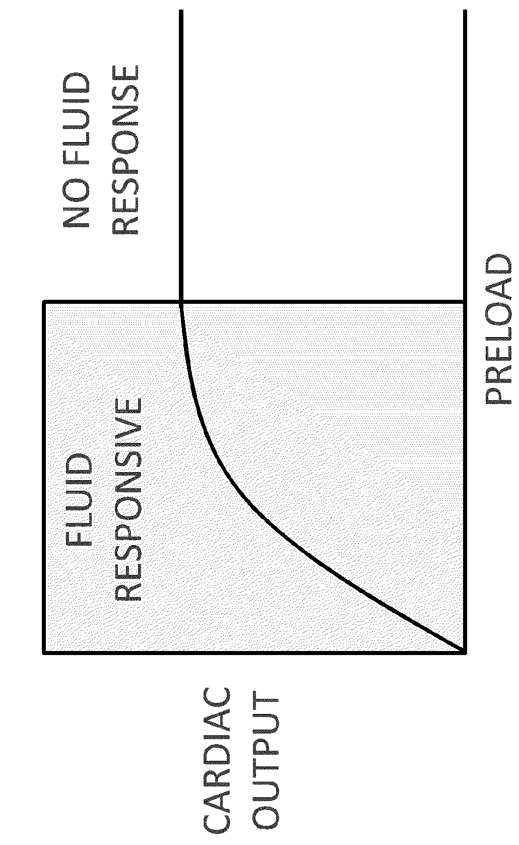
Perhaps most importantly, an accurate measure of preload at a point in time does not necessarily reflect fluid-responsiveness.
In healthy patients, an increase in preload (with volume challenge) results in a significant increase in stroke volume.
Furthermore, as a result of altered left ventricular compliance and function, the position of an acutely ill patient on their Frank-Starling curve cannot be predicted from their preload (LVEDV) alone.
Therefore, even a precise measurement of left ventricular preload does not determine if that left ventricle is fluid-responsive (i.e., if it will increase cardiac output in response to increased volume).
However, because of the changes in venous tone, intrathoracic pressures (positive end expiratory pressure, etc.), left and right ventricular compliance, and geometry that occurs in critically ill patients, it has been found that there is actually a poor relationship between CVP and right ventricular end-diastolic volume.
Furthermore, the right ventricular end-diastolic volume may not accurately reflect the patient's position on the Frank-Starling curve and, therefore, their preload reserve.
This device is highly invasive and requires a catheter to be introduced through a large vein such as the jugular, subclavian, or femoral vein.
The procedure is not without risk, and complications can be life threatening.
It can lead to arrhythmias, rupture of the pulmonary artery, thrombosis, infection, pneumothorax, bleeding, and other life-threatening complications.
Indeed, it was not long after the introduction of the pulmonary artery catheter that studies began to appear demonstrating that PAOP was a poor reflection of preload and more recent studies have demonstrated that pulmonary artery occlusion pressure (PAOP) is a poor predictor of preload and volume responsiveness.
While a number of studies have found the LVEDA to be a good predictor of fluid responsiveness, other studies have failed to replicate such findings.
A major limitation of echocardiography is that it provides a snapshot of ventricular function at a single period in time.
Mechanical insufflation decreases preload and increases afterload of the RV.
Cannulation for invasive vascular pressure monitoring is, however, associated with complications such as extravasation, thrombosis, and infection among other life threatening conditions.
Patients with invasive arterial monitoring require very close supervision, as there is a danger of severe bleeding if the arterial line becomes disconnected.
Peripheral vascular pressure monitoring devices are also known to be problematic in monitoring rapid changes in patients who are hemodynamically unstable.
As such, these devices may and do lead to erroneous cardiac output measurements during the administration of vasoactive drugs, during loss of circulating volume, e.g., hemorrhage, insufflation of the abdomen for laparoscopic surgery, pathophysiological diseases resulting in abnormal arterial pressure waves, and positional changes during surgery.
Conversely, drugs which have a vasodilation effect result in a decrease in resistance to blood flow and typically blood pressure falls which is interpreted as a reduction in flow, whereas blood flow typically increases as systemic resistance decreases as the heart is acting to pump against a reduced resistance.
Calibration is essential for absolute value accuracy and, in operating room conditions such calibration is complex to perform, is time consuming, needs to be repeated frequently, introduces chemical agents which may be toxic, and may be of limited accuracy in the presence of other drugs administered during patient treatment.
A disadvantage in monitoring SVC diameter variation as a dynamic indices of fluid responsiveness is that it requires transesophageal echocardiography which has all of the same disadvantages and limitations of other types of extravascular ultrasound—sensitivity to movement, user training and maintenance, and oftentimes difficult to visualize anatomical structures.
Such extravascular flow based measurements are, however, difficult to perform and require the presence of an operator that is capable of maintaining properly placement, alignment, and calibration of the ultrasound probe.
The ultrasound beam is directional and is sensitive to movement, which requires the operator to check the beam's focus and thus the device cannot be considered to be providing continuous monitoring without operator attendance.
In some circumstances, serious and prolonged elevation of PAP progresses to severe acute pulmonary hypertension, leading to life threatening complications including, but not limited to, refractory systemic arterial hypotension, severe hypoxemia, right ventricular (RV) dysfunction and failure, and ultimately resulting in cardiogenic and / or obstructive shock and death.
However, those data are not either specific enough or continuous to be useful in early diagnoses of acute pulmonary hypertension in intensive care units, nor for determining whether signs of acute pulmonary hypertension are hemodynamically significant.
Unfortunately, in most cases acute pulmonary hypertension remains under diagnosed or undiagnosed and treatment begins only after serious complications have developed.
Higher pressure in the pulmonary artery (behind the right heart) may overload the right ventricle, causing the right ventricle to dilate or even fail.
Even subtle dilation of the right ventricle may cause tricuspid valve insufficiency, that is, dysfunction of the valve between the right ventricle and the right atrium.
The insufficiency of the tricuspid valve may cause backward leakage during right ventricle systole (that is, contraction).

Method used


Image

  • Hemodynamic monitoring device and methods of using same
  • Hemodynamic monitoring device and methods of using same
  • Hemodynamic monitoring device and methods of using same

Examples


Embodiment Construction

[0053]Before explaining at least one embodiment of the presently disclosed and / or claimed inventive concept(s) in detail, it is to be understood that the presently disclosed and / or claimed inventive concept(s) is not limited in its application to the details of construction and the arrangement of the components or steps or methodologies set forth in the following description or illustrated in the drawings. The presently disclosed and / or claimed inventive concept(s) is capable of other embodiments or of being practiced or carried out in various ways. Also, it is to be understood that the phraseology and terminology employed herein is for the purpose of description and should not be regarded as limiting.

[0054]Unless otherwise defined herein, technical terms used in connection with the presently disclosed and / or claimed inventive concept(s) shall have the meanings that are commonly understood by those of ordinary skill in the art. Further, unless otherwise required by context, singular...


PUM


Abstract

Hemodynamic monitoring systems and methods are disclosed including a device comprising a first sensor configured to measure a velocity of blood flow in an adjacently-located portion of a superior vena cava of a mammalian patient using ultrasound waves; a second sensor configured to measure respiratory cycle data of the mammalian patient; and a computer configured to process the measured velocity of blood flow and the measured respiratory cycle data to provide hemodynamic parameters corresponding to the mammalian patient.

Description

INCORPORATION BY REFERENCE OF RELATED APPLICATIONS[0001]The present patent application claims priority to the provisional patent applications identified by U.S. Ser. No. 61 / 788,883 titled “HEMODYNAMIC MONITORING DEVICE AND METHODS OF USING SAME” filed on Mar. 15, 2013, U.S. Ser. No. 61 / 889,442 titled “HEMODYNAMIC MONITORING DEVICE AND METHODS OF USING SAME” filed on Oct. 10, 2013, and U.S. Ser. No. 61 / 952,679 titled “HEMODYNAMIC MONITORING DEVICE AND METHODS OF USING SAME” filed on Mar. 13, 2014, the entire contents of which are hereby incorporated herein by reference.BACKGROUND[0002]1. Field of the Inventive Concept(s)[0003]The presently disclosed and / or claimed inventive concept(s) relates generally to a hemodynamic monitoring device and methods of making and using same. The hemodynamic monitoring device and its use are particularly well-suited for the measurement of respiratory cycle data simultaneously with at least one of central venous blood pressure and intravascular blood fl...

Claims


Application Information

Patent Timeline
Patent Type & Authority Applications(United States)
IPC IPC(8): A61B8/06A61B5/00A61B5/0402A61B5/08A61B8/00A61B8/08A61B8/12
CPCA61B8/06A61B2562/0247A61B8/5207A61B5/7278A61B8/488A61B5/0803A61B5/0402A61B5/0809A61B8/5223A61B8/12A61B8/54A61B8/463A61B5/7425A61B5/7405A61B5/0816A61B5/02028A61B5/0261A61B5/08A61B8/0891A61B8/5284A61B5/349G16H50/30A61B5/318
Inventor NAVRATIL, MIROSLAVZADROBILEK, KARELBELOHLAVEK, JANKOVARNIK, TOMASMLCEK, MIKULASSANTAVY, MAREKVASEK, VLADIMIR
Owner NILUS MEDICAL
Why Eureka
Try Eureka