Magnetic resonance maps for analyzing tissue

a magnetic resonance map and tissue technology, applied in the field of magnetic resonance map, can solve the problems of limiting the validity of a progression free survival end point, low specificity of standard mr map sequence, premature discontinuation of effective adjuvant therapy,

Active Publication Date: 2016-04-21
RAMOT AT TEL AVIV UNIV LTD +1
View PDF12 Cites 23 Cited by
  • Summary
  • Abstract
  • Description
  • Claims
  • Application Information

AI Technical Summary

Benefits of technology

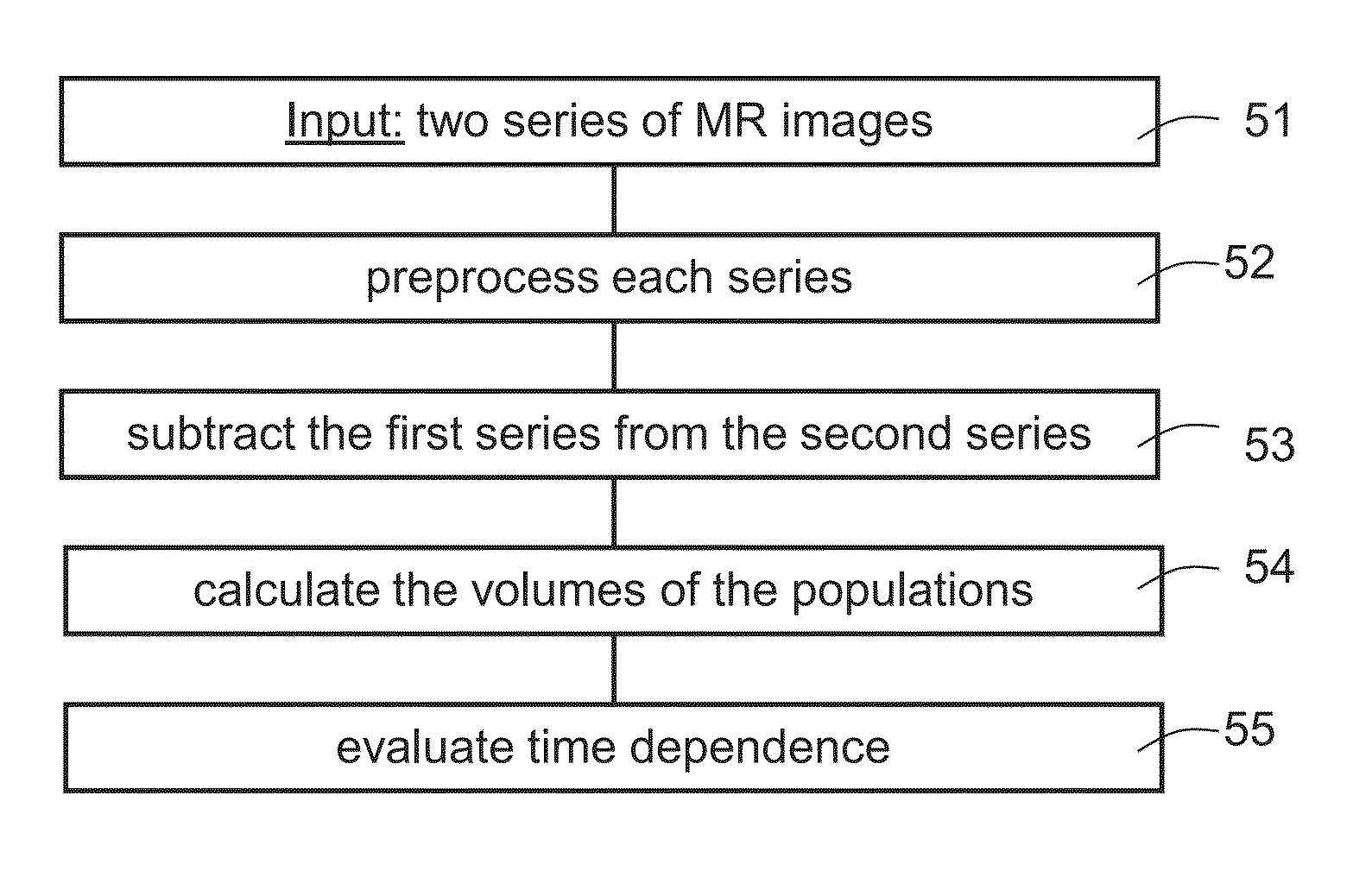
[0062]According to an aspect of some embodiments of the present invention there is provided a method for managing tumor treatment in patients, comprising: receiving a first and a second 3D MRI scan at the beginning and end of a predetermined time interval post contrast administration; forming a subtraction map from the first and the second 3D MRI scans by analyzing the scans to distinguish between two primary populations, a slow population, in which contrast clearance from the tissue is slower than contrast accumulation, and a fast population in which clearance is faster than accumulation; and providing an indication of distribution of the two primary populations, wherein the predetermined time period is at least twenty minutes; using the subtraction map and the indication to distinguish between tumor and non-tumor tissue and thereby to provide information for determination of further treatment.

Problems solved by technology

Although MRI provides high resolution and good sensitivity, standard MR sequences still suffer of low specificity especially in the case of tumors, e.g., brain tumors (during / after treatment).
This treatment related effect has implications for patient management and may result in premature discontinuation of effective adjuvant therapy.
This limits the validity of a progression free survival end point unless tissue-based confirmation of tumor progression is obtained.
Thus, treatment decisions, as to whether to operate on a patient with radiographic deterioration, continue chemotherapy or change to another non-surgical treatment is a day to day struggle involving interdisciplinary teams of neurosurgeons, neuro-oncologists and neuro-radiologists who are often unable to reach a unanimous interpretation of the patient status.
For glioblastoma patients treated by radiation and TMZ, conventional MRI is unable to provide a reliable distinction between tumor progression and treatment effects (also referred to as pseudoprogression).
For glioblastoma patients conventional MRI is unable to reliable depict the tumor or pseudoprogression after treatment with Bevacizumab.
Unfortunately, there was significant overlap between the two groups.
The prognosis of patients diagnosed with brain metastases is generally poor.
Among the reported complications of SRS is radiation-induced necrosis which, similarly to pseudoprogression, can be difficult to differentiate both clinically and radiologically from recurrent tumor at the treatment site.
Tumor progression, inflammation, infection and treatment-induced changes (such as radiotherapy-induced changes) can induce similar changes in enhancement which are impossible to differentiate.
Treatment decision, as whether to operate on a patient with radiographic deterioration, continue current treatment / follow-up or change to another non-surgical treatment is a daily struggle involving interdisciplinary teams of neurosurgeons, neuro-oncologists and neuro-radiologists which are often unable to reach unanimous interpretation of the patient's status.
The fact that increases in enhancing lesion volume in 20-30% of patients is due to treatment effects rather than tumor progression, prevents appropriate treatment in 50% of patients with brain tumors.
In both cases, the wrong choice of treatment significantly hampers the patient's quality of life and survival.
Conventional MRI is currently unable to provide reliable distinction between tumor progression and treatment effects such as pseudoprogression and radiation necrosis.
Still, most of these studies show some degree of overlap between the two disease entities.
In addition, using fast acquisition techniques has the disadvantages of low spatial resolution and high sensitivity to susceptibility artifacts.
Due to these limitations, in many cases the data is inconclusive and significant results are mostly obtained by averaging over the entire enhancing lesion while important information regarding the location and shape of small active tumor regions may be limited.

Method used


Image

  • Magnetic resonance maps for analyzing tissue
  • Magnetic resonance maps for analyzing tissue
  • Magnetic resonance maps for analyzing tissue

Examples


example 1

[0192]Referring now to FIG. 2A, an example of newly diagnosed GBM involves 49 year old male. The patient, with newly diagnosed GBM, underwent gross total resection at Sheba Medical Center followed by chemoradiation. The patient was recruited to the present study 1 week after chemoradiation with clinical deterioration. Conventional MRI showed a large partially enhancing lesion with low rCBV (FIG. 2A top). The maps (FIG. 2A below) showed that only part of the enhancing volume (on T1-MRI) consisted of morphologically active tumor (blue):

[0193]The patient was stabilized on steroids. The neuro-oncologist was determined to continue treatment with TMZ. The neurosurgeon was concerned surgery may be necessary. Referring now to FIG. 2B, the patient was rescanned 2.5 weeks later. Conventional MRI (including T1, T2, FLAIR, DWI and DSC PWI) showed no significant change. The maps (FIG. 2B below) showed significant increase in the active tumor volume with the addition of light blue regions surroun...

example 2

Newly Diagnosed GBM

[0198]A 50 year old male with newly diagnosed GBM underwent gross total resection at Sheba Medical Center followed by chemoradiation. 1 month after chemoradiation, increased enhancing volume was noted on contrast-enhanced T1-MRI with no clinical deterioration. PWI was inconclusive. The patient consulted with physicians at Sheba and Hadassah Medical Centers who could not advise whether he should be operated or continue TMZ treatment since they were unable to determine if the radiological deterioration was due to tumor progression or treatment effects. Referring now to FIG. 5, the patient was recruited to our study and his maps (FIG. 5A bottom) showed significant volume of active tumor (blue) surrounding the previous surgery site:

[0199]Therefore, it was decided to re-operate on the patient.

[0200]In this case, as per FIG. 5B, histology did not show a high density tumor and very little mitosis. But there were many clusters of small cells which raised suspicions of pro...

example 3

Recurrent GBM

[0205]Referring now to FIG. 8, a 58 year old male with recurrent GBM was recruited 7 month after diagnosis prior to initiation of Bevacizumab treatment. At diagnosis the patients was operated on and received chemoradiation followed by TMZ. Six months later, at progression, the patient underwent a second resection and was recruited to the present Bevacizumab study 1 month after the second surgery, prior to initiation of the Bevacizumab treatment. At this point his enhancing lesion consisted of nearly 50% tumor according to the maps. Three months after initiation of Bevacizumab treatment conventional MRI showed improvement but the maps showed significantly larger blue / tumor volume, consistent with clinical deterioration. Following this scan the patient was treated with re-irradiation (in parallel with continuing Bevacizumab) and was stabilized. The next follow-up showed no significant change on conventional MRI but significant treatment response in the maps (a large perce...


PUM

PropertyMeasurementUnit
timeaaaaaaaaaa
time t1aaaaaaaaaa
sizeaaaaaaaaaa

Abstract

Apparatus for operating MRI is disclosed. The apparatus comprises: a control for operating an MRI scanner to carry out an MRI scan; an input for receiving first and second MRI scans respectively at the beginning and end of a predetermined time interval post contrast administration; a subtraction map former for forming a subtraction map from the first and the second MRI scans by analyzing the scans to distinguish between a population in which contrast clearance from the tissue is slower than contrast accumulation, and a population in which clearance is faster than accumulation; and an output to provide an indication of distribution of the populations. The control is configured to carry out the first scan at least five minutes and no more than twenty minutes post contrast administration and to carry out the second scan such that the predetermined time period is at least twenty minutes.

Description

RELATED APPLICATION[0001]This application claims the benefit of priority of U.S. Provisional Patent Application No. 61 / 815,343 filed on Apr. 24, 2013, the contents of which are incorporated herein by reference in their entirety.FIELD AND BACKGROUND OF THE INVENTION[0002]The present invention relates to method and apparatus for producing magnetic resonance (MR) maps and, more particularly, but not exclusively to using such maps to identify and study tumors.[0003]MRI is considered the gold standard imaging modality for the brain. Although MRI provides high resolution and good sensitivity, standard MR sequences still suffer of low specificity especially in the case of tumors, e.g., brain tumors (during / after treatment).[0004]Gliomas are the most common malignant primary brain tumors in adults, with an annual incidence of 4-5 per 100,000 people (Central Brain Tumor Registry; Wen and Kesari, 2008).[0005]The standard of care for glioblastoma includes surgery, chemotherapy during and after...

Claims


Application Information

Patent Timeline
Patent Type & Authority Applications(United States)
IPC IPC(8): G01R33/28G01R33/56G01R33/48A61B5/055A61B5/00
CPCG01R33/281A61B5/055A61B5/0042A61B2576/026A61B5/4848G01R33/4806G01R33/5601A61B5/4842G01R33/5608A61B5/7275G06T2207/30096G06T7/0016G06T2207/10096G06T2207/30016G16H30/40G16H40/40G16H40/63A61K31/166A61K31/7068A61K2300/00A61K31/555
Inventor MARDOR, YAELZACH, LEORGUEZ, DAVIDLAST, DAVIDDANIELS, DIANNE
Owner RAMOT AT TEL AVIV UNIV LTD
Why Eureka
Try Eureka